Core Facilities

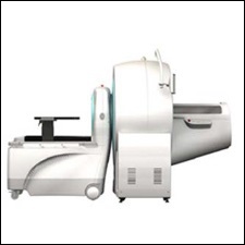
Siemens Inveon PET-CT Scanner
Description: Small animal PET-CT scanner
Manufacturer: Siemens Healthineers
Click here for manufacturer's website
Details
Please select a section for more details:
Capabilities
-
PET and CT subsystems, allows combined functional and anatomic imaging
-
implicit registration between PET and CT
Specifications
-
X-ray source voltage 20-130 kVp
-
Maximum field of view 10.8x10.8 cm2
-
Typical image resolution 50μm (best 15μm)
-
CCD detector: 4064x4064 pixels
Contact
Rao Gullapalli, PhD
Phone: 410-706-2694
rgullapalli@som.umaryland.edu
Su Xu, PhD
Phone: 410-706-5419
sxu@umm.edu
Mark Smith, PhD
Phone: 410-328-1320
mark.smith@som.umaryland.edu
Template for NIH "Facilities and Other Resources"
This template (to come) is for the small animal PET-CT scanner, which can be used in the "Facilities and Other Resources" page of NIH applications. Before using this template, please contact your collaborator to ensure the information is correct and up to date.
The University of Maryland houses the Core for Translational Research in Imaging @ Maryland (C-TRIM) which is operated by faculty associated with the Center for Advanced Imaging Research (CAIR) within the University of Maryland School of Medicine’s Department of Diagnostic Radiology & Nuclear Medicine. Within the core, both animal and human imaging facilities are available.
Core for Translational Research in Imaging @ Maryland (C-TRIM): The facilities of C-TRIM are located in two buildings. One is located in Howard Hall 6th floor where the 7 Tesla Bruker small Animal MRI, Siemens Inveon microPET/CT, SPECT, Xenogen, MR guided Focused Ultrasound (MRgFUS), fluorescence and bioluminescence imaging and wet lab for tissue processing are available. The other location is in the basement of the new Health Sciences Research Facility III (HSFIII) building where a 9.4 Tesla Bruker system is available. This location also hosts the human imaging arm of C-TRIM where a research dedicated 3.0 Tesla PRISMA FIT scanner and a state-of-the-art Siemens 3.0 Tesla mMR biograph system (combined PET/MR) are located. C-TRIM provides cross-sectional imaging services to various investigators on campus.
A small animal Inveon Micro-PET/CT from Siemens Molecular Imaging is available through a grant from NCRR and the Marlene and Stewart Greenebaum Cancer Center. The location is conveniently close to the animal imaging facilities. This equipment is used mainly to facilitate several metabolic, flow and anatomic studies pertaining to cancer and TBI projects. This system was chosen because of its versatility to operate as independent single units or as a combined unit. The PET aspect of this unit provides high spatial resolution, sensitivity and count rate performance and has transmission imaging capability. The CT aspect of this instrument will provide high resolution anatomical images with a large field of view and will allow for PET attenuation and scatter correction.
Two SurgiVet MR-conditional animal anesthesia systems and an SA Instruments model 1030 gating and monitoring system for use with a clinical MR scanner are available for delivery of isoflurane anesthesia and to monitor physiological parameters (ECG, respiration, temperature, heart rate, and blood oxygen saturation).
