Image File-Types & Image Handling
The Core Lab de-identifies, standardizes, and archives images in a proprietary searchable database compatible with DICOM format (preferred) JPG, PNG, TIF, etc.. Images are transferred via a free, HIPAA compliant portal compatible with most web browsers.
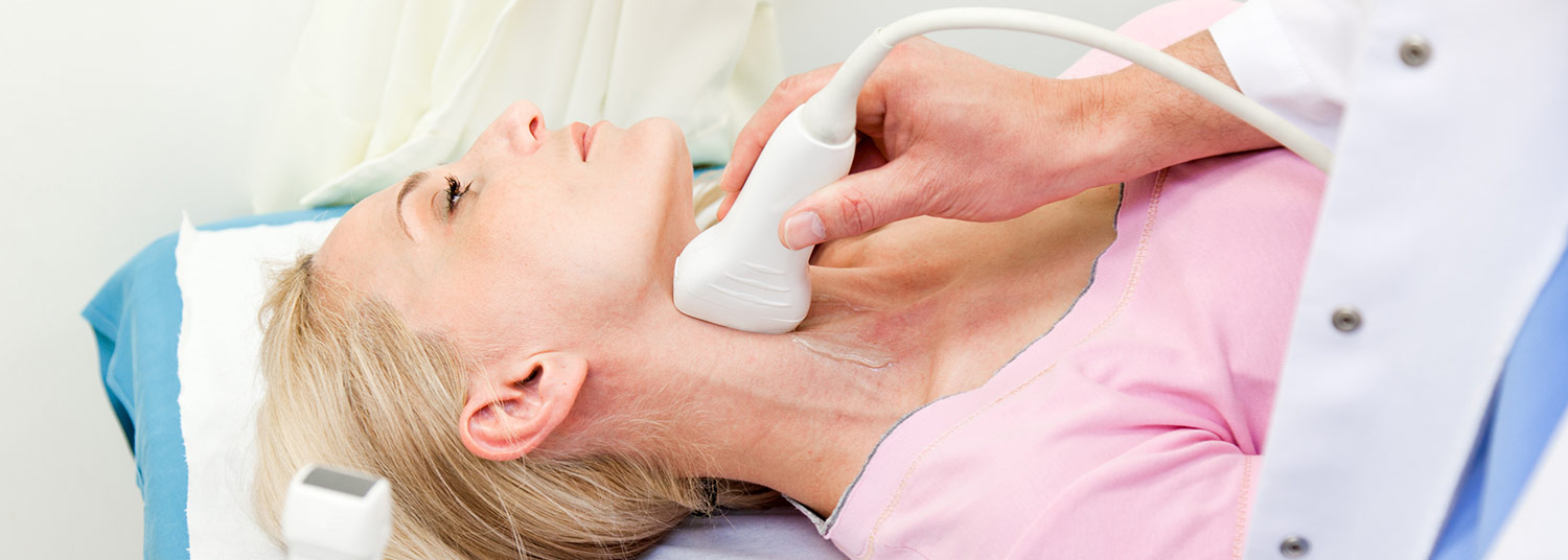
Duplex Ultrasound
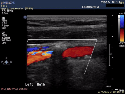
Arterial B-mode and Doppler ultrasonography
Standard B-mode and Doppler imaging used to determine degree of stenosis or other luminal or wall abnormalities in the carotid, upper/lower extremity, aorta, mesenteric, or renal arteries.
Venous B-mode and Doppler ultrasonography
Standard B-mode and Doppler imaging used to determine obstruction or reflux in the lower extremity veins, as well as detection of stenosis, thrombosis, and other abnormalities in the upper/lower extremity veins, inferior vena cava, et cetera.
Atherosclerotic plaque geometry and composition
Lesion analysis of 2D and 3D geometry and composition using proprietary software (Grayscale Median, Tissue Composition).
Plaque Biomechanics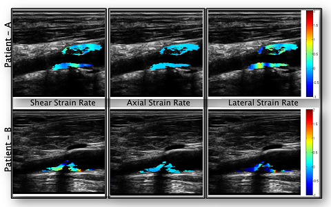
Technique that uses 2D sine-ultrasonography to measure plaque strain.
Dialysis access assessment
Evaluation for vein mapping, maturation, or complications within dialysis accesses.
3D ultrasonography
Technique that uses commercial/proprietary hardware and software to convert standard 2D grayscale ultrasound images into a volumetric dataset, which can be retrospectively converted into a 3D image.
Contrast-enhanced ultrasound
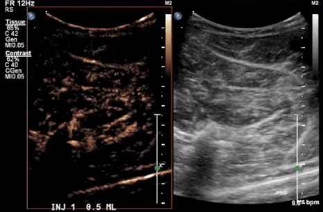
For Peripheral Artery Disease and aortic endografts. Uses a contrast medium to enhance the contrast of the imaging of the vessel when compared to the surrounding tissues.
Intima-media thickness
Measurement of the tunica intima and tunica media, the two innermost layers of the arterial wall via ultrasound.
Transcranial Doppler
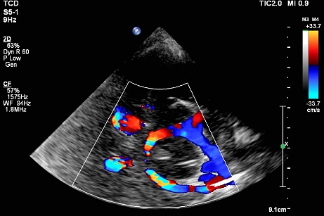
Transcranial ultrasound technique that can detect and measure impaired cerebrovascular function via breathholdling index, Co2 TCD, Pulsatility Index and Resistivity Index. Can also be used post-trauma to detect intracranial bleeding and vasospasm.
Standard TCD
Measures direction and velocity of blood flow through the blood vessels of the brain.
Bubble TCD
Contrast-enhanced TCD technique used to detect patent foramen ovale.
Computed Tomography (CT)
Combines x-ray imaging slices and computer processing to create cross-sectional images of the body.
Can be used to measure:
- Aortic aneurysm morphology and wall stress/strain
- Aortic dissection morphology and wall biomechanics
- Atherosclerotic plaque geometry and composition
- Lesion analysis of 2D and 3D geometry and composition using proprietary software (Elucid vascuCAP)
- Assessment of intracranial vasculature, Circle of Willis integrity and brain infarction. 3D quantification (number and volume) of stroke.
Near Infared Spectroscopy
Uses absorption of near-infrared light (700-900nm) through biologic tissues to determine tissue oxygenation in the Brain and in the Muscles. Functional NIRS used to assess change in brain perfusion while performing various cognitive tasks.
Magnetic Resonance Imaging (MRI)
Diffusion weighted and FLAIR imaging
Assessment of new ischemic strokes.
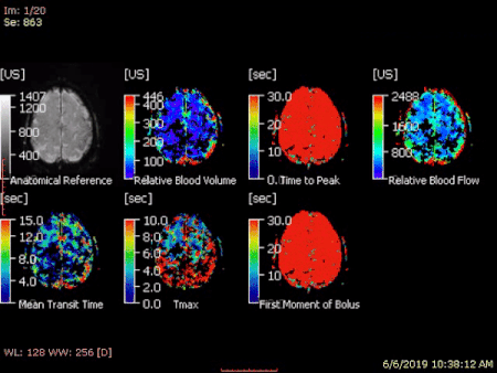
Perfusion weighted imaging
Quantifies brain perfusion (Time to Peak, Cerebral Blood Flow, Cerebral Blood Volume, etc.).
Atherosclerotic plaque geometry and composition
2D and 3D analysis of lesion morphology.
Catheter Based Angiography
Quantification of arterial anatomy and luminal lesions displayed on catheter angiography of ALL vascular branches in the body including:
- Brain
- Neck
- Chest
- Abdomen
- Upper extremities
- Lower extremities
Infrared Thermography
Infrared thermography is being used to detect differences in skin temperature on the forehead as a potential screening methodology for patients with carotid stenosis.
Functional Near-Infrared Spectroscopy (fNIRS)
Functional near-infrared spectroscopy (fNIRS) is a non-invasive imaging technique that uses near-infrared light to image changes in oxy- and deoxy-hemoglobin. Sensors are placed on the skin to detect changes in the optical absorption of oxygenated and deoxygenated hemoglobin in the tissue as activities are being performed. Thus, fNIRS can quantify regional brain activation in real-time and during tasks. fNIRS has been shown to correlate with functional MRI (fMRI), but has the added benefits of being non-invasive, not requiring a contrast agent, and avoiding risks associated with MR imaging. Our lab utilizes the Artinis Brite System.