UM School of Medicine Researchers Awarded $3 Million National Cancer Institute Grant for Personalized Imaging to Improve Radiation Treatment
September 17, 2021
Study Could Improve the Precision of Radiotherapy to Reduce Side Effects to Healthy Lung Tissue and Surrounding Organs
Researchers at the University of Maryland School of Medicine (UMSOM) have received more than $3 million in federal funding from the National Cancer Institute to conduct a study to develop a new precision-based medicine approach to treat lung cancer patients with radiation therapy.
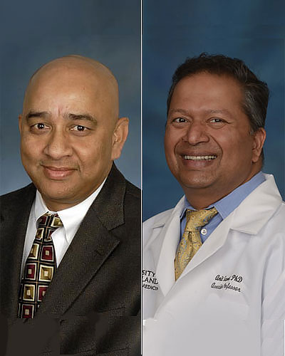
The R01 grant was awarded in July 2021 to Rao P. Gullapalli, PhD, MBA, Professor and Associate Vice Chair for Research in the Department of Diagnostic Radiology and Nuclear Medicine, and Amit Sawant, PhD, Professor and recently appointed Vice Chair for Medical Physics in the Department of Radiation Oncology. They will serve as co-principal investigators on the 5-year project, which involves an academic–industrial partnership with Robin Medical, Inc., based in Baltimore, MD.
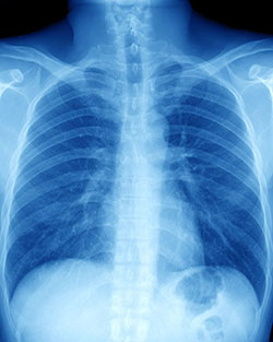
Building on previous collaborative research studies, the researchers will use four-dimensional (4D) imaging with magnetic resonance imaging (MRI) and computed tomography (CT) to help refine and improve “motion management,” a type of radiation technology that senses and accounts for lung motion during radiation therapy.
The added fourth dimension is time, and the researchers plan to develop a system that uses the two imaging modalities in real time to account for the movement of lungs as a patient inhales and exhales during a procedure.
 “When lung cancer patients undergo radiation treatments, respiratory motion can cause substantial changes, from one breathing cycle to the next, in terms of shifting the precise position of their tumors and neighboring critical organs,” said Dr. Sawant. “This is a longstanding challenge that increases the potential for radiation being misdirected to normal lung tissue and nearby organs.”
As a result of unanticipated respiratory motion, patients undergoing radiotherapy for lung cancer can experience damage to normal lung tissue and organs, reducing their quality of life and, potentially, causing post-treatment complications. The researchers plan to develop a patient-specific model that allows them to create a “3D movie” of the breathing anatomy to correct for motion effects in real time while each patient is under treatment.
“We expect that by combining 4D MRI and 4D CT, we will be able to pinpoint the motion and location of the tumor relative to every organ in the chest cavity, throughout the delivery of radiation,” said Dr. Gullapalli. “This will allow clinicians to deliver radiotherapy with more confidence and fewer side effects.”
The multi-stage project will include:
- Development and adaptation of imaging techniques;
- Creation of the patient-specific model and sensors;
- Production and team evaluation of a prototype; and
- Assessment of the systems’ utility using clinical data from patients undergoing treatment for lung cancer.
 “This study is especially exciting because of the cross-departmental collaboration with our colleagues in Radiology,” said William F. Regine, MD, Professor and Isadore and Fanny Schneider Foxman Chair of Radiation Oncology. “Imaging is an integral part of successful cancer therapy, and the proposed system is likely to enhance our joint efforts to tailor treatments to individual patients with lung cancer.”
The research team plans to conduct a clinical study in 44 lung cancer patients to compare the performance of the new motion management 4-D model to the current standard of care, which uses a less sophisticated technology for motion management. The goal is to translate the new approach into clinical practice to enable safer administration of highly potent and clinically effective forms of radiation therapy for lung cancer patients.
 “Dr. Gullapalli is a recognized expert in developing MR imaging techniques. Together with Dr. Sawant’s groundbreaking work in image-guided radiation therapy, the two scientists are well positioned to meet with success in this initiative,” said Elias R. Melhem, MD, Professor and Dean John M. Dennis Chair of Diagnostic Radiology and Nuclear Medicine.
The combining of two advanced imaging techniques will help personalize the imaging approach to build patient-specific motion models that can map the motion and location of the tumor and every organ in the chest cavity, throughout the delivery of radiation treatment. With such precise delivery of radiation, clinicians can better delivery radiotherapy to destroy malignant tissue with fewer side effects to healthy tissues.
“This is a stellar example of interdepartmental collaboration to advance the field of precision medicine,” said E. Albert Reece, MD, PhD, MBA, Executive Vice President for Medical Affairs, UM Baltimore, and the John Z. and Akiko K. Bowers Distinguished Professor and Dean, University of Maryland School of Medicine. “It is exciting to imagine the positive outcomes this may provide cancer patients in the future in terms of fewer side effects and more effective radiation treatments.”
About the University of Maryland School of Medicine
Now in its third century, the University of Maryland School of Medicine was chartered in 1807 as the first public medical school in the United States. It continues today as one of the fastest growing, top-tier biomedical research enterprises in the world -- with 46 academic departments, centers, institutes, and programs, and a faculty of more than 3,000 physicians, scientists, and allied health professionals, including members of the National Academy of Medicine and the National Academy of Sciences, and a distinguished two-time winner of the Albert E. Lasker Award in Medical Research. With an operating budget of more than $1.2 billion, the School of Medicine works closely in partnership with the University of Maryland Medical Center and Medical System to provide research-intensive, academic and clinically based care for nearly 2 million patients each year. The School of Medicine has nearly $600 million in extramural funding, with most of its academic departments highly ranked among all medical schools in the nation in research funding. As one of the seven professional schools that make up the University of Maryland, Baltimore campus, the School of Medicine has a total population of nearly 9,000 faculty and staff, including 2,500 students, trainees, residents, and fellows. The combined School of Medicine and Medical System (“University of Maryland Medicine”) has an annual budget of over $6 billion and an economic impact of nearly $20 billion on the state and local community. The School of Medicine, which ranks as the 8th highest among public medical schools in research productivity (according to the Association of American Medical Colleges profile) is an innovator in translational medicine, with 606 active patents and 52 start-up companies. In the latest U.S. News & World Report ranking of the Best Medical Schools, published in 2021, the UM School of Medicine is ranked #9 among the 92 public medical schools in the U.S., and in the top 15 percent (#27) of all 192 public and private U.S. medical schools. The School of Medicine works locally, nationally, and globally, with research and treatment facilities in 36 countries around the world. Visit medschool.umaryland.edu
Contact
Office of Public Affairs
655 West Baltimore Street
Bressler Research Building 14-002
Baltimore, Maryland 21201-1559
Contact Media Relations
(410) 706-5260
Related stories
Wednesday, July 31, 2019
Inconsistent Reporting of Methodologies Makes Most Radiation Biology Studies Impossible to Replicate
Nearly 80 percent of radiation oncology studies funded by the National Institutes of Health involve investigating the effects that radiation has on tumor cells and healthy tissue in pre-clinical settings, such as experiments done in cell cultures or mice. A majority of these radiation biology studies, however, have serious flaws in how their irradiation methodology is described, which makes them very difficult to replicate, according to a new finding from the University of Maryland School of Medicine (UMSOM).
Friday, July 21, 2017
University Of Maryland School Of Medicine Scientist Receives Top Award for His Pioneering Work on Improving Radiation Therapy Outcome
Søren M. Bentzen, PhD, DMSc, a professor in the Department of Epidemiology and Public Health (EPH) at the University of Maryland School of Medicine (UM SOM), has been awarded a gold medal from the American Society for Radiation Oncology (ASTRO). This is the highest honor bestowed upon ASTRO members. It recognizes a distinguished scientist who has made major contributions to the field of radiation oncology.