Brain Sectioning - Protocol Method 2
PROTOCOL METHOD 2 IS THE CURRENT METHOD USED FOR ALL BRAIN TISSUE DONATIONS, WHERE POSSIBLE.
General Information
When possible, all brains should be chilled/cooled in wet ice for at least one half hour prior to sectioning to enhance the ease and quality of sectioning.
The preferred method of freezing the individual sections is in isopentane/dry ice at -30 to -40 degrees Centigrade. The second method of choice is in liquid nitrogen. The third method of choice is freezing the samples on a tray in a -80 degree freezer.
10 percent formalin is used for all fixed sections.
MEDULLA
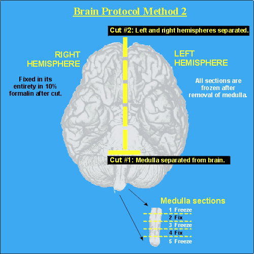
First, the medulla is removed from the brainstem by transecting at its juncture with the distal pons(Cut #1). The medulla is sectioned in a coronal plane into five samples of 2-3mm thickness beginning at the pontine junction. These five samples are assigned a sequential identifier from 1 to 5. Sections 1,3,5 are frozen; sections 2 and 4 are fixed in 10% formalin. After removal of the medulla, the entire brain is sectioned into its left and right hemispheres (Cut #2). The right hemisphere is fixed in its entirety in 10% formalin.
LEFT HEMISPHERE: CEREBRUM
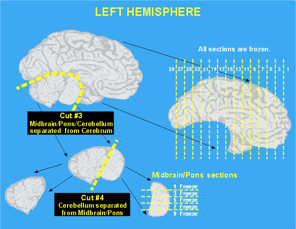
A cut is made just posterior to the cerebral peduncle and the midbrain/pons/cerebellum are removed as a unit from the left hemisphere (Cut #3). The remaining cerebrum is sectioned coronally, at approximate 1 cm intervals beginning from the frontal pole apex and proceeding caudally. As each section is isolated, it is gently rinsed with water, blotted dry, assigned a sequential numeric identifier (odd numbers only!), and placed in the freezing bath. The handling of sections is best aided by the use of a plastic spatula. Each frozen section is placed into individual plastic bags appropriately labeled and sealed. All bags are then stored in a -80 degree Centigrade freezer prior to shipping. Frozen sections of the cerebrum are identified as sections 1,3,5,7,9...
LEFT HEMISPHERE: MIDBRAIN/PONS
The midbrain/pons (upper brainstem) is separated from the cerebellum (Cut #4). The midbrain/pons is placed on a flat cutting board, mesial surface down, and sectioned into four or five sections at approximate 0.3 to 0.4 cm intervals beginning at the midbrain and moving caudally. These sections are assigned a sequential identifier (odd numbers only!). Frozen sections of the midbrain/pons are identified as sections 1,3,5...
LEFT HEMISPHERE: CEREBELLUM
The remaining cerebellum is placed in a vertical plane (its normal anatomic position) and sectioned at 0.5 to 0.6 cm intervals beginning from the medial surface (vermis) and moving laterally. Each resulting section is assigned a sequential identifier (odd numbers only!). Frozen sections of the cerebellum are identified as sections 1,3,5,7,9...
RIGHT HEMISPHERE
The right hemisphere is fixed in its entirety in 10 percent formalin and is sectioned similarly to the left hemisphere. Fixed sections of the cerebrum (right hemisphere) are identified as sections 2,4,6,8,10... Fixed sections of the midbrain/pons (right hemisphere) are identified as sections 2,4,6... Fixed sections of the cerebellum (right hemisphere) are identified as sections 2,4,6,8,10...
THE ABOVE PROTOCOL MAY BE MODIFIED IN A CERTAIN NUMBER OF CASES DUE TO THE NATURE OF THE INJURY OR DIAGNOSIS.


