Brain Sectioning - Protocol Method 1
In the past, Protocol Method 1 was the first method used by the Brain and Tissue Bank for brain tissue donations. This method is no longer the current method used for tissue donations.
CEREBRUM
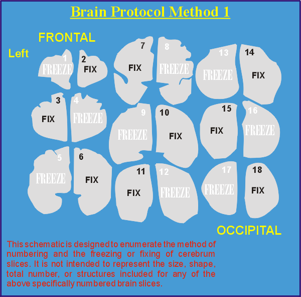
The brain is placed on a horizontal surface, dorsum down, with gyrus rectus parallel to the surface. A single coronal section, perpendicular to the surface, is made at a point caudal to the mammillary bodies, but anterior to the rostal origin of the crus cerebri. The brainstem/cerebellum portion is removed from the caudal half of the cerebrum by transecting the midbrain at its junction with the rostral pons. A plexiglass cutting guide is used to obtain one centimeter thick serial, coronal sections of the cerebrum. Each coronal section is then cut in the sagittal plane through its midline, creating two hemisections. The cerebral hemisections are fixed and frozen in an alternating sequence as shown.
BRAINSTEM/CEREBELLUM
Sectioning of the remaining brainstem/cerebellum is initiated by transecting the medulla from the pons in a strict transverse plane, perpendicular to the long axis of the lower brainstem. The intact pons/cerebellum is sectioned in a transverse plane at one centimeter intervals and hemisected. The pons/cerebellum is fixed and frozen in the same alternating scheme as the cerebrum.
MEDULLA
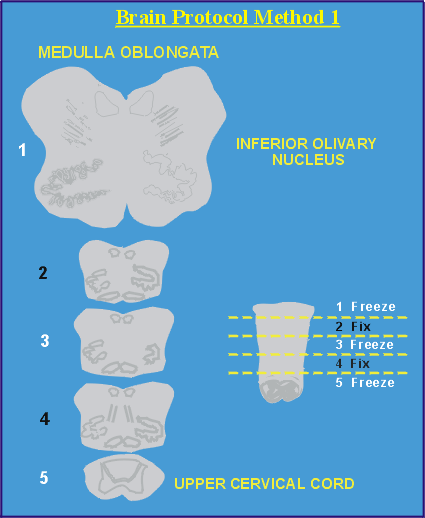
The medulla is cut transversely at the following four levels to create 5 sections: mid-olive, 4-5 mm distal, caudal pyramidal decussation and 4-5 mm distal. Alternate sections of the medulla are fixed and frozen.


