
Cauda Equina Syndrome (CES)
A recent pearl discussed CES. This is a very challenging diagnosis to make, especially on initial presentation
The 5 “classic” characteristic features are
- Bilateral radiculopathy
- Saddle anesthesia
- Altered bladder function
- Loss of anal tone
- Sexual dysfunction
Not all symptoms will be present in a given patient and there is no sign/symptom combination that either reliably diagnoses or excludes CES.
To illustrate how difficult this diagnosis is to make, a study looked at the predictive abilities of Neurosurgical residents.
Positive MRI for CES was accurately predicted by senior neurosurgical residents in approximately 50% of patients suspected of CES based on history and physical findings. As clinical certainty only becomes apparent with the classic symptoms (which are generally late findings) waiting to initiate MRI will delay decompressive surgery and can lead to worsened functional outcomes. This leads to increased MRI demand with more negative MRIs. Not surprisingly, only ~20% of MRI scans for suspected CES are positive.
Show References
A True Tracheostomy Emergency
- Patients with a tracheostomy often present to the ED for evaluation of a potential complication.
- Consider a tracheoarterial fistula in any patient with a tracheostomy who presents with brisk bleeding.
- Most occur within 3 to 4 weeks following tracheostomy placement, and the most common location is the innominate artery.
- Up to 50% of patients will present with a sentinel bleed - an episode of brisk bleeding that has usually stopped at the time of presentation.
- For patients who present with active hemorrhage, overinflate the tracheostomy cuff in an attempt to tamponade the bleeding.
- If that does not stop the bleeding, remove the tracheostomy and compress the artery against the poterior sternum with your finger.
Show References
| Mechanism of Action | Tranexamic Acid (TXA) is an antifibrinolytic agent that is a competitive inhibitor of plasminogen activation, and a non-competitive inhibitor of plasmin Inhibits the breakdown of fibrin mesh allowing clot formation
|
| When is it Indicated? | Epistaxis/Oral Bleeds/Fistula Bleeds
|
| Trauma
| |
| Adverse Reactions |
|
Back pain with lower extremity symptoms can be concerning for cauda equina. Some pointers regarding the H&P:
- Symptoms develop within less than 24 hours in 90% of patients
- Urinary retention develops before incontinence, but up to 30% of patients will have neither.
- Saddle anesthesia or hypoesthesia is present in 81% of patients. Perineal numbness may be patchy, mild, and unilateral initially, making it difficult to elicit.
None of these symptoms independently predicts cauda equina syndrome with an accuracy greater than 65%.
Bottom Line: do not depend on any one finding to reliably exclude or confirm cauda equina.
Show References
Despite ongoing research and efforts to improve our care of patients with ARDS, it remains an entity with high morbidity and mortality. Early recognition of the disease process and appropriate management by emergency physicians can have profound effects on the patient's course, especially in centers where ICU boarding continues to be an issue.
Recognition of ARDS (Berlin criteria)
- Acute in onset
- Bilateral infiltrates on chest imaging not due to cardiac failure/volume overload
- PaO2 : FiO2 < 300 despite PEEP of at least 5cmH2O
- This is the standard ED patient who gets intubated with multifocal pneumonia and has continued hypoxemia
*An ABG should be obtained in the ED if physicians are unable to wean down FiO2 from high settings, if oxygenation by pulse ox is marginal, or if the patient is in a shock state.
Tenets of ARDS Management:
- Low tidal volume ventilation (6-8ml/kg ideal body weight*)
- Maintain plateau pressures (Pplat) < 30 cmH2O
- Driving pressure (Pplat – PEEP) < 15 cmH2O
- Goal PaO2 > 55-60
- Permissive hypercapnia to pH >7.2
*IBW Males = 50 + 2.3 x [Height (in) - 60] / IBW Females = 45.5 + 2.3 x [Height (in) - 60]
Strategies for Refractory Hypoxemia in the ED: You can't prone the patient, but what else can you do?
1. Escalate PEEP in stepwise fashion
- ex: 2cmH20 every 10 minutes
- can use ARDSnet PEEP/FiO2 table as guide
2. Recruitment maneuvers
- "20 of PEEP for 20 seconds" or "30 for 30"
- if patient is "PEEP responsive," leave PEEP on a higher setting than when you started (ex: 10 instead of 5, 16 instead of 10)
- Risk of barotrauma with higher PEEPs and hypotension in underresuscitated or hemodynamically unstable patients due to decreased venous return
3. Appropriate sedation and neuromuscular blockade
- promotes patient synchrony with lung protective settings
- can result in improved oxygenation by itself
4. Inhaled pulmonary vasodilators (inhaled prostaglandins, nitric oxide) if known or suspected right heart failure or pulmonary hypertension
Bottom Line: Emergency physicians are the first line of defense against ARDS. Early recognition of the disease process and appropriate management is important to improve outcomes AND to help ICU physicians triage which patients need to be emergently proned or even who should potentially be referred for ECMO.
Show References
In patients with lower back pain, there is good evidence that muscle relaxants reduce pain as compared to placebo and that different types are equally effective. However, the high incidence of significant side effects such as dizziness and sedation limits their use. Muscle relaxants may be beneficial in an every bedtime capacity thereby limiting side effects.
If cyclobenzaprine is used during daytime hours, a lower dose schedule may work as well as a higher dose with somewhat less somnolence (5 mg three times a day vs 10 mg three times a day. In general, muscle relaxants should only be used when patients cannot tolerate NSAIDs but can tolerate the side effect profile.
We commonly add muscle relaxants to NSAIDs hoping for a larger analgesic effect. However, combination therapy does not appear to be better than monotherapy.
Adding cyclobenzaprine to high-dose ibuprofen does not seem to provide additional pain relief in the first 48 hours in ED patients with acute myofascial strain. Among an ED population with acute non radicular low back pain, a randomized trial found that adding cyclobenzaprine/other muscle relaxants to Naproxen did not improve functional outcomes or pain at one week or 3 months compared to naproxen alone.
Take home: Consider the limited usefulness use of muscle relaxants in ED patients with back pain
Show References
The hyperoxia-hyperventilation test (aka 100% Oxygen Challenge test) is used to differentiate the cause of central cyanosis in the sick neonate. The majority of neonatal cyanosis is caused by either cardiac or respiratory pathology.
Classically the test is performed as follows:
1. An ABG is obtained with the neonate breathing room air
2. The patient is placed on 100% FiO2 for 10 minutes
3. A repeat ABG is performed looking for an increase in PaO2 to >150 mmHg
- If the hypoxia is secondary to a respiratory cause, the PaO2 should increase to >150 mmHg.
- If the hypoxia is secondary to a congenital cardiac lesion (i.e. secondary to a right-to-left cardiac shunt) the PaO2 is not expected to rise significantly.
In practice, many physicians instead use pulse oximetry and monitor the SpO2 pre and post administration of 10 minutes of 100% FiO2.
- If after 10min of 100% FiO2, if SpO2 is not ? 95% (some resources use 85%) then the central cyanosis is likely secondary to intracardiac shunt.
- When this occurs, presume the sick neonate is symptomatic from a congenital cardiac lesion and initiate prostaglandin E-1 (PGE1) at 0.05-0.01 mcg/kg/min. Use caution as PGE1 may cause apnea.
The primary tenet of poisoning treatment is to separate the patient from the poison. Gastric decontamination has been the cornerstone of poisoning treatment throughout history and methods include induced emesis, nasogastric suctioning, EGD or gastrostomy retrieval, activated charcoal, and whole bowel irrigation. Current guidelines for gastic decontamination are limited to few clinical situations. The detection of residual life threatening poisons in the stomach would be of value in predicting who might benefit from gastric decontamination in overdose.
Plain radiographs have variable sensitvity in detecting radioopaque pills. Computed tomography (CT) has been successful and gained wide acceptance in the detection of drug in body packers. In a recent study, authors studied the usefulness of non-contrast abdominal computed tomography for detection of residual drugs in the stomach in patients presenting over 60 minutes from acute drug overdose:
- 140 patients were included in this study
- Median ingested drug amounts were 28 tablets or capsules
- Median time until CT scan was performed after drug ingestion was 4 hours
- Multiple types of drugs were ingested in 53.6%
- Sustained-release drugs were ingested in 17.1 %
- Gastric lavage and WBI were performed on 32.9% patients
- Drugs were detected in 25.7% in the non-contrast CT scan performed over 60 min after ingestion.
- Total duration of hospital stay was significantly longer in the “presence of drugs” group
BOTTOM LINE:
Non-contrast CT may help to predict which patients would benefit from gastric decontamination in acute life-threatening drug poisonings.
Show References
Does This Patient Have Pericardial Tamponade?
- Echocardiography is critical for the identification of a pericardial effusion and rapid diagnosis of pericardial tamponade.
- Common echocardiography findings that suggest tamponade include diastolic right ventricular collapse, systolic right atrial collapse, a plethoric IVC with minimal respiratory variation, and potentially exaggerated respiratory cycle changes in mitral and triscupid inflow velocities.
- Of these, systolic right atrial collapse is the earliest echocardiographic sign of tamponade, with a sensitivity ranging from 50% to 100%.
- Of the 4 standard echo views, systolic right atrial collapse can best be viewed in the apical 4-chamber and subxiphoid views.
Show References
Hyponatremia is the most common electrolyte abnormality in hospitalized patients, affecting approximately 15-30% of patients. Children have historically been given hypotonic maintenance IV fluids based off of theoretical calculations from the 1950s. Multiple studies have shown complications related to iatrogenic hyponatremia, including increased length of hospital stay, seizures and death.
The American Academy of pediatrics completed a systematic review and developed an updated clinical practice guideline:
Patient's age 28 days to 18 years requiring maintenance IV fluids should receive isotonic solutions with the appropriate amount KCl and dextrose.
Show References
Physostigmine is a cholinergic agent that can be administered to reverse delirium associated with anticholinergic toxicity. However, it is infrequenly used since the reports of cardiac arrest in patients with TCA overdose.
A recently published study reviewed 161 articles – involving 2299 patients – to determine the adverse effects and their frequency after the administration of physostigmine.
Findings
Adverse effects were observed in 415 patients (18.1%)
- In patients with anticholinergic overdose: 7.7%
- In patients with non-anticholinergic agent overdose: 20.6%
Specific adverse effects
- Hypersalivation: 206 (9%)
- Nausea/vomiting: 96 (4.2%)
- Seizure: 14 (0.61%)
- Symptomatic bradycardia: 8 (0.35%) – including 3 with bradyasystolic arrest
- Asymptomatic bradycardia: 4 (0.17%)
- Ventricular fibrillation: 1 (0.04%) patient had a history of coronary artery disease
- Cardiac arrest: 4 (0.17%)
- Death: 5 (0.22%)
Of 394 TCA overdose, adverse effects occurred in 14 patients (3.6%)
Conclusion
- Adverse effects from physostigmine occurs infrequently.
- However, inappropriate dosing or use of physostigmine can result in cholinergic toxicity.
- For isolated anticholinergic toxicity (e.g. antihistamine overdose): physostigmine dosing: 0.5 mg (dilute in 5 – 10 mL normal saline) IV over 2 -5 minutes. May repeat every 5-10 minute to max dose total of 2 mg. (patient needs to be on cardiac monitor with atropine at bedside)
- Therapeutic goal: reversal of delirium
- Avoid physostigmine in the presence of QRS widening (cardiac Na-channel blockade) and patients with history of underlying coronary artery disease.
Show References
Neutropenic enterocolitis can occur in immunosuppressed patients, classically those being treated for malignancy (hematologic much more commonly than solid tumor). When involving the cecum specifically, it is known as "typhlitis."
It should be considered in any febrile neutropenic patients with abdominal pain or other symptoms of GI discomfort (diarrhea, vomiting, lower GI bleeding), and can be confirmed with CT imaging.
A recent study found that invasive fungal disease, most often candidemia, occurred in 20% of febrile neutropenic patients with CT-confirmed enteritis, a rate that increased to 30% if the patient was in septic shock.
Take Home:
1. Have a lower threshold for abdominal CT imaging in your patients with febrile neutropenia and abdominal pain/GI symptoms, especially if they are critically ill.
2. Consider addition of IV antifungal therapy if they are hemodynamically unstable with enterocolitis on CT.
Show References
Spurling’s maneuver and modified Spurling’s maneuver aka neck compression test.
This maneuver is highly specific for the presence of cervical root compression
Can be used to reproduce radicular pain/symptoms.
Perform this maneuver with caution as it should not be performed in patients who have potential cervical spine instability.
Keeping the patient’s head in a neutral position pressing down on the top of the head. If this fails to reproduce the patient's pain, the test is repeated with the head extended, rotated and tilted to the affected side (the modified Spurling’s maneuver).
Reproduction of symptoms (limb pain or paresthesias) beyond the shoulder is considered positive. Neck pain alone is nonspecific and constitutes a negative test.
The test has a high specificity (0.89 to 1.00) but low sensitivity (0.38 to 0.97).
Meaning a positive test is helpful but a negative test does not rule out radicular pain.
This test should be used in conjunction with a thorough history and physical examination (strength, sensation and reflex testing)
https://www.youtube.com/watch?v=17QWqbXjSpc
Show References
Management of Acute Variceal Bleeding
- Patients with an acute UGIB secondary to esophageal or gastric varices frequently present in extremis.
- The initial resuscitation of patients with a variceal bleed should focus on the administration of antibiotics, packed red blood cells (PRBC), vasoactive agents, and emergent endoscopy.
- Antibiotics have been shown to reduce recurrent bleeding and mortality. A third-generation cephalosporin (e.g., ceftriaxone) is commonly recommended as the initial antibiotic of choice.
- Utilize a restrictive PRBC transfusion strategy to target a Hb between 7 to 8 g/dL.
- Vasoactive agents (e.g., octreotide) reduce portal pressure through splanchnic vasoconstriction and have been shown to reduce acute bleeding and the need for transfusion.
Show References
Yes.
Serum creatinine decreases with age with the decrease in lean body mass. However, the number of functioning glemeruli and kidney function decrease with age as well, making the creatinine an unreliable indicator of renal function in older adults.
The solution? Calculate the creatinine clearance (CrCl) (or GFR) for a more accurate assessment of the renal function. You can use simple equations such as the Cockroft-Gault equation which incorporate the body weight and age.
CrCl (mL/min) = (140-age) x lean body weight (kg) x (0.85 if female)
serum creatinine (mg/dL) x 72
Show References
Show Additional Information
Show References
Most clinicians are familiar with use of methylene blue for the treatment of methemoglobinemia, as a urinary analgesic, anti-infective, and anti-spasmodic agent, or for its use in endoscopy as a gastrointestinal dye, but this compound also has a role as a rescue antidote in life threatening poisonings causing refractory shock states and other shock states.
- Nitric Oxide plays an important role in the regulation of vascular tone.
- Metylene blue inhibits the NO-cGMP pathway which decreases vasodilitation and increases responsiveness to vasopressors.
- Several case reports document hemodynamic improvement in recalcitrant shock states form calcium channel and beta blockers despite multiple therapies including vasopressors, glucagon, high dose insulin, and fat emulsion therapy.
- Dosing is 1-2 mg/kg (0.1-0.2 ml/kg) of 1% solution given IV over 5 minutes folllowed by continuous infusion.
Bottom Line:
Methylene blue should be considered when standard treatment of distributive shock fails.
Show Additional Information

Show References
Historically, there has been very limited data regarding the epidemiology of OHCA in pregnant females. Two recently-published studies tried to shed some light on the issue.
Both Maurin et al.1 and Lipowicz et al.2 looked at all-cause out-of-hospital maternal cardiac arrest (MCA) data in terms of numbers and management, in Paris and Toronto respectively, from 2009/2010 to 2014. Collectively, they found:
- MCA was relatively rare: 0.8 MCA per 1000 OHCA (Maurin) and 1.71 MCA per 100,000 pregnant females (Lipowicz)
- Low incidence of bystander CPR in witnessed MCA (33% and 0%)
- Adherence to PMCS guidelines was poor
- Maternal survival was lower than what has been previously quoted for in-hospital CA: 12.5 and 16.7% compared to 40-50%3,4
A few reminders from the 2015 AHA guidelines for the management of cardiac arrest in pregnancy:
- Hand location for chest compressions should be in the center of the chest as for nonpregnant patients (previous recommendations had been to shift upward to accommodate for the gravid uterus but there is no data to support this).
- Chest compressions should be performed with the patient supine, using manual lateral uterine displacement for aortocaval decompression. Left lateral tilt position is no longer recommended due to poorer quality of cardiac compressions, the lack of full aortocaval decompression, and further complication of other procedures such as airway management.
- IV or IO access should be obtained above the diaphragm, to ensure no interference to flow to the heart by the gravid uterus.
- Rate and depth of chest compressions, ACLS drugs and doses, and defibrillation all remain the same as in nonpregnant OHCA patients.
- NB: As opposed to nonpregnant patients periarrest, oxygen saturation in the pregnant female should be maintained at 95% or greater, or PaO2 > 70mmHg, to ensure appropriate oxygen delivery to the fetus. The goal PCO2 is ~28-32 mmHg, to facilitate fetal CO2 removal.6
- If advanced airway is pursued, the most experienced provider should perform intubation due to the higher intrinsic difficulties, more rapid decompensation, and propensity for airway trauma and bleeding in the pregnant female.
- Perimortem c-section should occur within the first 5 minutes of cardiac arrest / arrival to the ED in ongoing arrest.
Bottom Line: Although maternal cardiac arrest is relatively rare, survival in OHCA is lower than perhaps previously thought. Areas to improve include public education on the importance of bystander CPR in pregnant females, and appropriate physician adherence to PMCS recommendations, with decreased on-scene time by EMS in order to decrease time to PMCS.
Show Additional Information
Show References
Hook of hamate fracture
Often missed fracture despite classic history
A frequent athletic injury
Seen in stick sports (golf, baseball, hockey)
Typically caused by a direct blow (grounding a gold club)
https://upload.orthobullets.com/topic/6035/images/hamate_baseball.jpg
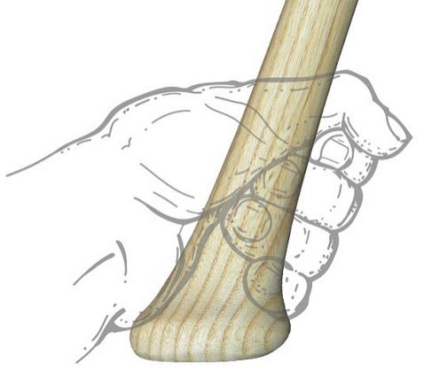
Patient presents with hypothenar pain and pain with tight gripping
https://upload.orthobullets.com/topic/6035/images/hamate_golf.jpg

Presentation may be subacute with longstanding wrist or palmer pain
Physical exam: Tender to palpation over hook of hamate
Specialized test: hook of hamate pull test
Supinated hand held in ulnar deviation. Ask patient to actively flex 4th and 5th digits against resistance at DIP.
https://www.youtube.com/watch?v=A-mjRnC1yWQ
Radiology: Consider adding carpal tunnel view to standard wrist series if diagnosis is suspected
CT sometimes needed to image the fracture
Tx: Immobilize in a short arm splint
Show References
Therapeutic use or overdose of tramadol has been associated with seizure. However, it is unknown if there are any specific predisposing factor that increases a patient’s risk of seizure after tramadol use/overdose.
In a recently published study, eighty patient data with single ingestion of tramadol were reviewed.
- 52.5% of the patient developed seizure
- 11% developed serotonin syndrome
Risk of seizure
- Higher risk of seizure were found in Asian patients (OR=7.1, 95% CI: 1.9 – 27.3) and patients with abuse/misuse of tramadol (OR=3.2, 95% CI: 1.2-8.3)
- Patients who presented with opioid toxidrome were less likely to develop seizure (OR=0.12, 95% CI: 0.02 – 0.71)
- Acute overdose and age were not associated with increased risk of seizure.
Conclusion
In this small study, Asian patients and patients with abuse/misuse were at higher risk of developing seizure compared to patients who overdose tramadol.