
Search
Subscribe to daily UMEM Pearls (FREE!) via:
1-20 of 384 results with category "Neurology"
The 2025 American Headache Society guidelines give the greater occipital nerve (GON) block a level A recommendation for ED migraine treatment, boldly calling it a “must offer” therapy for migraine patients requiring parenteral treatment.
- Mechanism:
- The GON, a branch of the C2 spinal nerve, innervates the posterior scalp and converges with trigeminal afferents at the C2 dorsal horn. Blocking the GON dampens trigeminal signaling at this convergence point, reducing acute migraine pain. Since central sensitization drives migraine chronicity, benefits often outlast the local anesthetic, persisting for weeks to months.
- Efficacy:
- A meta-analysis of over 400 patients showed a mean reduction of 3.6 headache days/month and lower pain severity scores versus controls, with greater benefit from scheduled, recurring blocks. For acute migraine, one large study found 82% of patients reported moderate-to-significant relief.
- How to:
- Positioning:
- Seat the patient with the head flexed slightly forward. Avoid any lateral or rotational movement, which may distort landmarks.
- Landmark-based:
- Palpate the external occipital protuberance and mastoid process. The GON runs about one-third of the distance (2-4 cm) between them, just medial to the occipital artery (which is often palpable). Prep the skin, advance the needle while aspirating until you hit bone, withdraw slightly, then inject 1-3 mL of 1% lidocaine or 0.5% bupivacaine with a fanning technique.
- Ultrasound-guided (more precise):
- Place a linear probe with sterile cover transversely over the occipital protuberance, then slide down to the C2 spinous process, which has a characteristic bifid appearance. Moving laterally from here, identify the GON between the semispinalis capitis and obliquus capitis inferior fascial plane, medial to the occipital artery (consider color Doppler to confirm). Inject 1-3 mL of anesthetic into this fascial plane, avoiding injection into the nerve fascicles themselves.
- Adjuncts?
- Dexamethasone mixed with local anesthetic has evidence of benefit with cervicogenic headache and occipital neuralgia, but has no proven added benefit for migraine specifically.
- Positioning:
Bottom Line: Consider adding the GON block to your toolkit for migraine treatment in the ED.
Show References
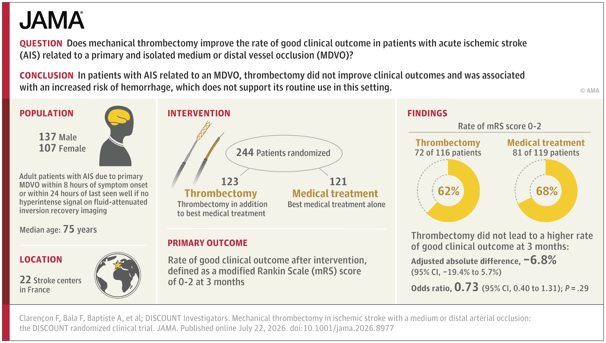
Bottom Line: In patients with acute ischemic stroke due to medium or distal vessel occlusion, mechanical thrombectomy did not improve functional outcomes at 3 months and was associated with an increased risk of symptomatic intracranial hemorrhage.
Show Additional Information
Show References
Lumbar puncture (LP) carries a relatively small risk of hemorrhagic complications, including traumatic tap (a lab finding) and spinal hematoma (an imaging finding).
In a cohort study of 83,000 LPs, the risk of spinal hematoma was not significantly increased in patients with documented coagulopathy (0.23% versus 0.20%), with coagulopathy defined by Plt < 150,000, INR > 1.4, aPTT > 39s.
Data on platelet transfusion and anticoagulant reversal prior to LP are limited to small retrospective cohort studies and case series. However, the limited data on this subject show no significant difference in major bleeding complications in patients who did or did not receive reversal prior to LP.
Although the risk of iatrogenic spinal hematoma may be low, current guidelines generally support the following practices:
- Plt < 50,000: Platelet transfusion is recommended prior to LP. Some studies suggest 40,000 is also a safe threshold.
- INR > 1.4: Consider correction with Vit K or PCC prior to LP
- aPTT > 40s: Consider correction based on cause (factor deficiency or heparin effect) prior to LP.
- Aspirin: Not considered a contraindication, no reversal needed
- DAPT: No clear guidelines. One small study demonstrated no increased risk of spinal hematoma.
- DOACs: Consider reversal if last dose < 24 hours and LP is emergently indicated.
- Resumption of AC: Generally considered safe after 12-24 hours if no evidence of bleeding or neurological complications.
Show References
Cerebral venous sinus thrombosis (CVST) is an emergent diagnosis frequently missed on standard brain imaging in the ED, with studies reporting miss rates up to 30–73% on noncontrast CT alone. Diagnostic delays average 4–10 days from initial presentation in confirmed cases. CTV and MRV both have very high sensitivity for detection of CVST.
When to Suspect CVST
- Unexplained focal neurologic deficits: Hemiparesis, aphasia, or new seizures without corresponding arterial infarct, mass effect, or hemorrhage on noncontrast CT/CTA.
- Signs of increased ICP without mass lesions or traumatic findings
- Hypercoagulable states: OCP use, malignancy, peripartum status, thrombophilia
- High risk CT findings:
- Skull fracture traversing a dural sinus (SSS, transverse)
- Empty delta sign
- Atypical hemorrhage patterns:
- Multifocal cortical subarachnoid hemorrhage (especially posterior-predominant).
- Nontraumatic-appearing cortical SAH or bilateral thalamic hemorrhages.
- Hemorrhagic venous infarcts (“thumbprint edema”).
Summary: Consider adding CTV in patients with strong thrombotic risk factors, atypical/multifocal hemorrhage patterns, or focal deficits unexplained by CT/CTA.
Show References
While this study is imperfect and may not be measuring patient important outcomes, it does fit with other literature on the topic of intensive blood pressure control in patients with acute ischemic stroke. These patients were randomized to aggressive blood pressure control (SBP 130-140 within 1 hour of TPA administration continued for 72 hours) or the standard SBP <180. Repeat imaging was performed to assess the degree of cerebral swelling that each group developed. There was no difference in swelling between the two groups.
Take away is aggressive blood pressure management in this group of ischemic stroke patients does not seem to be beneficial.
Show References
The concept of positioning the head of bed flat in a patient with a neurologic catastrophe seems like a recipe for badness. For most neurologic emergencies, elevating the head of the bed (HOB) to 30° is standard to help control intracranial pressure and reduce aspiration risk. However, emerging evidence indicates that acute large vessel occlusion (LVO) stroke patients—particularly before thrombectomy—may be an important exception.
The ZODIAC trial, published in June of this year, was a prospective, randomized, multicenter study comparing 0° (flat) versus 30° HOB positioning in patients with confirmed LVO stroke awaiting endovascular thrombectomy. The rationale stems from physiologic studies, including transcranial Doppler ultrasonography, showing that flat positioning can improve cerebral perfusion to ischemic tissue.
The primary outcome was early neurologic deterioration (>2-point worsening in NIHSS prior to thrombectomy). Safety endpoints included hospital-acquired pneumonia and all-cause mortality at 3 months.
In the trial’s 92 enrolled patients, flat positioning markedly reduced early neurologic deterioration, which occurred in 2.2% in the 0° group versus 55.3% in the 30° group. There were no significant differences in pneumonia or 3-month all-cause mortality. The authors also found a statistically insignificant improvement in 90-day functional outcomes in the 0° group. Due to the magnitude of benefit, the study was stopped early at interim analysis.
This technique represents a simple, cost-free, and practical method of preventing neurologic decline ahead of definitive management for LVO. This may be especially beneficial for LVO patients who require interhospital transfer to a thrombectomy-capable center.
Bottom Line: For patients with LVO stroke awaiting thrombectomy, flat (0°) head positioning is safe and significantly reduces early neurologic decline by improving blood flow to ischemic brain tissue.
Show References
Acute ischemic strokes involving the posterior circulation have significantly lower rates of hemorrhagic transformation after thrombolysis compared to anterior circulation strokes, a difference attributed to smaller infarct sizes and greater “ischemic tolerance” in the posterior circulation.
Given this lower hemorrhage risk, the EXPECTS trial evaluated the safety and efficacy of extending the thrombolysis window to 4.5–24 hours in patients with acute posterior circulation ischemic stroke who were not candidates for endovascular thrombectomy. This randomized controlled trial, conducted across 30 sites in China, enrolled 234 adults with mainly mild posterior circulation stroke (median NIHSS 3) and no evidence of extensive infarction on CT. Participants were randomized to receive either intravenous alteplase or standard medical care within the 4.5–24 hour window after symptom onset.
The primary outcome of functional independence at 90 days (modified Rankin Scale 0–2) was achieved in 89.6% of the alteplase group versus 72.6% of the standard care group. Rates of symptomatic intracranial hemorrhage were low and similar between groups (1.7% alteplase vs. 0.9% standard care), and 90-day mortality was lower in the alteplase group (5.2% vs. 8.5%).
These findings support extending the therapeutic window for intravenous thrombolysis in posterior circulation stroke beyond 4.5 hours when thrombectomy is not an option. However, the trial’s limitations, including a study population predominantly with mild strokes, exclusion of patients with extensive infarction, and enrollment limited to Chinese centers, warrant further investigation in larger and more diverse populations.
Bottom Line: Within limitations, emerging evidence supports an extended thrombolytic window beyond 4.5 hours for patients with posterior circulation strokes who are ineligible for thrombectomy.
Show References
Author: Matthew Jackson, MD
The Ket-Mid Study (1) is a recent RCT out of a tertiary center in a largely rural region of Egypt that evaluated combined ketamine (2 mg/kg) and midazolam (0.2 mg/kg) (ket-mid) to midazolam plus placebo (pla-mid) as the first line seizure abortive therapy. Children in the ket-mid group achieved 76% termination at 5 minutes compared to 21% of those in the pla-mid group (p<0.001). Adverse outcomes were rare, though the pla-mid group did have more hypotension.
Notably, the population included differed from that typically seen in the US. These kids were seizing for a long time, with a median seizure duration before IV meds of 34 minutes, and the benefit of ketamine on subgroup analysis seemed to shrink for those that were treated in less than 30 minutes. CNS infections were also much more common, accounting for the underlying pathology in 11% of cases. Important exclusion criteria also included trauma, inborn errors of metabolism, underlying CNS malignancy, and others not always known to EMS or ED clinicians at the time therapy begins.
Show References
How likely is your TIA pt to go on to have a stroke? This study in JAMA gives us some answers. While EM providers aren’t as interested in 5 and 10 year prediction, it provides more evidence TIAs need to be evaluated and are risk factors for future events.
“In this systematic review and meta-analysis of 171?068 patients with TIA or minor stroke from 38 studies, the risk of subsequent stroke was 5.9% within 1 year, 12.8% within 5 years, and 19.8% within 10 years.”
Show References
Anticoagulation is the mainstay of treatment of cerebral venous sinus thrombosis, irrespective of whether associated venous hemorrhage is present. Anticoagulant selection is variable, with physicians opting for unfractionated heparin (UFH) about 72% of the time in one international study. However, recent evidence favors the use of low-molecular-weight heparin (LMWH), with meta-analytic data showing trends towards lower mortality rates and improved functional outcomes in LMWH cohorts. UFH is often viewed more favorably due to the ability to rapidly discontinue the infusion in the event of major bleeding; however, risk of major bleeding complications were actually found to be lower in patients treated with LMWH compared to UFH. Further, LMWH has many pharmacological and practical benefits compared to UFH, including more predictable pharmacokinetics, reduced risk of heparin-induced thrombocytopenia (HIT), lack of need for frequent aPTT monitoring, ease of administration (daily subcutaneous injection), and ease of transition to outpatient therapy.
Takeaway: Consider LMWH (1.5mg/kg subcutaneously once daily) as first-line treatment for CVST in patients with acceptable renal function.
Show References
Multiple trials have aimed to assess the effect of blood pressure control in the prehospital setting for patients with suspected acute stroke. The INTERACT-4 trial was a multicenter, prospective, randomized, open-label, blind endpoint trial conducted in China, in which 2404 hypertensive patients with suspected acute stroke (based on FAST score >2, symptom onset <2h, SBP >150mmHg) were randomized to receive urapidil versus usual care in the prehospital setting. The primary outcome was modified Rankin score (mRs) distribution at 90 days. Overall, no significant difference in functional outcomes at 90 days were observed in the urapidil versus usual care groups (OR 1.00, 95% CI 0.87-1.15). However, when analyzed by stroke type, improved functional outcomes and lower rates of rebleeding were seen in patients with hemorrhagic stroke (46.5% of all enrolled patients), while worsened functional outcomes and mortality were seen in patients with ischemic stroke. These observations are consistent with standard practices of intensive blood pressure reduction in patients found to have ICH, versus the “permissive hypertension” approach to patients found to have cerebrovascular occlusion. The results of this trial are not practice-changing, but do highlight the importance of prompt stroke recognition, streamlined hospital workflows for expedited diagnostics (CT), and timely initiation of antihypertensive therapy in ICH patients.
Bottom line: Prehospital blood pressure reduction was not shown to improve clinical outcomes in hypertensive patients suspected to have acute undifferentiated stroke.
Show References
Intrathecal baclofen pumps are increasingly used to manage spasticity in patients with conditions such as cerebral palsy, spinal cord injury, multiple sclerosis, traumatic brain injury, and other dystonias. The most common causes of baclofen pump dysfunction include pump-related issues (e.g., programming errors, battery failure), catheter problems (e.g., extra-thecal dislodgement, kinking, leaks), and medication depletion (e.g., overdue or insufficient refills). Symptoms of dysfunction can be nonspecific, ranging from mild (spasticity, dysphoria, dysesthesias) to severe (e.g., rigidity, rhabdomyolysis, seizures, fever, autonomic dysfunction, cardiomyopathy).
Once dysfunction is recognized, management involves stabilizing vital functions (ABCs, temperature management, fluids), administering multimodal antispasmodics (enteral or parenteral baclofen, benzodiazepines, dexmedetomidine, tizanidine), and performing pump interrogation, often in collaboration with neurology or PM&R specialists. Restoration of intrathecal flow is the preferred and definitive therapy; however, patients with severe withdrawal may require aggressive temporizing measures including intubation. Some authors describe intrathecal baclofen administration via lumbar puncture as a rescue measure for severe cases with limited access to definitive care. Imaging with plain radiographs, fluoroscopy, or CT may be indicated in select cases where there is concern for catheter displacement or kinking, and some patients may require surgical revision.
Takeaway: Consider baclofen withdrawal in patients on chronic baclofen therapy who present with nonspecific symptoms that may mimic conditions such as alcohol withdrawal, delirium, sympathomimetic toxicity, neuroleptic malignant syndrome, serotonin syndrome, thyrotoxicosis, rhabdomyolysis, sepsis, or status epilepticus. In cases of intrathecal pump dysfunction, the definitive treatment is restoration of baclofen flow, so involve consultants early for pump interrogation while temporizing with supportive measures.
Show References
- Antimicrobial medications can be associated with neurological adverse reactions.
- An individual’s risk is influenced by their age, weight, nutritional status, the medications they are taking concurrently, and pharmacological properties (dosage, half-life, CNS permeability).
- Encephalopathy
- Seen with beta-lactams, fluoroquinolones, clarithromycin, and sulfamethoxazole-trimethoprim.
- Most commonly with cefepime.
- Higher risk in elderly, renal dysfunction, and preexisting CNS disease.
- Seizures
- Beta-lactams block GABA receptors.
- Highest risk with cefepime and imipenem.
- Peripheral neuropathy
- Associated with metronidazole, fluoroquinolones, linezolid, chloramphenicol, and isoniazid.
- Most cases are dose dependent.
- Some cases are irreversible.
- Ototoxicity
- Aminoglycosides cause cochlear NMDA receptor excitotoxicity.
- Weakness
- Fluoroquinolones, macrolides, and aminoglycosides inhibit acetylcholine release and bind neuromuscular junction receptors.
- Should be avoided in myasthenia gravis and Lambert-Eaton syndrome.
- Movement disorders
- Tremors – sulfamethoxazole-trimethoprim
- Dyskinesia, dystonic reactions – fluoroquinolones, chloramphenicol
- Cerebellar syndrome – metronidazole, aminoglycosides
Bottom Line: Recognition of antibiotic associated neurotoxicity reduces unnecessary workup and serious adverse effects.
Show References
Ventriculoperitoneal (VP) shunts are common. Unfortunately shunt complications are also common!
There are 3 major categories of shunt complications:
- Mechanical Failure - obstruction, fracture, disconnection, migration, perforation
- Functional Failure - overdrainage, slit ventricle syndrome, pseudocyst, ascites, meastasis
- Infection - may occur in up to 10% of patients (bacterial, fungal, parasitic)
Shunt series are helpful, but are NOT 100% sensitive. If you have a clinical concern for a shunt complication, make sure to involve neurosurgery.
Show Additional Information
Show References
Your patient presents with a large traumatic subdural hematoma with midline shift and clinical evidence of herniation. Your nearest neurosurgeon is several hours away, what do you do?
Initial resuscitation should follow ATLS. Treatment of intracranial hypertension and herniation includes elevating the head of bed, administering osmotic therapies, optimizing analgesia/sedation, and hyperventilation. If all measures have been exhausted and there is a delay to definitive neurosurgical intervention, an emergency department burr hole may be considered.
Indications:
- GCS < 8, dilated and nonreactive pupil(s), posturing suggestive of uncal or transtentorial herniation
- Radiographic evidence of an extra-axial (subdural/epidural) hematoma causing midline shift and brainstem compression
- Lack of timely neurosurgical intervention
- Procedure will not delay transfer to definitive care
Contraindications:
- Neurosurgical intervention available within reasonable time frame
- Skull fracture at site of planned burr hole
Equipment:
- Razor
- Surgical marker
- Sterile prep and drape
- Syringe, needle, lidocaine
- Scalpel, forceps, retractor, sharp hook, scissors
- Hand drill, hex wrench, drill bit with guard
- Sterile saline, gauze, dressing
Transtemporal Approach:
- Measure skull thickness on CT for depth of drill guard.
- Position patient supine and elevate the ipsilateral shoulder with a shoulder roll. Utilize tape or have assistant hold the head in place.
- Shave the hair.
- Mark the point 2 cm superior and 2 cm anterior to the tragus.
- Sterile prep and drape.
- Inject local anesthetic and then make a 3 cm vertical skin incision down to the periosteum. Dissect and use a retractor to expose the skull.
- Drill with steady pressure perpendicular to the skull. Irrigate with sterile saline to remove bone fragments.
- Once the skull is penetrated:
- If an epidural hematoma, blood should be released. Can use sterile saline to facilitate drainage of clotted blood.
- If a subdural hematoma, use a sharp hook to tent the dura and make a small cruciate incision.
- Place loose sterile dressing.
- Transfer to definitive care.
Additional Points:
- Neurosurgery consultation before performing this procedure is recommended.
- Antibiotic prophylaxis with gram-positive coverage is recommended.
- In extenuating circumstances, this may be considered without CT confirmation of the location of the extra-axial hematoma. However, there is risk of a negative exploratory burr hole due to a hematoma not in the temporal location or due to a false localizing sign.
Show References
Study Question: A recent study investigated whether a history of concussion caused greater disturbances in cerebral blood flow and cerebral white matter after subsequent concussions.
Background: Researchers used changes in blood flow in the cingulate cortex and white matter microstructure in the corpus callosum as evidence of underlying brain injury.
Population: 228 athletes with an average age of 20. Divided into 2 groups, recent and non-recent concussion.
61 athletes had a recent (uncomplicated) concussion and 167 did not. Within the first group, 36 had a history of concussion. Within the second group, 73 had a history of concussion.
Note: researchers used “self-reported” history of concussion in study.
Intervention: Researchers took up to 5 MRI scans of each recently concussed athlete. This encompassed the acute phase of injury (1 to 7 days post-injury), the subacute phase (8 to 14 days), medical clearance to return to sport, one month post return and one year post return.
The sport concussion assessment tool (SCAT) was also used to evaluate effects of history of concussion on symptoms, cognition and balance.
Results: One year after a recent concussion, those athletes with a history of concussion had sharper declines in blood flow within one area of the cingulate cortex compared to those without a history of prior concussions.
Athletes with a history of concussion had an average cerebral blood flow of 40 mL per minute, per 100 grams of brain tissue.
Athletes without a history of concussion had an average cerebral blood flow of 53 mL per minute, per 100g of brain tissue.
In the weeks following concussion, those athletes with a prior history of concussion had microstructural changes in the corpus callosum.
Effects were seen in the absence of differences in SCAT domains or time to return to sport.
Conclusion: Athletes with a history of concussion experience identifiable injury to their brains as evidenced by changes in blow flow and white matter microstructure. Athletes “cleared” for return to play following concussion may be at greater risk of subtle patterns of brain injury versus their peers.
Show References
- The landscape of acute ischemic stroke treatment changed dramatically with endovascular thrombectomy (EVT).
- However, few patients with basilar artery occlusions were included in major EVT trials.
- Basilar artery occlusion accounts for 10% of large vessel occlusions and can result in devastating neurological deficits.
- The recently published BASICS trial evaluated the efficacy of EVT within 6 hours of symptom onset in 300 patients with basilar artery occlusion strokes.
- 44.2% of the EVT group had a good outcome compared to 37.7% of the medical treatment group (p=0.19).
- Good outcome was defined as modified Rankin scale of 0 (no symptoms) to 3 (moderate disability but able to walk without assistance) at 90 days.
- Symptomatic intracranial hemorrhage was higher in the EVT group (4.5% vs. 0.7%, p=0.06).
- History of AFib was more common in the EVT group (28.6% vs. 15.1%).
- It is important to note that this study did not use advanced neuroimaging for patient selection unlike in landmark EVT trials of anterior circulation large vessel occlusion strokes.
Bottom Line: There is no significant difference between endovascular thrombectomy and medical management for basilar artery occlusion strokes within 6 hours of symptom onset.
Show References
- Post-lumbar puncture (LP) headache, reported in up to 33% of patients, is due to a persistent CSF leak causing intracranial hypotension.
- A recent review by Cognat et al. looked to answer several frequently asked clinical questions:
- Who is at decreased risk of post-LP headache?
- Infants and children have a similar prevalence compared to adults.
- Older patients have a lower risk, with an incidence of <5% in those over 60 years old.
- Does needle choice minimize the risk of post-LP headache?
- Atraumatic non-cutting ("Whiteacre" or "Sprotte") needles have lower rates (RR 0.4, 0.34-0.47).
- The use of atraumatic needles does not affect the rate of success, success on first attempt, or duration of the LP.
- Does performing the LP in a specific way prevent post-LP headache?
- LPs performed in the lateral decubitus position and at a higher intervertebral space have a lower incidence.
- Difficult LPs (e.g. multiple attempts, traumatic tap) do not appear to affect the rate.
- The volume of CSF removed does not affect the rate.
- Do any treatments after the LP reduce post-LP headache occurrence?
- Bed rest after LP does not reduce and may in fact worsen the likelihood.
- Fluids and caffeine do not prevent post-LP headaches.
- Who is at decreased risk of post-LP headache?
Bottom Line: The use of atraumatic needles is most effective in reducing the risk of post-LP headaches. These needles are easy to use and have similar rate of success as cutting needles.
Show References
- Functional neurological disorders (FND) are unintentional and involuntary.
- Imaging and electrophysiological studies have shown cerebral dysfunctions in attention and perception, which may explain why symptoms often improve with distraction.
- Diagnosis requires demonstration of inconsistency and/or incongruency with recognized neurological or medical conditions.
- No clinical sign alone is diagnostic.
- Patients may have comorbidities such as multiple sclerosis, stroke, or epilepsy.
- Hoover’s sign and drift without pronation have been described as positive signs for FND.
- These can also be seen in patients with pain, neglect, or apraxia.
- Avoid maneuvers that may harm the patient, such as dropping their arm onto their face.
- A high-pitched tuning fork applied to the nostrils is an effective stimulus to assess responsiveness.
- Avoid using terms like non-organic, psychogenic, or pseudoseizure.
- When counseling a patient, avoid only explaining what conditions they do not have or attributing symptoms to psychological problems or stress.
- Instead, name the diagnosis, explain that their symptoms are real and common, and emphasize that symptoms are potentially reversible.
- Early diagnosis of FND is associated with improved physical and psychological outcomes.
Bottom Line: Functional neurological disorders (FND) are commonly encountered in the ED. A thorough neurological exam may reveal positive signs suggestive of FND. Early diagnosis and referral to specialists may improve outcomes.
Show References
- Alteplase administration in acute ischemic stroke (AIS) has been associated with errors.
- A recent study looked at the types of errors that occurred at regional hospitals and Comprehensive Stroke Centers.
- 19.8% (133/676) of patients had an error associated with alteplase administration with the majority occurring at regional hospitals without stroke certification.
- The most common error was over-dosage of alteplase, occurring in 5% (36) of patients.
- Other common errors included under-dosage, infusion errors, and apparent contraindications.
- The most common contributing factor leading to the error was incorrect calculation.
- Administration error was associated with higher rate of hemorrhagic conversion (12.7% vs. 7.1%, p=0.04).
Bottom Line: Alteplase administration in acute ischemic stroke is associated with errors, most commonly with over-dosage of the medication.