
University of Maryland School of Medicine logo
Emergency Medicine
All Departments
UMSOM Assistant
UMEM Educational Pearls
  • Home
  • Departments
  • Emergency Medicine
  • CME
  • UMEM Educational Pearls
  • CME
  • Courses and Conferences
  • Free Open Access Medical Education
  • UMEM Educational Pearls

Search

1-20 of 93 results by Mimi Lu

Previous |  1 |  2 |  3 |  4 |  5 |  Next

Title: Pelvic injury (submitted by Cheyenne Falat, MD)

Category: Pediatrics

Keywords: avulsion fracture, orthopedics, pelvic injury, trauma (PubMed Search)

Posted: 2/15/2020 by Mimi Lu, MD (Updated: 2/15/2020)

Question

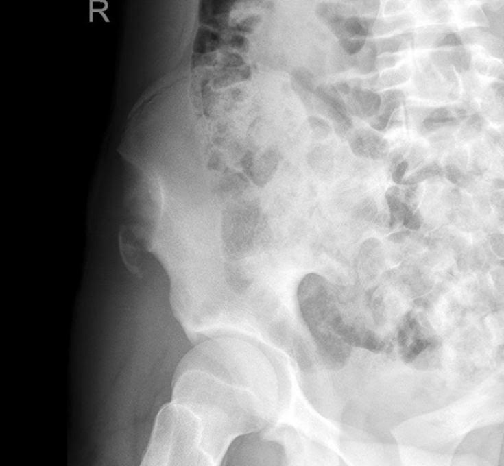
A 15 y.o. female presents to your emergency department with sudden onset hip pain after winding up to kick a soccer ball during her game today.  You see a well-developed female in obvious discomfort, with tenderness to palpation over her lateral hip and pain with passive ROM at the hip.  You obtain this x-ray.  What is your diagnosis?


Show Answer

Answer: Avulsion fracture of the anterior superior iliac spine. 


Injury: 

  • An avulsion fracture of the anterior superior iliac spine is an uncommon injury seen almost exclusively in adolescent athletes. 

  • They mostly result from a sudden, forceful, and/or unbalanced contraction of the attached musculotendinous unit (sartorius muscle or tensor fascia lata) during the starting phase of running, jumping, kicking, etc. 

  • The most implicated sports include soccer, running, and ice hockey. 

History/Exam: 

  • The patient may report sudden shooting pain referred to the involved tuberosity.   

  • Physical exam may include localized edema and tenderness to palpation and pain on passive ROM at the hip. 

Anatomy: 

  • The iliac crest is the weakest component of the pelvic ring during adolescence.  

  • The avulsed fragment is usually displaced distally and laterally. 

Treatment: 

  • Conservative therapy includes 2-3 weeks of limited activity and walking with partial weight bearing restrictions and crutches.  May also include bed rest with the affected lower extremity positioned with the hip and knee in flexion to ensure minimal tension of the muscles attaching to the ASIS.  This is considered for minimally displaced fractures in younger children.  

  • Surgical management generally consists of ORIF with a lag screw, and is generally recommended for patients with fracture fragments > 3cm or severely displaced fragments that cause compression of the lateral cutaneous nerve resulting in meralgia parasthetica.

Show References

Rossi F, Dragoni S. Acute avulsion fractures of the pelvis in adolescent competitive athletes: prevalence, location and sports distribution of 203 cases collected. Skeletal Radiol. 2001;30(3):127-31.


Kautzner J, Trc T, Havlas V. Comparison of conservative against surgical treatment of anterior-superior iliac spine avulsion fractures in children and adolescents. Int Orthop. 2014;38(7):1495-8. 


Title: Crystalloid fluid choice in Pediatric Sepsis

Category: Pediatrics

Keywords: lactated ringer, LR, normal saline, NS (PubMed Search)

Posted: 10/25/2019 by Mimi Lu, MD

  • Resuscitation with crystalloid fluid is a cornerstone of pediatric septic shock treatment.
  • Recent publication from the adult literature have suggested that balance crystalloid solutions may be better than 0.9% normal saline (NS) for select conditions.
  • Lactated Ringer's (LR) is a common balance crystalloid solution often used for fluid resuscitation and critically ill patients.
  • However whether resuscitation with balance fluids is associated with improved outcomes compared to NS in pediatric sepsis is unclear.
  • A matched retrospective cohort study of 12,529 pediatric patient with severe sepsis/septic shock at 382 US hospitals compared outcomes with versus without LR as a part of the initial resuscitation.
  • Outcomes includesd: 30-day hospital mortality, acute kidney injury, new dialysis, and length of stay.
  • After matching, mortality was not different between LR and NS groups. There were no differences in secondary outcomes except longer hospital length of stay in the LR groups.
  • The PRoMPT BOLUS randomized control trial pilot was a feasibility study designed to study the comparative effectiveness of LR versus NS fluid resuscitation for pediatic septic shock.  Completion of a more robust study may help provide answers to these ongoing questions. 

Bottom line: Balance fluid resuscitation with LR was not associated with improved outcomes compared to NS and pediatric sepsis. Selective LR use necessitates a prospective trial to definitively determine comparative effects among crystalloids.

Show References

1. Weiss SL, Keele L, Balanmuth F, Vendetti N, Ross R, Fitzgerald JC, Gerber JS. Crystalloid Fluid Choice and Clinical Outcomes in Pediatric Sepsis: A Matched Retrospective Cohort Study. J Pdatr.207 Mar:182:304-310.

2. Balamuth F, Kittick M, McBride P, Woodford AL, Vestal N, Casper TC, Metheney M, Smith K, Atkin NJ, Baren JM, Dean JM, Kuppermann N, Weiss SL.  Pragmatic Pediatric Trial of Balanced versus Normal Saline Fluid in Sepsis: The PRoMPT BOLUS Randomized Controlled Trial Pilot Feasibility Study. Acad Emerg Med. 2019 Jun 10


Title: UTI Calculator

Category: Pediatrics

Keywords: UTIcalc, SBI, serious bacterial infection, febrile infant, urinary tract infection (PubMed Search)

Posted: 9/13/2019 by Mimi Lu, MD

Question:  In febrile children younger than 2 years, what combination of clinical and laboratory variables best predicts the probability of a urinary tract infection?

Given that urinary tract infections (UTI) are the most common source of serious or invasive bacterial infections in young febrile infants, early identification and treatment has the potential to reduce poor outcomes.  Wouldn't it be great if there was an easy way to identify patients at highest risk?

Researchers from the Children’s Hospital of Pittsburgh formulated a calculator (UTICalc) that first estimates the probability of urinary tract infection (UTI) based on clinical variables and then updates that probability based on laboratory results.

  • The nested case-control study of 2,070 children aged 2 to 23 months with a documented temperature of 38°C or higher
  • In contrast with the American Academy of Pediatrics algorithm, the clinical model in UTICalc reduced testing by 8.1% (95% CI, 4.2%-12.0%) AND decreased the number of missed UTIs.

Bottom line:

The UTICalc calculator can be used to guide to tailor testing and treatment in children with suspected urinary tract infection with the hope of improving outcomes for children with UTI by reducing the number of treatment delays.

Go ahead and give it a click!! https://uticalc.pitt.edu/

Show References

Shaikh N, Hoberman A, Hum SW, Alberty A, Muniz G, Kurs-Lasky M, Landsittel D, Shope T. Development and Validation of a Calculator for Estimating the Probability of Urinary Tract Infection in Young Febrile Children. JAMA Pediatr. 2018 Jun 1;172(6):550-556.


Title: Rock Paper Scissors OK ! (submitted by Leen Ablaihed, MBBS, MHA)

Category: Pediatrics

Keywords: NV exam, neurovascular, upper extremity injury, orthopedics, hand, fracture, supracondylar (PubMed Search)

Posted: 8/23/2019 by Mimi Lu, MD (Updated: 8/23/2019)

  • The assessment of peripheral nerves in children with upper limb injuries can be challenging. 
  • Neurovascular deficit was not documented in 25% of children presenting with upper extremity injury
  • BOAST (British Orthopedic Association Standards for Trauma) guidelines state that each of the Median, Ulnar, Radial, Anterior Interosseous Nerve exams must be individually documented in any supracondylar fracture
  • Dawson described an easy way to test and document your exam. Have the child play “Rock, Paper, Scissors, Ok”
    • Rock: tests the Median nerve
    • Paper: tests the Radial nerve
    • Scissors: tests the Ulnar nerve
    • Ok: tests the Anterior Interosseous nerve
  • This method increased proper documentation and reduced missed nerve injuries in upper extremity fractures.
  • Dr. Sarah Edwards and Dr. Hannah Lock created an easy infographic in the link below and found near 100% increase in NV documentation in their ED. Their poster won the prize for best infographic at the 2018 Emergency Medicine Educators' Conference (EMEC)
  • https://www.peminfographics.com/infographics/rock-paper-scissors-ok


Show References

  • Dawson AW. Rock-Paper-Scissors. Injury 2003 Jan;34(1):61-3
  • Marsh AG, Robertson JS, Goodman A, Boyle J, Huntly JS. Introduction of a simple guideline to improve neurological assessment of paediatric patients presenting with upper limb fractures.  Emerg Med J. 2016 Apr;(33)4:273-7


Title: Urethral prolapse in prepubertal females (submitted by Megan Kirk, MD)

Category: Pediatrics

Posted: 5/24/2019 by Mimi Lu, MD

Presentation:

- Prepubertal females are especially susceptible to urethral prolapse

- Can present incidentally is a painless mass found during bathing or on exam

- More commonly presents as urogenital bleeding, dysuria, or (rarely) urinary retention


Evaluation:

- Appears as a partial or circumferential "donut" of bright red, often friable prolapsed mucosa

- Typically occurs in the setting of UTI, cough, or constipation

- Need to rule out complications: UTI, urethral necrosis, and urinary retention

Treatment:

- Medical management start with sitz baths twice daily and addressing causative factors (treatment constipation, UTI, etc.)

- Can add either topical corticosteroid (hydrocortisone) or estrogen (Estrace or Premarin 0.01% twice daily)

- Urology follow-up necessary as many will require surgical resection of prolapsed mucosa


Title: Identifying children at risk for Persistent Post-Concussive Symptoms (submitted by Aleta Hong, MD)

Category: Pediatrics

Keywords: closed head injury, concussion, CHI (PubMed Search)

Posted: 4/12/2019 by Mimi Lu, MD

Over 630,000 children visit the ED every year with a diagnosis of concussion

Predictors of persistent post-concussive symptoms (PPCS):

  • female sex
  • age over 13 years
  • previous concussive symptoms lasting over 1 week
  • headache
  • sensistivity to noise
  • fatigue
  • slow response to questions.

Appromixately 1/3 of pediatric patients will have PPCS lasting over 2 weeks

Likelihood of PPCS increases to >50% in those with risk factors identified in the ED

Every state has a youth concussion law. The basic tenants are a) immediate removal from play b) written clearance from health professional to return to play c) education for athlete, parents, coaches.

Show References

  • Babcock, L, Kurowski, BG. Identifying childeren and adolescents at risk for persistent postconcussion symptoms. JAMA. 2016. 315(10): 987-988.
  • Kruse AJ, Nugent, AS, Peterson AR. Using sideline concussion tests in the emergency department. Open Access EM. 2018(10): 113-121. 
  • Tarimala, A, Singichetti, B. Initial emergency department visit and follow-up care for concussions among children with Medicaid. J. of Pediatrics. 2018(206): 178-183.


Title: The Hyperoxia Test for the Cyanotic Infant (submitted by Nicholas Fern, MBBS)

Category: Pediatrics

Keywords: CCHD, congenital cardiac lesions, congenital heart disease (PubMed Search)

Posted: 2/23/2019 by Mimi Lu, MD

The hyperoxia-hyperventilation test (aka 100% Oxygen Challenge test) is used to differentiate the cause of central cyanosis in the sick neonate. The majority of neonatal cyanosis is caused by either cardiac or respiratory pathology.

Classically the test is performed as follows:

1. An ABG is obtained with the neonate breathing room air

2. The patient is placed on 100% FiO2 for 10 minutes

3. A repeat ABG is performed looking for an increase in PaO2 to >150 mmHg

 -   If the hypoxia is secondary to a respiratory cause, the PaO2 should increase to >150 mmHg.

-    If the hypoxia is secondary to a congenital cardiac lesion (i.e. secondary to a right-to-left cardiac shunt) the PaO2 is not expected to rise significantly. 

In practice, many physicians instead use pulse oximetry and monitor the SpO2 pre and post administration of 10 minutes of 100% FiO2.

-          If after 10min of 100% FiO2, if SpO2 is not ? 95% (some resources use 85%) then the central cyanosis is likely secondary to intracardiac shunt.

-          When this occurs, presume the sick neonate is symptomatic from a congenital cardiac lesion and initiate prostaglandin E-1 (PGE1) at 0.05-0.01 mcg/kg/min. Use caution as PGE1 may cause apnea.


Title: Acute Disseminated Encephalomyelitis (submitted by Musidapa Rouzi, MD)

Category: Pediatrics

Keywords: ADEM (PubMed Search)

Posted: 1/11/2019 by Mimi Lu, MD

Acute Disseminated Encephalomyelitis (ADEM) is primarily a pediatric disease and can cause a wide variety of neurologic symptoms. As such, should always be in the differential for pediatric patient presenting with vague neurologic symptoms including altered mental status. It is an immune-mediated, demyelinating disease that can affect any part of the CNS; usually preceding a viral illness or rarely, immunizations.

The average age of onset is 5-8 years of age with no gender predilection. It usually has a prodromal. That includes headache, fever, malaise, back pain etc. Neurological symptoms can vary and may present with ataxia, altered mental status, seizures, focal symptoms, behavioral changes or coma.

MRI is the primary modality to diagnose this condition. Other possible indicators may be mild pleocytosis with lymphocyte predominance, and elevated inflammatory markers such as ESR, CRP. These findings, however, are neither sensitive nor specific.

First-line treatment for ADEM is systemic corticosteroids, typically 20-30 mg/kg of methylprednisolone for 2-5 days, followed by oral prednisone 1-2 mg/kg for 1-2 weeks then 3-6-week taper. For steroid refractory cases, IVIG and plasmapheresis may be considered.

ADEM usually has a favorable long-term prognosis in the majority of patients. However, some may experience residual neurological deficits including ataxia, blindness, clumsiness, etc.

Take home points:

  • Always keep ADEM on the differential for any pediatric patient presenting with any neurologic symptoms
  • MRI is the diagnostic modality of choice.
  • If ADEM diagnosed, start treatment early in conjunction with pediatric neurology.


Show References

Gray, M.P. & Goralick, M.H. (2016).  Acute disseminated encephalomyelitis (6th ed, volume 32). Milkwaukee, WI


Title: Isolated vomiting and head injury in children

Category: Pediatrics

Keywords: PECARN, traumatic brain injury, head injury, concussion (PubMed Search)

Posted: 11/9/2018 by Mimi Lu, MD (Updated: 11/9/2018)

5 year old previously healthy male referred to the ED for vomiting after he fell 2.5 feet while jumping from the couch.  No other injurys noted and no other pain reported. He denies a headache and parents report he is acting baseline. His exam is reassuring (no, really....)
 
What would you do next?  Which Clinical Decision Rule (CDR) do you use?  PECARN? CHALICE? CATCH?
What if he vomited 3 times? 5 times?
 
A secondary analysis of the Australasian Paediatric Head Injury Rule Study attempted to determine the prevalence of traumatic brain injuries in children who vomit after head injury and identify variables from published CDRs that increased risk.  Vomited characteristics were correlated with CDR predictors and the presence of clinically important traumatic brain injury (ciTBI) or traumatic brain injury on computed tomography (TBI-CT).
 
Of the 19 920 children enrolled, 3389 (17.0%) had any vomiting. With isolated vomiting, only 1 (0.3%; 95% CI 0.0%-0.9%) had ciTBI and 2 (0.6%; 95% CI 0.0%-1.4%) had TBI-CT. Predictors of increased risk of ciTBI with vomiting included: signs of skull fracture, altered mental status, headache, and acting abnormally.

Bottom Line:

TBI-CT and ciTBI are uncommon in children presenting with head injury with isolated vomiting  (vomiting without other CDR predictors) and observation without imaging appears appropriate.


Show References


Borland ML, Dalziel SR, Phillips N, Dalton S, Lyttle MD, Bressan S, Oakley E, Hearps SJC, Kochar A, Furyk J, Cheek JA, Neutze J, Babl FE; Paediatric Research in Emergency Department International Collaborative group. Vomiting With Head Trauma and Risk of Traumatic Brain Injury. Pediatrics. 2018 Apr;141(4).


Title: Infantile spasms - subtle findings, profound diagnosis (submitted by John Peffer, MD)

Category: Pediatrics

Keywords: West syndrome, seizures (PubMed Search)

Posted: 9/21/2018 by Mimi Lu, MD

Originally described a Dr. West in 1841 – it is a rare (~1200 cases annually)  seizure disorder in young kids, generally less than 1 year old.  Very subtle appearance, often with only bending forward or ‘jerking’ of the extremities as opposed to Brief Resolved Unexplained Event (BRUE) or tonic-clonic in description.  The spasms can be thought of as a syndrome, where 70% of those have an undiagnosed rare metabolic/genetic disease.

A prompt evaluation, including labs, EEG, MRI, metabolic and genetic studies is vital in helping to establish a diagnosis which can have a profound impact on the patients prognosis. Examples might include Tuberous Sclerosis, Pyridoxine Dependent Seizures among over 50 others.

Bottom line: In pediatric patients less than 1 year old who present to the Emergency Department with a description of spasm-like episodes, consider Infantile Spasms on the differential, and consult your friendly neighborhood Pediatric Neurologist for help in determining a proper disposition.

Show References

http://www.childneurologyfoundation.org/disorders/infantile-spasms/

https://www.ninds.nih.gov/Disorders/All-Disorders/Infantile-Spasms-Information-Page

https://www.ncbi.nlm.nih.gov/pmc/articles/PMC1464162/

https://www.healthychildren.org/English/health-issues/conditions/head-neck-nervous-system/Pages/Infantile-Spasms-What-Parents-Need-to-Know.aspx


Title: CDC Guideline on Mild Traumatic Brain Injury Among Children

Category: Pediatrics

Keywords: Concussion, minor head injury, traumatic brain injury, mTBI (PubMed Search)

Posted: 9/14/2018 by Mimi Lu, MD

The Centers for Disease Control and Prevention recently released guidelines on the diagnosis and management of mild traumatic brain injury (mTBI**) among children. From 2005-2009, children made almost 3 million ED visits for mTBI. Based on a systemic review of the literature, the guideline includes 19 sets of recommendations on the diagnosis, prognosis, and management/treatment of pediatric mTBI.

Key Recommendations:

1. Do not routinely image patients to diagnose mTBI (utilize clinical decision rules to identify children at low risk and high risk for intracranial injury (ICI), e.g. PECARN)

2. Use validated, age-appropriate symptoms scales to diagnose mTBI

3. Assess evidence-based risk factors for prolonged recovery.  No single factor is strongly predictive of outcome.

4. Provide patients with instructions on return to activity customized with their symptoms (see CDC Resources below)

5.  Counsel patients to return gradually to non-sports activities after no more than 2-3 days of rest.


A wealth for information and tools for provder and families can be found at:

www.cdc.gov/HEADSUP (including evaluation forms and care plans for providers)

www.cdc.gov/traumaticbraininjury/PediatricmTBIGuideline.html

Show Additional Information

**Although concussion, minor head injury, and mBI are frequently used interchangeably, they have different connotations which allows for misinterpretation and confusion. The guideline recommends the clinical use of the single term mild traumatic brain injury. This is defined as "an acute brain injury resulting from mechanical injury to the head from external physical forces including: (1) 1 or more of the following: Confusion or disorientation, loss of consciousness for 30 minutes or less, posttraumatic amnesia for less than 24 hours, and/or other transient neurologic abnormality such as focal signs, symptoms, or seizure; (2) Glasgow Coma Scale score of 13-15 after 30 minutes post injury or later upon presentation for healthcare


Show References

Diagnosis and management of mild traumatic brain injury in children: A systemic review. Lumba-Brown A, Yeates KO, Sarmiento K, Breiding MJ, Haegerich TM, Gioia GA, Turner M, Benzel EC, Suskuer SJ, Giza CC, Joseph M,Broomand C, Weissa B, Gordon W Wright DW, Moser RS, McAvoy K, Ewing-Cobbs L, Duaime AC, Putukian M, Holhouse B, Paulk D, Wade SL, Herig SA, HalsteadM, Keenan H, Choe M, Christia CW, Gusiewic K, Raksin PB, Gregory A, Mucha A, Taylor HG, Callahan JM, DeWtt J, Collins MW, Kirkwood MW, Ragheb J, Ellenbogen RG, Spinks TJ, Ganiats TG, Sabelhaus LJ, Altenhofen K, Hoffman , Getchius T, Gronseh G,Donnell Z, O'Connor RE, Timmons SD JAMA Pediatr 2018 Sept 4.


Title: Fluids in Pediatric Diabetic Ketoacidosis (with contribution by Alexis Salerno, MD)

Category: Pediatrics

Keywords: DKA, cerebral edema, PECARN (PubMed Search)

Posted: 6/22/2018 by Mimi Lu, MD

Children with diabetic ketoacidosis (DKA) may have brain injuries ranging from mild to severe. The debate over the contribution from intravenous fluids towards poor neurologic outcomes has been ongoing for decades. 

PECARN's large multicenter randomized, controlled trial examined the effects of the rate of administration and the sodium chloride content of intravenous fluids on neurologic outcomes in children with diabetic ketoacidosis may finally put the controversy to rest. There was no difference on significant neurologic outcomes based on the rate (fast vs slow) or concentration (0.9% vs 0.45%) of IV fluid administration.

Clinically apparent brain injury occurred in 12 of 1389 episodes (0.9%) of children in DKA.

Any change in the mental or neurological status of the patient should be concerning for life threatening edema and should be treated with mannitol 1g/kg IV bolus or hypertonic saline (3%) 5-10 mL/kg IV over 30 minutes.

BOTTOM LINE:

"Neither the rate of administration nor the sodium chloride content of intravenous fluids significantly influenced neurologic outcomes in children with diabetic ketoacidosis"

Show References

Kuppermann N, Ghetti S, Schunk JE, Stoner MJ, Rewers A, McManemy JK, Myers SR, Nigrovic LE, Garro A, Brown KM, Quayle KS, Trainor JL, Tzimenatos L, Bennett JE, DePiero AD, Kwok MY, Perry CS 3rd, Olsen CS, Casper TC, Dean JM, Glaser NS; PECARN DKA FLUID Study Group. Clinical Trial of Fluid Infusion Rates for Pediatric Diabetic Ketoacidosis. N Engl J Med. 2018 Jun 14;378(24):2275-2287.

Long, B; Koyfman, A. Emergency medicine myths: cerebral edema in pediatric diabetic ketoacidosis and intravenous fluids. J. Emerg. Med; 2017:53(2),212-221.


Title: Conjunctivitis-otitis syndrome

Category: Pediatrics

Keywords: augmentin, conjunctivitis, AOM, otitis media (PubMed Search)

Posted: 6/8/2018 by Mimi Lu, MD

Although conjuncitivitis outside of the neonatal period is commonly caused by viruses, there are times when antibiotics are warranted due to bacterial infections, such as conjuncitivits-otitis syndrome.

  • up to 25% of patients with conjunctivitis have concurrent otitis media (even in the abscence of ear pain) and up to 73% of patients with purulent conjunctivitis
  • Non-typeable H. influenzae is the most common recovered bacteria.
  • For these patients, systemic (oral) antibiotics are recommended and the topical ophthalmic antibiotics are NOT necessary.
  • Antibiotics should cover beta-lactamase producing organisms, e.g. high dose amoxicillin-clavulanic acid (45 mg/kg BID; 600 mg/5mL concentration which is formulated to have less clavulanic acid

Bottom line: Every patient with conjunctivitis should have an examination of his/her TMs, as your management may change.

Show References

Teoh DL, Reynolds S. Diagnosis and management of pediatric conjunctivitis. Pediatric Emergency Care: 2003; 19(1), pp. 48-55.

Bodor FF, Marchant CD, Shurin PA, Barenkamp SJ. Bacterial etiology of conjunctivitis-otitis media syndrome. Pediatrics: 1985; 76(1), pp.26-28.

Bodor FF. Conjunctivitis-Otitis Syndrome. Pediatrics: 1982; 69(6), 695-698.


Title: Mucositis... when the shoe doesn't fit (submitted by Alexis Salerno, MD)

Category: Pediatrics

Keywords: Kawasaki's disease, SJS, TEN, dermatitis (PubMed Search)

Posted: 2/9/2018 by Mimi Lu, MD

Question

Case:  5 year old presents to the ED with 2 weeks of fever. She has extensive cracked, bleeding lips and a rash on her hands and feet. She was recently diagnosed with “walking pneumonia” and hand, foot and mouth disease this week. Her pediatrician sent her in for further workup after she was found to have an elevated CRP on outpatient labs. A similar picture appears in the link below:

http://www.eblue.org/cms/attachment/2024057003/2043959646/gr1_lrg.jpg.

What's the diagnosis?


Show Answer

Answer: Mycoplasma pneumoniae-induced rash and mucositis

The diagnosis of Mycoplasma pneumonia-induced rash and mucositis (MIRM) was recently termed in the 2015 Journal of American Academy of Dermatology. It is characterized by mucocutaneous eruptions with prominent mucosal involvement.  94% of patients in the reviewed cases had extensive oral lesions that can range from erosions, ulcers or vesiculobullous lesions. 82% of patients had ocular involvement characterized by purulent bilateral conjunctivitis. In 63% of cases, patients were found to have urogenital lesions.  Almost all of these patients had prodromal symptoms of cough and fever preceding the eruption by 1 week.  The disease was found to be most prominent with young (11.9 ± 8.8 years) and with a 66% male predominance.  The treatment is antibiotics such as azithromycin and oral corticosteroids with a minority of patients requiring IVIG. These patients have a good prognosis.  

Bottom Line: Consider MIRM in patients with extensive mucosal disease that do not completely fit the criteria of Kawasaki’s or Stevens-Johnson Syndrome/ Toxic Epidermal Necrolysis.


Reference:

Canavan TN, Mathes EF, Frieden I, Shinkai K. Mycoplasma pneumoniae-induced rash and mucositis as a syndrome distinct from Stevens-Johnson syndrome and erythema multiforme: a systematic review. J Am Acad Dermatol. 2015

Feb;72(2):239-45.


Title: The Pediatric Tongue Laceration (submitted by J. David Gatz, MD)

Category: Pediatrics

Posted: 1/12/2018 by Mimi Lu, MD

Tongue laceration is a common injury in children - occurring in the setting of falls and seizures. The most common location is the anterior dorsal portion of the tongue. Priorities are to evaluate for airway compromise (swelling, hematoma, bleeding) and retained foreign bodies (teeth fragments, etc). The vast majority of lacerations DO NOT require repair and do well with routine dental hygiene and antiseptic mouth wash. While there is no clear consensus for indications to repair, considerations include uncontrolled bleeding, airway compromise, wounds greater than 2 cm, and wounds that gape while the tongue is still in the mouth. Use large absorbable sutures (like 4-0 chromic gut). Check out this great video from EM:RAP - https://youtu.be/h14KyO8JlZE

Title: Are viral upper respiratory tract infections an important risk factor for the development of acute otitis media? (submitted by Brian Parker, MD)

Category: Pediatrics

Keywords: URI, AOM, wait-and-see, antibiotic stewardship (PubMed Search)

Posted: 11/24/2017 by Mimi Lu, MD

As we are approaching the winter in the northern hemisphere, the number of visits for ear pain or respiratory symptoms are expected to increase.  The occurrence of acute otitis media (AOM) will also increase, but are these two disease processes related?

Drs. Heikkinen and Chonmaitree published a systematic review of previously reported studies regarding the correlation of these two disease processes (1).  As far back as 1990, studies have shown that up to 94% of pediatric patients diagnosed with AOM have concomitant upper respiratory infection (URI) type symptoms at time of diagnosis (2).   The viral infections most commonly associated with AOM are respiratory syncytial virus, influenza virus, and adenovirus (3).

The most commonly taught risk factors for developing AOM include young age, male gender, multiple siblings, day care attendance, and passive smoking.  These factors are also related to the development of upper respiratory symptoms, and the development of AOM should be thought of as a complication of the upper respiratory infection (4). 

Koivunen et al noted the highest incidence of AOM at day 3 after the onset of an URI, and the median time to diagnosis was day 4 (5). If you see a patient in day 2-4 of an URI, who has started to develop an ear effusion, but not clinical AOM, you may want to consider a “Wait-to-see” treatment option if the patient meets treatment criteria (https://em.umaryland.edu/educational_pearls/2049/).

Show References

(1) Heikkinen T, Chonmaitree T. Importance of Respiratory Viruses in Acute Otitis Media. Clinical Microbiology Reviews. 2003;16(2):230-241.

 (2) Arola M, Ruuskanen O, Ziegler T, Mertsola J, Näntö-Salonen K, Putto-Laurila A, Viljanen MK, Halonen P. Pediatrics. 1990 Dec; 86(6):848-55.
 
(3) Henderson FW, Collier AM, Sanyal MA, Watkins JM, Fairclough DL, Clyde WA Jr, Denny FW, N Engl J Med. 1982 Jun 10; 306(23):1377-83.
 
(4) Daly KA, Brown JE, Lindgren BR, Meland MH, Le CT, Giebink GS. Pediatrics. 1999 Jun; 103(6 Pt 1):1158-66.
 
(5) Koivunen, P., T. Kontiokari, M. Niemelä, T. Pokka, and M. Uhari. 1999. Time to development of acute otitis media during an upper respiratory tract infection in children. Pediatr. Infect. Dis. J. 18:303-305.


Title: Pediatric ARDS continued...

Category: Pediatrics

Keywords: ARDS, oxygenation index, OI, PALICC, acute lung injury, respiratory distress, PARDS (PubMed Search)

Posted: 10/27/2017 by Mimi Lu, MD

Some pediatric practitioners have adopted the oxygenation index (OI) ([FiO2 × mean airway pressure (Paw) × 100]/ PaO2) or oxygen saturation index (OSI) ([FiO2 × Paw × 100]/ SpO2) to assess hypoxemia in children instead of P/F ratios because of the less standardized approach to positive pressure ventilation in children relative to adults. 

OI can be used in pediatric patients to define severity of Acute Respiratory Distress Syndrome (ARDS) in patients receiving invasive mechanical ventilation and assess for potential ECMO treatment. 

In contrast, the P/F ratio should be used to diagnose Pediatric ARDS for patients receiving noninvasive continuous positive airway pressure [CPAP] or bilevel positive airway pressure [BiPAP]) with a minimum CPAP of 5 cm H2O.

Oxygen Index (OI) = FiO2 x MAP x 100
                                 ---------------------
                                         PaO2

  • Mild ARDS: 4 ≤ OI ≤ 8
  • Moderate ARDS: 8 ≤ OI < 16
  • Severe ARDS: OI ≥ 16
  • OI < 25: good outcome
  • OI 25-40: >40% mortality
  • OI > 40: Consider ECMO

Show References

1) Trachsel D, McCrindle BW, Nakagawa S, Bohn D. Oxygenation index predicts outcome in children wtih acute hypoxemic respiratory failure. Am J Respir Crit Care Med. 2005 Jul 15;172(2):206-11.
 
2) Pediatric Acute Respiratory Distress Syndrome: Consensus Recommendations from the Pediatric Acute Lung Injury Consensus Conference.  Pediatric Acute Lung Injury Consensus Conference Group. Pediatr Crit Care Med. 2015 Jun;16(5):428-39


Title: Blunt Renal Trauma in pediatrics (submitted by Elizabeth England, MD)

Category: Pediatrics

Keywords: Trauma, hematuria, kidney injury (PubMed Search)

Posted: 10/13/2017 by Mimi Lu, MD

Pediatric patients are at a higher risk of blunt renal injury due to multiple anatomic features, include relatively less protective perinephric fat and surrounding musculature, and larger size of the kidneys in relation to the abdomen compared to their adult counterparts (1). For this reason, it is important to keep a high clinical suspicion for renal injury in the pediatric patient with blunt abdominal trauma, particularly in those with lower rib fractures, direct injury, flank ecchymosis and/or tenderness, rapid deceleration injury, or other significant traumatic mechanism (2). Despite the risk of radiation exposure, the preferred imaging modality for the diagnosis of renal injury in pediatric patients is computed tomography (similar to adults). Studies evaluating the utility of renal ultrasound have demonstrated poor sensitivity with a decreased likelihood of diagnosing low-grade injuries. While ultrasound may be a useful screening tool to evaluate for severe injury, it should not be used to rule out traumatic injury (1). Take home point: Keep a high suspicion for renal injury in pediatric patients with blunt abdominal trauma and confirm the diagnosis with computed tomography of the abdomen and pelvis with contrast.

Show References

(1) Fraser, J.D., Aguayo, P., Ostlie, D.J. et al. Pediatr Surg Int (2009) 25: 125. https://doi.org/10.1007/s00383-008-2316-4 (2) Gerstenbluth RE, Spirnak JP, Elder JS. Sports participation and high grade renal injuries in children. J Urol 2002; 168:2575.


Title: Pediatric Acute Respiratory Distress Syndrome (ARDS)

Category: Pediatrics

Keywords: ARDS, oxygenation index, OI, PALICC, acute lung injury (PubMed Search)

Posted: 9/22/2017 by Mimi Lu, MD (Updated: 10/27/2017)

Since the first description of acute respiratory distress syndrome (ARDS), various consensus conferences (including American-European Consensus Conference (AECC) and the Berlin Conference) have produced definitions focused on adult lung injury but have limitations when applied to children. 

This prompted the organization of the Pediatric Acute Lung Injury Consensus Conference (PALICC), comprised of  27 experts, representing 21 academic institutions and eight countries.  The goals of the conference were 1) to define pediatric ARDS (PARDS); 2) to offer recommendations regarding therapeutic support; and 3) to identify priorities for future research in PARDS.

Although there were several recommendations from the group, some notable ones, in contrast to the Berlin definition focused on adults, include: 1) use the Oxygenation Index (or, if an arterial blood gas is not available, the Oxygenation Severity Index) rather than the P/F ratio; 2) elimination of the requirement for “bilateral” pulmonary infiltrates (may be unilateral or bilateral) 3) elimination of  specific age criteria for PARDS.

Tune in next month for pearls on management for children with PARDS...

Show References

Pediatric Acute Respiratory Distress Syndrome: Consensus Recommendations from the Pediatric Acute Lung Injury Consensus Conference.  Pediatric Acute Lung Injury Consensus Conference Group. Pediatr Crit Care Med. 2015 Jun;16(5):428-39

Collaborators: Jouvet P, Thomas NJ, Wilson DF, Erickson S, Khemani R, Zimmerman J, Dahmer M, Flori H, Quasney M, Sapru A, Cheifetz IM, Rimensberger PC, Kneyber M, Tamburro RF, Curley MA, Nadkarni V, Valentine S, Emeriaud G, Newth C, Carroll CL, Essouri S, Dalton H, Macrae D, Lopez-Cruces Y, Quasney M, Santschi M, Watson RS, Bembea M.


Title: What about Anaphylaxis in kids? (submitted by Yitschok Applebaum, MD)

Category: Pediatrics

Keywords: allergic reaction, anaphylaxis, auto-injector, epi-pen (PubMed Search)

Posted: 8/25/2017 by Mimi Lu, MD (Updated: 8/25/2017)

What if you were out in public and a 1 year old child (est 10 kg) suddenly develops anaphylaxis but you only have an epinephrine auto-injector with the “adult” dose of 0.3 mg.  Is it safe to give?

Anaphylaxis is a life threatening emergency with mortality of up to 2% [1]. Early recognition is imperative and administration of timely Epinephrine is the single most important intervention [2]. While providers may be hesitant to administer epinephrine in older patients due to fear of precipitating adverse cardiovascular events, they may also hesitate in younger patients due to fear of overdose. 

Iimmediate administration with any dose available is recommended because:

  • the risks of untreated anaphylaxis are greater than the risk of over-treating with epinephrine.
  • 20% of Anaphylaxis patients require a second dose of Epinephrine [3].
  • The recommended IM dose of 0.01mg /kg was determined arbitrarily.
  • The vast majority of epinephrine overdoses are via IV injection at doses 100 - 1000 fold the recommended  IV dose [4]

Bottom line:

There are no absolute contraindications (including age) for epinephrine in patients with anaphylaxis.  Give the initial dose IM into the anterolateral thigh.

Show References

1-  Bock SA, Muñoz-Furlong A, Sampson HA. Fatalities due to anaphylactic reactions to foods. J Allergy Clin Immunol. 2001 Jan. 107(1):191-3. [Medline].

2-  Sheikh A, Shehata YA, Brown SG, Simons FE. Adrenaline for the treatment of anaphylaxis: cochrane systematic review. Allergy 2009; 64:204.

3-  Oren E, Banerji A, Clark S, Camargo CA Jr. Food-induced anaphylaxis and repeated epinephrine treatments. Ann Allergy Asthma Immunol 2007; 99:429.

4-  Wood JP, Traub SJ, Lipinski C. Safety of epinephrine for anaphylaxis in the emergency setting. World Journal of Emergency Medicine. 2013;4(4):245-251. doi:10.5847/wjem.j.issn.1920-8642.2013.04.001.


Previous |  1 |  2 |  3 |  4 |  5 |  Next
University of Maryland School of Medicine logo
  • Departments
  • Programs
  • Research Centers
  • Institutes
  • Graduate Education
  • CIBR Cores
  • Offices of the Dean
    • University of Maryland, Baltimore
    • University of Maryland Medical Center
    • Medical Alumni Association of the University of Maryland
    • Health Sciences and Human Services Library
University of Maryland School of Medicine
655 W. Baltimore Street
Baltimore, MD 21201
Contact Us

© University of Maryland School of Medicine

    • Webmaster
    • Web Accessibility
    • Site Index
    • UMB Hotline
    • Employment
    • Map