
Search
Subscribe to daily UMEM Pearls (FREE!) via:
41-60 of 384 results with category "Neurology"
- The ACEP clinical policy on the evaluation and management of acute nontraumatic headache in the ED was recently updated.
- Similar to prior policies, it focuses on the diagnosis of subarachnoid hemorrhage (SAH) due to the disproportionate amount of literature in comparison to other high risk etiologies.
- In summary:
-
- Are there risk-stratification strategies that reliably identify the need for emergent neuroimaging?
- The Ottawa SAH Rule has a high sensitivity but low specificity for patients presenting with a normal neurological exam and peak headache intensity within 1 hour of symptom onset (Level B recommendation).
- Caution in application of this rule, as use in the incorrect population may increase unnecessary testing.
- Are nonopioids preferred to opioids for treatment of acute primary headache?
- Preferentially use nonopioid medications in the treatment of acute primary headaches in ED patients (Level A recommendation).
- Consider discharge medication and education to reduce headache recurrence and repeat ED visit.
- Does a normal noncontrast head CT performed within 6 hours of headache onset preclude the need for further diagnostic workup for SAH?
- Noncontrast head CT using at least a 3rd generation scanner performed within 6 hours of headache onset can be used to rule out nontraumatic SAH (Level B recommendation).
- If clinical suspicion remains high despite the negative noncontrast head CT, further evaluation may be pursued.
- In a patient who is still considered to be at risk for SAH after a negative noncontrast head CT, is CTA as effective as LP to rule out SAH?
- Use shared decision making to select the best modality for each patient after weighing the potential for false-positive CTA and the pros/cons associated with LP (Level C recommendation).
- Are there risk-stratification strategies that reliably identify the need for emergent neuroimaging?
- This clinical policy does not address the evaluation of other potential etiologies for acute headache, including in the pregnant woman and postpartum woman.
Show References
- Elevated BP is common with acute ICH and is associated with hematoma expansion and worse outcome.
- Early BP lowering in ICH appear to be safe, though did not improve outcomes in the two largest trials INTERACT2 and ATACH-II.
- A preplanned pooled analysis of 3829 patients from these 2 trials found:
- Every 10 mmHg reduction in SBP was associated with a 10% increase in odds of better functional recovery.
- Reduced variability of SBP was associated with improved outcomes.
- The association between BP variability and outcomes in ICH has been observed in several other recent studies.
Bottom Line: Reduced SBP variability is associated with improved outcomes in ICH.
Show References
- Symptoms/signs that suggest serious underlying conditions causing headaches are summarized by the mnemonic SNOOP:
- S - Systemic symptoms/signs/disease
- e.g. fever, weight loss, HIV, malignancy, pregnancy
- N - Neurologic symptoms/signs
- e.g. altered mental status, diplopia, pulsatile tinnitus, loss of consciousness
- O - Onset sudden, abrupt, thunderclap
- i.e. pain reaches maximal intensity instantly after onset
- O - Older age of onset, especially > 50 years
- P - Pattern change
- e.g. change in frequency, severity, clinical features, precipitated by Valsalva, aggravated by postural change
- S - Systemic symptoms/signs/disease
- Consider structural pathologies, vascular disorders, infectious and inflammatory conditions in the evaluation of secondary headache syndromes.
Show References
Primary headaches (not secondary to a life-threatening disease) can be challenging to manage. Remember the following pearls:
-
Things that DO NOT work: IV fluids, 5-HT3 Antagonists (aka Zofran), diphenhydramine (aka Benadryl), opioids
-
Things that KINDA work: oxygen for all headaches, sphenopalatine ganglion block (4% lido spray)
- Things that REALLY work: ketorolac, metoclopramide, prochlorperazine, triptans and ergots, oxygen for cluster headaches
-
Things that PREVENT recurrence: dexamethasone for migraine headaches
- Benzodiazepines are first-line treatment for status epilepticus.
- Guidelines for the treatment of status epilepticus recommend dosing as:
- 10 mg midazolam IM for patients > 40 kg or 5 mg midazolam IM for patients 13-40 kg
- 0.1 mg/kg lorazepam IV (max 4 mg/dose), can repeat x 1
- 0.15-0.2 mg/kg diazepam IV (max 10 mg/dose), can repeat x 1
- The recent Established Status Epilepticus Treatment Trial (ESETT) compared the treatment of patients who did not respond to benzodiazepines.
- Overall, 29.8% of the first dose of benzodiazepines given in the ED met minimum dose recommendations.
- Dosing for patients < 40 kg more frequently met minimum dose recommendations.
- This study found a pattern of multiple, small doses instead of a single full dose of benzodiazepine as recommended by guidelines.
Bottom Line: Underdosing of benzodiazepines in status epilepticus may contribute to treatment failure.
Show References
Show References
- Hypertonic saline and mannitol are commonly used for management of acute intracranial hypertension and cerebral herniation.
- The choice of medication is often limited by venous access.
- 23.4% NaCl has been shown to decrease intracranial pressure in patients refractory to mannitol.
- It requires administration through a central line to avoid sclerosis of the peripheral veins and tissue necrosis with extravasation.
- Intraosseous (IO) access provides a more rapid route for 23.4% NaCl administration.
- No complications were observed relating to IO insertion site.
- Transient hypotension occurred in more patients who received 23.4% NaCl via IO vs. central line.
Bottom Line: Use of IO allows more rapid administration of 23.4% NaCl with no immediate serious complications.
Show References
- Misdiagnosis of neurologic emergencies can result in serious neurologic dysfunction or death.
- A recent retrospective analysis using AHRQ databases looked at >3 million adults discharged from the ED with diagnoses of atraumatic headache or back pain.
- A serious neurologic condition or death occurred within 30 days after ED discharge in:
- 0.5% of patients with nonspecific diagnosis of headache
- 0.2% of patients with nonspecific diagnosis of back pain
- The frequency of adverse outcome was highest between days 1 and 3 after ED discharge.
- The most frequent adverse outcome was ischemic stroke (18.1%) for headache and intraspinal abscess (44%) for back pain.
- Age ≥ 85, male sex, non-Hispanic white, comorbidities such as neurologic disorders, HIV/AIDS, and malignancy were associated with higher incidence of adverse outcome.
Bottom Line: The rate of serious neurologic conditions missed at an initial ED visit is low. However, the potential harm of misdiagnosis can be substantial.
Show References
Back pain with lower extremity symptoms can be concerning for cauda equina. Some pointers regarding the H&P:
- Symptoms develop within less than 24 hours in 90% of patients
- Urinary retention develops before incontinence, but up to 30% of patients will have neither.
- Saddle anesthesia or hypoesthesia is present in 81% of patients. Perineal numbness may be patchy, mild, and unilateral initially, making it difficult to elicit.
None of these symptoms independently predicts cauda equina syndrome with an accuracy greater than 65%.
Bottom Line: do not depend on any one finding to reliably exclude or confirm cauda equina.
Show References
Show Additional Information
Show References
- Intracerebral hemorrhage (ICH) volume is a predictor of mortality and clinical outcome.
- Communicating ICH volume to neurosurgical and neurocritical care consultants can help direct treatment decisions.
- ICH volume can be estimated using the ABC/2 formula:
- Select the CT slice with the largest area of the hemorrhage (reference slice)
- A = Measure the largest diameter
- B = Measure the largest diameter perpendicular to A
- C = Multiply the number of CT slices with the hemorrhage by the slice thickness
- Slices with 25-75% of the hematoma volume compared to the reference slice count as 1/2 slice
- Slices with <25% of the hematoma volume compared to the reference slice do not count
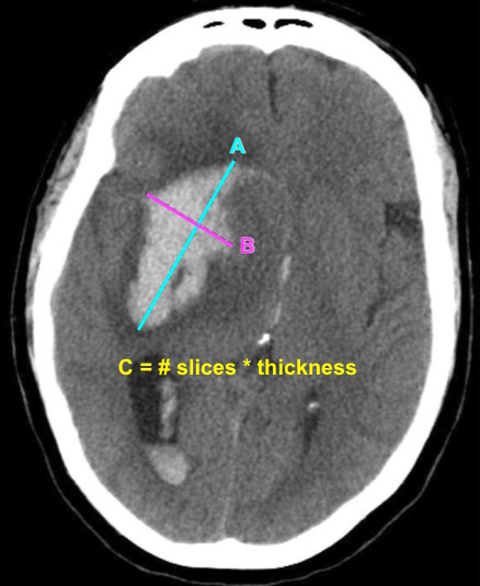
- A recent study by Dsouza et al. found that EM attendings as well as EM trainees were reliable in estimating ICH volume using ABC/2 compared to radiologists.
Bottom Line: EPs can reliably estimate ICH volume using the ABC/2 formula. Communicating ICH volume to neurosurgical and neurocritical care consultants can help direct treatment decisions.
Show References
A previous pearl discussed medication-overuse headache (MOH).
MOH is also known as analgesic rebound headache, drug-induced headache or medication-misuse headache.
It is defined as headache… occurring on 15** or more days per month in a patient with a preexisting headache disorder who has been overusing one or more acute treatment drugs for headache with symptoms for three or more months.
The diagnosis is clinical, and requires a hx of chronic daily headache with analgesic use more than 2-3d per week.
The diagnosis of MOH is supported if headache frequency increases in response to increasing medication use, and/or improves when the overused medication is withdrawn.
The headache may improve transiently with analgesics and returns as the medication wears off. The clinical improvement after wash out is not rapid however, patients may undergo a period where their headaches will get worse. This period could last in the order of a few months in some cases.
The meds can be dc’d cold turkey or tapered depending on clinical scenario.
Greatest in middle aged persons. The prevalence rages from 1% to 2% with a 3:1 female to male ratio.
Migraine is the most common associated primary headache disorder.
** Each medication class has a specific threshold.
Triptans, ergot alkaloids, combination analgesics, or opioids on ten or more days per month constitute medication overuse.
Use of simple analgesics, including aspirin, acetaminophen and NSAIDS on 15 or more days per month constitutes medication overuse.
Caffeine intake of more than 200mg per day increases the risk of MOH.
Consider MOH in patients in the appropriate clinical scenario as sometimes doing less is more!
- Lumbar punctures (LPs) are a common ED procedure with variable reported success rates.
- A recent systematic review and meta-analysis looked at 12 studies comprising 957 adult and pediatric patients comparing pre-procedural ultrasound-assisted LPs with traditional landmark-based technique.
- Some studies utilized ultrasound-assistance in all LPs, others selected patients who were anticipated to be difficult LPs.
- No studies assessed dynamic ultrasound-guided LPs.
- Overall, ultrasound-assisted LP was 90.0% successful compared with landmark-based LP that was 81.4% successful (OR 2.22, 95% CI = 1.03 - 4.77).
- Ultrasound-assisted LP was also associated with reduced rate of traumatic LPs, shorter time to successful LP, and reduced patient pain scores.
Bottom Line: Consider using pre-procedural ultrasound-assistance for all lumbar punctures.
Show References
Diplopia can be a challenging complaint to address in the ED. Although not all patients will require imaging, use the simplified table below to help guide the imaging study needed:
| Clinical Situation | Suspected Diagnosis | Imaging Study | |
| Diplopia + cerebellar signs and symptoms | Brainstem pathology | MRI brain | |
| 6th CN palsy + papilledema | Increased intracranial pressure (e.g. idiopathic intracranial hypertension or cerebral venous thrombosis) | CT/CTV brain | |
| 3rd CN palsy (especially involving the pupil) | Compressive lesion (aneurysm of posterior communicating or internal carotid artery) | CT/CTA brain | |
| Diplopia + thyroid disease + decreased visual acuity | Optic nerve compression | CT orbits | |
| Intranuclear ophthalmoplegia | Multiple sclerosis | MRI brain | |
| Diplopia + facial or head trauma | Fracture causing CN disruption | CT head (dry) | |
| Diplopia + multiple CN involvement (3,4,6) + numbness over V1 and V2 of trigeminal nerve (CN5) +/- proptosis | Unilateral, decreased visual acuity | Orbital apex pathology | CT orbits with contrast |
| Uni- or bi-lateral, normal visual acuity | Cavernous sinus thrombosis | CT/CTV brain | |
C.N.: cranial nerve
Show References
- The Canadian C-Spine Rule (CCR) has been shown to decrease the use of cervical spine imaging in low-risk trauma patients.
- While developed for use by physicians, CCR has also been validated in ED triage nurses with moderate interrater reliability (kappa 0.78) by Stiell et al. in 2010.
- Stiell’s group has since implemented the use of CCR by ED triage nurses at 9 teaching hospitals in Ontario with a combined annual volume of approximately 670,000 ED visits.
- 180 certified nurses evaluated 1408 patients.
- 806 (57.2%) arrived with c-spine immobilization.
- 602 (42.8%) had neck pain but no immobilization.
- Overall, nurses removed immobilization in 331 (41.4%) patients and applied immobilization in 203 (14.4%) patients.
- Diagnostic imaging was performed in 612 (43.4%) patients and found 16 (1.1%) clinically important and 3 (0.6%) clinically unimportant injuries.
- There were no missed c-spine injuries to the knowledge of the authors as the study hospitals were closely connected with the regional spine centers.
- Time from nursing assessment to discharge decreased by 26.0% (3.4h vs. 4.6h)
Bottom Line: ED triage nurses can safely use the Canadian C-Spine Rule. This approach can improve patient care and decrease length of stay in the ED.
Show References
Manifestations due to neurosyphilis present as one of 3 categories: stroke due to arteritis, masses in the brain (granulomata), and chronic meningitis.
Although serum VDRL/TPPA tests will be positive in almost all patients, it’s important to remember that the diagnosis requires the presence of ALL of the following criteria:
1. positive treponemal (e.g. FTA-ABS, TP-PA) AND nontreponemal (e.g. VDRL, RPR) serum test results
2. positive CSF VDRL OR positive CSF FTA-ABS test result
3. one CSF laboratory test abnormality, such as pleocytosis (cell count >20/μL) or high protein level (>0.5 g/L)
4. clinical symptoms
This is important because the treatment of neurosyphilis is distinctly different from other forms, as it requires admission for IV antibiotics for at least 10 days.
Bonus Pearl: CSF RPR is unreliable as it is more likely to be falsely positive than other specific CSF testing.
Show References
Does using a combination of aspirin and clopidogrel decrease your patient’s risk of recurrent stroke after a minor ischemic stroke or high risk TIA event?
- The recent international Platelet-Oriented Inhibition in New TIA and Minor Stroke (POINT) trial compared 4881 patients receiving aspirin/clopidogrel vs. aspirin/placebo within 12 hours of symptom onset.
- Patients who received DAPT had a lower rate of major ischemic events at 90 days compared to aspirin/placebo (5.0% vs. 6.5%, p=0.02).
- However, patients who received DAPT had a higher rate of major hemorrhage compared to aspirin/placebo (0.9% vs. 0.4%, p=0.02).
- A similar Chinese study, the Clopidogrel in High-Risk Patients with Acute Nondisabling Cerebrovascular Events (CHANCE) trial, compared 5170 patients receiving DAPT vs. aspirin/placebo within 24 hours also found lower rate of stroke (8.2% vs. 11.7%, p<0.001) but similar rates of moderate/severe hemorrhage (0.3% vs. 0.3%, p=0.73).
- Major differences between these two trials are the population studied and the duration of DAPT, as POINT utilized DAPT for 90 days while CHANCE utilized DAPT for 21 days.
Bottom Line: The use of DAPT in minor ischemic stroke and high risk TIA reduces the risk of recurrent stroke. However, the duration of DAPT may affect the risk of major hemorrhage.
Show Additional Information
Show References
Show References
Show References
- Anticoagulation is the mainstay for treatment of acute cerebral venous thrombosis (CVT) to prevent clot propagation, recanalize occluded veins and sinuses, and prevent new venous thrombosis.
- A recent meta-analysis of 4 RCTs compared the efficacy and safety of low molecular weight heparin (LMWH) vs. unfractionated heparin (UFH) for the treatment of CVT.
- All studies were small, with 20 to 66 patients each.
- Treatment with LMWH compared with UFH had similar mortality (OR 0.21; 95% CI 0.02-2.44; p=0.21) and disability (OR 0.5; 95% CI 0.11-2.23; p=0.36).
Bottom Line: LMWH appear to be similar in efficacy and safety compared with UFH for the management of CVT.