
The search for an objective reliable test for mild traumatic brain injury found an early promising result last week.
May be arriving in your hospital in the near future.
A handheld sideline version is sure to follow
The FDA approved the first blood test for concussion/mild TBI
Called the Banyan BTI (Brain Trauma Indicator)
This test measures 2 neural protein biomarkers released into the blood following mild TBI
The FDA approved this test within 6 months after reviewing data on just under 2,000 blood samples.
They concluded the Banyan BTI can predict the absence of cranial CT lesions with an accuracy greater than 99% and may reduce imaging in up to a 1/3rd
Be optimistic but consider the small sample size and remember that this test looks for biomarkers and may miss subtle cases where proteins didn’t leak. This test is NOT ready to be used for return to play decisions. It takes 3 to 4 hours to result and costs about $150. Other biomarkers are being investigated and may prove to be better
https://www.fda.gov/newsevents/newsroom/pressannouncements/ucm596531.htm
Abnormal ocular movement (e.g. nystagmus) can often be observed in select CNS pathology.
Certain drugs/toxin overdose can also induce nystagmus.
- Anti-epileptics: carbamazepine, lamotrigine, topiramate, phenytoin
- Ethanol
- Ketamine, phencyclidine (PCP), dextromethorphan – vertical or rotary nystagmus
- Serotonergic syndrome/5-HT agonists – opsoclonus
- Monoamine oxidase inhibitors – ping-pong nystagmus
- Lithium
- Scorpion envenomation
In an "unknown" intoxication, physical exam findings such as nystagmus may help narrow the identity of the suspected ingestion/overdose.
-Nonischemic cardiomyopathy, classically seen in post-menopausal women preceded by an emotional or physical stressor
-Named for characteristic appearance on echocardiography and ventriculography with apical ballooning and contraction of the basilar segments of the LV – looks like a Japanese octopus trap or “takotsubo" (pot with narrow neck and round bottom)
-Clinical presentation usually similar to ACS with chest pain, dyspnea, syncope, and EKG changes not easily distinguished from ischemia (ST elevations – 43.7%, ST depressions, TW inversions, repol abnormalities) and elevation in cardiac biomarkers (though peak is typically much lower than in true ACS)
** Diagnosis of exclusion – only after normal (or near-normal) coronary angiography **
-Care is supportive and prognosis is excellent with full and early recovery in almost all patients (majority have normalization of LVEF within 1 week)
-Supportive care may include inotropes, vasopressors, IABP, and/or VA ECMO in profound cardiogenic shock
** LVOT Obstruction **
-occurs in 10-25% of patients with Takotsubo’s cardiomyopathy
-LV mid and apical hypokinesis with associated hypercontractility of basal segments of the LV predisposes to LV outflow tract obstruction
-Important to recognize as it is managed differently:
-may be worsened by hypovolemia, inotropes, and/or systemic vasodilatation
-mainstay of treatment is avoidance of the above triggers/exacerbating factors while increasing afterload
*phenylephrine is agent of choice +/- beta blockade
Take Home Points:
***Diagnosis of exclusion!!! Presentation very similar to ACS and ACS MUST be ruled out
* Treatment is supportive and similar to usual care for cardiogenic shock. Can be severe and require mechanical circulatory support!
*10-25% have LVOT obstruction. Manage with phenylephrine +/- beta blockade
Show References
Question
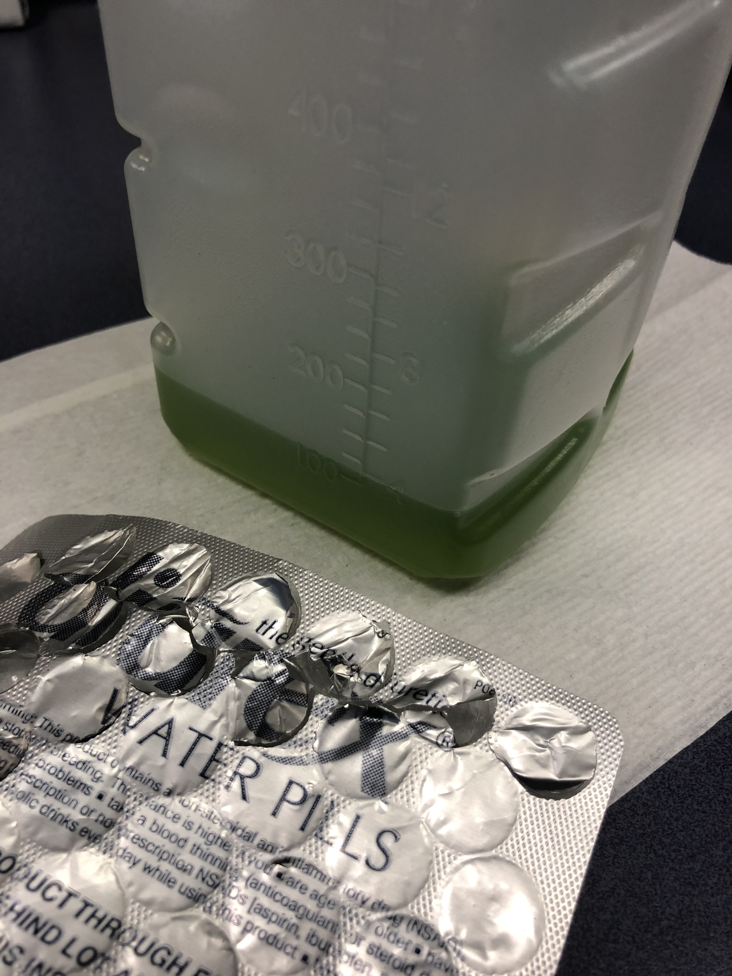
75 y/o M is brought in by EMS after he fell off the light rail and hit his head. In the ED he is A&Ox3, and is asking for a urinal. Two minutes later the tech comes running to show you the following:
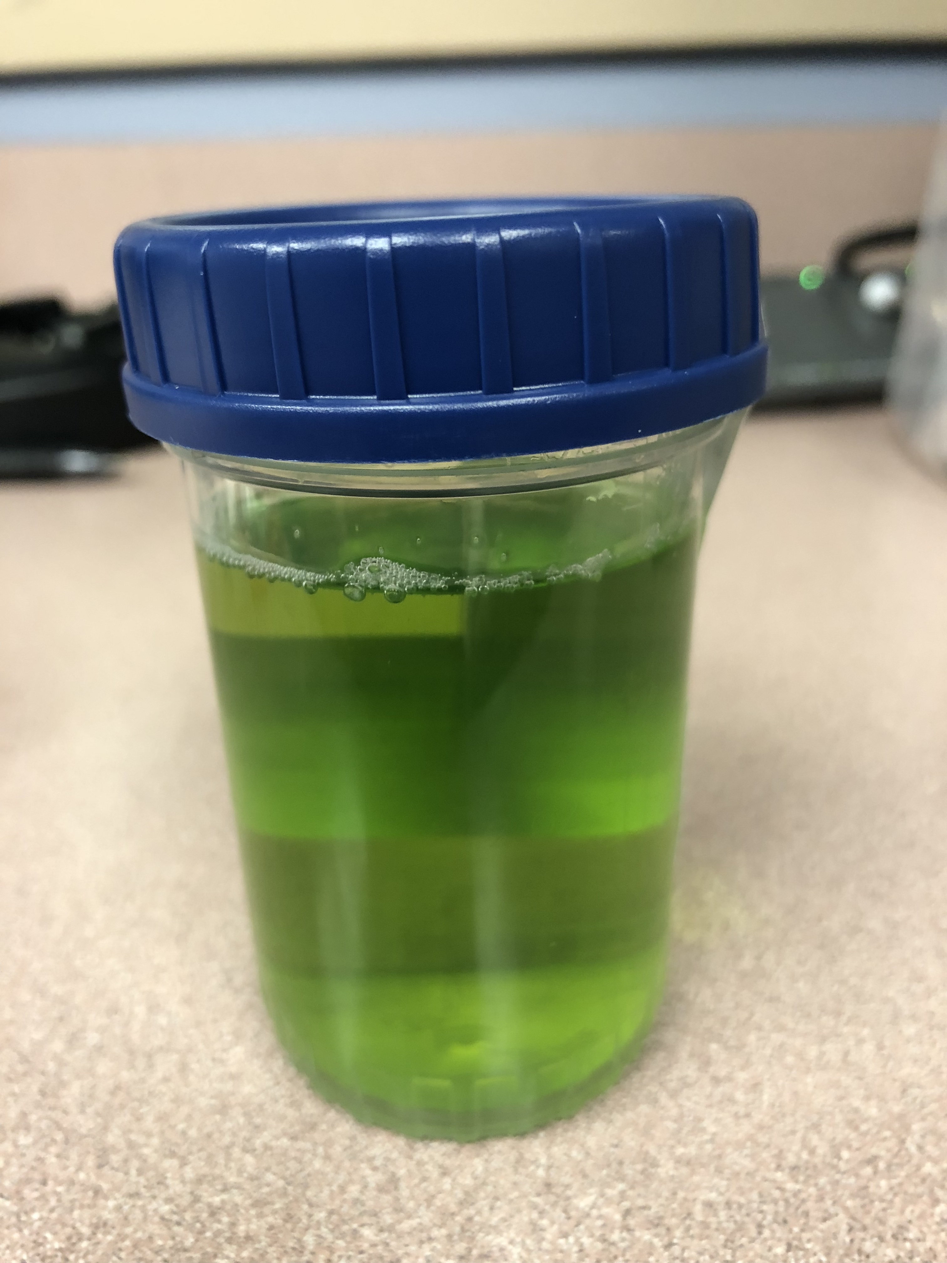
What is the cause of this patients Jolly Rancher Green Apple looking urine sample?
Show Answer
Question
Patient: 11 month old with trouble breathing and color change after a family member sprayed air freshener. Symptoms have since resolved.
What are you concerned about in the attached xrays?
Show Answer
Show References
Attachments
Bupropion (Wellbutrin, Zyban) is unique monocyclic antidepressant and smoking cessation agent that is structurally similar to amphetamines. Bupropion blocks dopamine and norepinephrine reuptake and antagonizes acetylcholine at nicotinic receptors.
- One of the most common causes of drug-induced seizures.
- Sinus tachycardia is the most frequently seen cardiac effects with overdose.
- QTc prolongation and ventricular dysrhythmias can occur in severe overdose. New evidence supports this is not related to cardiac sodium channel block but likely due to blockade of the delayed rectifying (ikr) potassium channel and gap junction inhibition in the myocardium simulating effects class IA effect.
Bottom line:
Bupropion is a common cause of drug induced seizures but in severe overdose can also cause prolonged QTc and wide complex ventricular dysrhythmia that may be responsive to sodium bicarbonate. All patients with an overdose of bupropion should have an ECG performed and cardiac monitoring to watch for conduction delays and life-threatening arrhythmias.
Show References
- Greater occipital nerve (GON) block with local anesthetics is an alternate treatment option for headaches.
- Zhang et al. conducted a systematic review and meta-analysis of 7 randomized controlled trials assessing the efficacy of GON block for migraine.
- Pooled outcome suggests that GON block:
- Reduces pain intensity (mean difference -1.24 [-1.98, -0.49], p=0.001)
- Decreases analgesia medication consumption (mean difference -1.10 [-2.07, -0.14], p=0.02)
- Has no significant impact on headache duration (mean difference -6.96 [-14.09, 0.18], p=0.06)
Show References
Femoral neck stress fractures
Adults>kids
Represents 5% of all stress fractures
Usually due to repetitive abductor muscle contraction
As with all stress fractures can occur in 2 types
1) Insufficiency type (normal physiologic stress on abnormal bone)
2) Fatigue type (abnormal/excessive physiologic stress on normal bone)
2 locations on interest:
1) Compression side (inferior femoral neck)
2) Tension side (superior femoral neck)
History: Insidious onset of groin or lateral hip pain associated with weight bearing
Exam: Antalgic gait, pain with hip log roll and with FABER (hip flexion, Abduction and external rotation test)
Treatment:
Compression side: reduced weight bearing and activity modification
Tension side: Non weight bearing (due to high risk of progression to displacement with limited weight bearing) AND surgical consultation for elective pinning to prevent displacement. If displaced, will require ORIF
Show References
Question
Case: 5 year old presents to the ED with 2 weeks of fever. She has extensive cracked, bleeding lips and a rash on her hands and feet. She was recently diagnosed with “walking pneumonia” and hand, foot and mouth disease this week. Her pediatrician sent her in for further workup after she was found to have an elevated CRP on outpatient labs. A similar picture appears in the link below:
What's the diagnosis?
Show Answer
Hyperoxia and the Post-Arrest Patient
- Current post-arrest guideilnes recommend titrating supplemental O2 to avoid hypoxia and limit exposure to hyperoxia.
- Importantly, these recommendations are based primarily on retrospective studies that have used ABG values within the first 24 hours following ROSC.
- The latest study to evaluate the impact of hyperoxia following cardiac arrest was just published in Circulation.
- This study is a prospective, cohort study that evaluated the association between early hyperoxia and poor neurologic outcome in adults following cardiac arrest. (ABGs were obtained at 1 hour and 6 hours following ROSC)
- Of 280 patients, 38% were exposed to early hyperoxia (defined as a PaO2 > 300 mm Hg)
- Take Home Points
- Early hyperoxia was found to be an independent predictor of poor neurologic outcome at hospital discharge.
- One hour longer duration of hyperoxia was associated with a 3% increase in the risk of poor neurologic outcome
- SaO2 could not reliably exclude the presence of hyperoxia.
Show References
15% of older adults presenting to ED for dizziness have serious etiologies; 4-6% are stroke-related and sensitivity of CT for identifying stroke or intracranial lesion in dizziness is poor (16%), so if CNS etiology suspected, seek neuro consult or MRI (83% sensitivity)
Show References
Debating between cefepime or piperacillin/tazobactam for your septic patient? Use this table to help you decide.
|
|
| Cefepime | Piperacillin/Tazobactam |
| Gram Negative Spectrum | Pseudomonas aeruginosa | Yes | Yes |
| Aerobic gram negative organisms | E. coli Klebsiella sp. Proteus mirabilis M catarrhalis H. influenza | E. coli Klebsiella sp. Proteus mirabilis M. catarrhalis H. influenza | |
| Anerobic gram negative organisms | No | B. fragilis
| |
| Gram Positive Spectrum | MRSA | No | No |
| Aerobic gram positive organisms | MSSA CoNS Group A Strep S. pneumoniae
| MSSA CoNS Group A Strep S. pneumoniae E. faecalis | |
| Anaerobic gram positive organisms | P. acnes Peptostreptococci | P. acnes Peptostreptococci Clostridium sp. | |
| Infection Site Concerns | CNS Penetration | Yes | No1 |
| Urine Penetration | Yes | Yes | |
| Lung Penetration | Yes | Low2 | |
| Dosing Frequency (Normal Renal Function) | Q8h | Q6h | |
1. Tazobactam CNS penetration is limited, thus limiting antipseudomonal activity in the CNS
2. Low pulmonary penetration, may not achieve therapeutic levels in patients with critical illness
Show Additional Information
Show References
Question
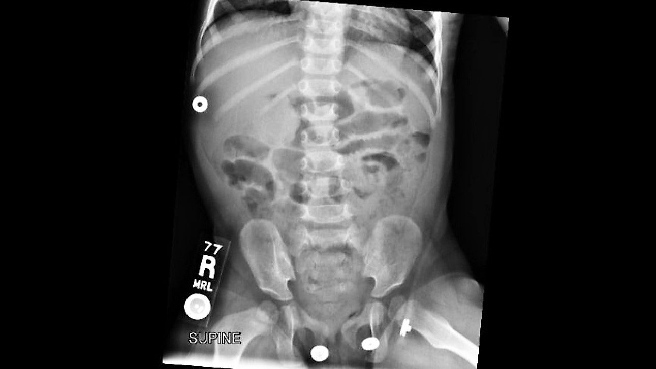 Your patient is an18 months old female with intermittent abdominal pain for the last 4-5 days. She has history of constipation and soy allergy, seen at an outside hospital three days ago for the same. She had an xray and was discharged home with instructions for at home clean out with diagnosis of constipation.
Mother is bringing her to your ED because the pain is back. The laxatives helped somewhat, but her symptoms have returned. She reports that the patient cries spontaneously, lasting 1-2 minutes, then completely resolves. These episodes happen at multiple times during the day.
ROS: Decreased appetite and energy, but NO fevers, vomiting, diarrhea, bloody stool, abdominal distension, hematuria, or lethargy.
Show Answer
Show References
Attachments
Question
47 year old woman presents with cough, headache, weakness, and low grade fever. Her symptoms have been present for several days. Vital signs are temperature 99.9 F, HR 96, RR 16, BP 140/88, Pulse Ox 98%. Physical exam is nonfocal. She is Influenza negative. She is treated with Ibuprofen and oral fluids. Upon discharge she mentions she is having difficulty hearing and feels dizzy. Upon further questioning she admits to ringing in her ears. What tests should you order?
Show Answer
Show References
As hospital volumes increase and ED patient boarding becomes more commonplace, emergency physicians may find themselves managing critically ill patients beyond the initial resuscitation.
The benefit of glucocorticoids in critically ill patients with septic shock has remained a topic of controversy for decades due to conflicting studies, including the 2002 Annane trial and the 2008 CORTICUS trial, which had opposing results when it came to the mortality benefit of steroids.
The results of the eagerly-awaited ADRENAL trial, a multicenter randomized controlled trial investigating the benefit of steroids in septic shock, were released earlier this month:
- 3658 patients from 69 different medical and surgical ICUs
- Adults with septic shock requiring mechanical ventilation (including noninvasive) and vasopressors/inotropes for at least 4 hours
- Continuous infusion hydrocortisone 200mg/day vs placebo for 7 days or until ICU discharge, if shorter
- No mortality benefit at 90 days (primary outcome) or at 28 days (secondary outcome)
- Other secondary outcomes:
- Hydrocortisone group = Shorter ICU LOS, shorter duration of shock, shorter duration of initial mechanical ventilation, fewer # of patients receiving a blood transfusion
- No difference in: mortality at 28 days, hospital LOS, recurrence of shock, total vent-free days, mean volume of blood transfused in patients receiving blood products, use of renal replacement therapy, development of new bacteremia/fungemia
Take Home Points:
1. Administration of standard daily dose hydrocortisone by infusion does not seem to affect mortality in septic shock.
2. Emergency providers should continue to consider stress-dose steroids in patients with shock and a high risk of adrenal insufficiency (e.g., chronic steroid therapy, genetic disorders, infectious adrenalitis, etc).
Show References
Dental Avulsion in the field/sporting event
- Only replace avulsed secondary teeth
- Handle the tooth by the crown only
- Rinse tooth with cold running water gently (the root should not be wiped)
- Immediate attempt to reimplant permanent tooth into socket by 1st capable person:
* Time is tooth: Each minute tooth is out of socket reduces tooth viability by 1%
* Best chance of success if reimplant done within 5–15 min*? Poor tooth viability if avulsed for >1 hr
- If unsuccessful, place tooth in a transport solution (from most to least desirable):
- Hanks balanced salt solution (HBSS)
* Balanced pH culture media available commercially in the Save-A-Tooth kit
* Effective hours after avulsion
- Cold milk:
* Best alternative storage medium
* Place tooth in a container of milk that is then packed in ice (prevents dilution)
- Saliva:
* Store in a container of parent or child's saliva
- Never use tap water or dry transport
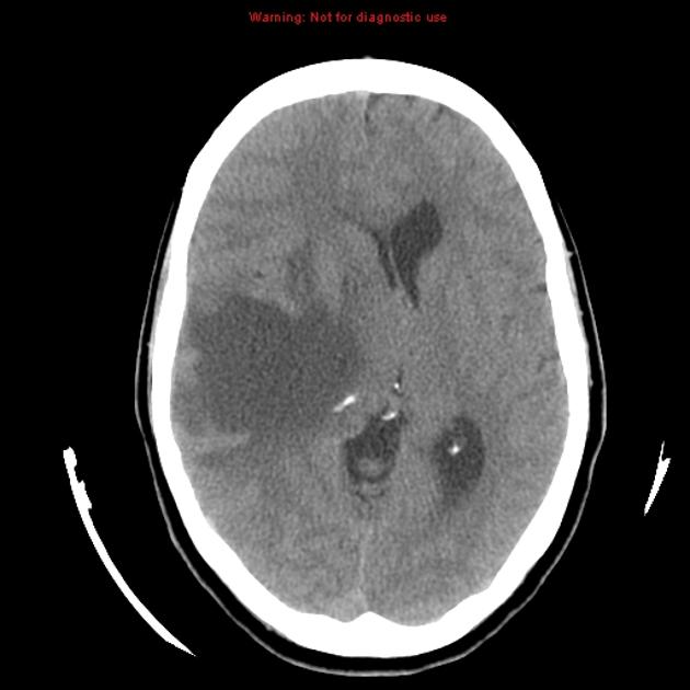
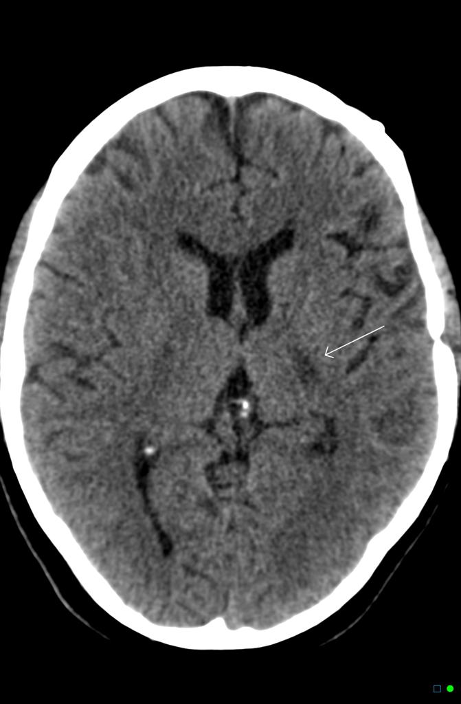
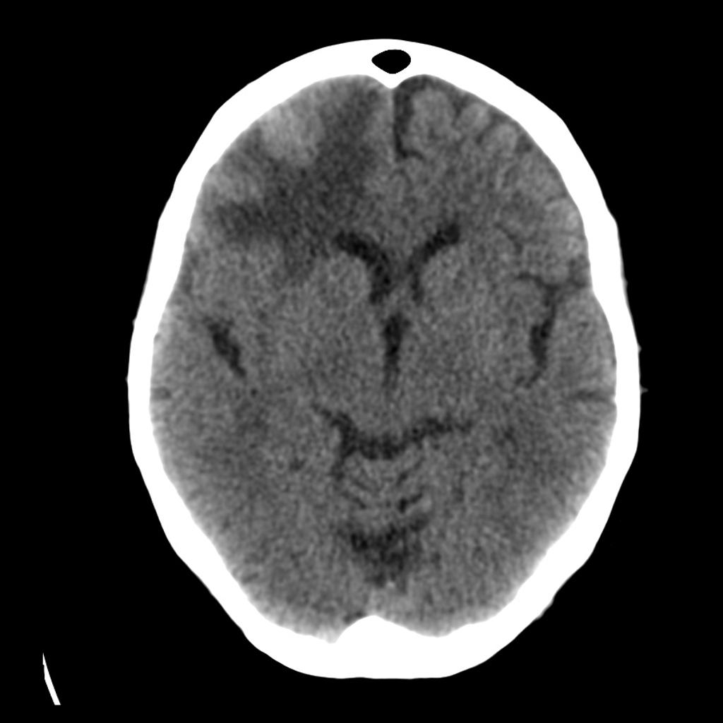
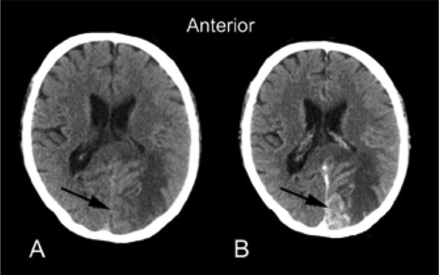
- Hyperattenuation = bright = dense (blood)
- Hypoattenuation = dark = radiolucent (fluid, air, lipid, scar)
- Masses that are darker + increased volume or mass effect = edema (image 1)
- Masses that are darker + decreased volume = scar tissue or atrophy (image 2)
- Masses that are bright + edema = hemorrhage (image 3)
- Adding IV contrast improves detection of tumors: abnormal enhancement from disruption of blood brain barrier, necrosis or increased vascularity. (Image 4)
Show References
Bottom line: Oral morphine was not superior to ibuprofen and both drugs decreased pain with no difference in efficacy. Morphine was associated with more adverse events.
Show References
Acute liver failure carries a high morbidity without liver transplantation. Liver support systems can act as “bridge” until an organ becomes available for the transplant procedure or until the liver recovers from injury. Artificial liver support systems temporally provide liver detoxification utilizing albumin as scavenger molecule to clear the toxins without providing synthetic functions of the liver (coagulation factors). One of the most widely used devices is the Molecular Adsorbent Recirculating System (MARS).This system has 3 different fluid compartments: blood circuit, albumin with charcoal and anion exchange column, and a dialysate circuit that removes protein bound and water soluble toxins with albumin.
- Mars has been used in several case reports to treat acetaminophen, Amanita phalloides,Phenytoin, lamotrigine, theophylline, and calcilum channel blockers poisonings.
- All the extracorporeal liver assist devices are able to remove biological substances (ammonia, urea, creatinine, bilirubin, bile acids, amino acids, cytokines, vasoactive agents) but the real impact on the patient's clinical course has still to be determined.
Bottom Line
MARS therapy could be a potentially promising life saving treatment for patients with acute poisoning from drugs that have high protein-binding capacity and are metabolized by the liver, especially when concomitment liver failure. Consider consultation and transfer of patients to liver center.
Show References
Concussion – Where are we now?
The Sport Concussion Assessment Tool 5th edition (SCAT 5) was released in 2017
It is a standardized tool to assist health care professionals in the evaluation of sport associated concussions
It should be used for those 13 years and older (there is a child version for younger athletes)
Print and bring to the sideline for your next coverage event!
http://bjsm.bmj.com/content/bjsports/early/2017/04/26/bjsports-2017-097506SCAT5.full.pdf
Some points to consider:
It should take at least 10 minutes to complete. Any less and you may not be performing the test correctly
The SCAT5 is the standard tool used in concussion assessment in the NCAA and NFL and other professional sports
Some symptoms of concussion appear over time. For example, an athlete may have zero or minimal symptoms immediately after yet be considerably symptomatic in 10 to 15 minutes.
-Follow up screening evaluations are essential even in those with a negative initial sideline screening test
The SCAT5 should be used immediately after injury
-Utility decreases post injury after days 3-5
-The included symptom checklist has utility in tracking recovery
-Attempt to perform in an environment free of distractions (crowd noise, bad weather)
The clinical utility of the SCAT5 can be enhanced by adding assessment of other factors such as reaction time, balance assessment, video-observable signs (if available) and oculomotor screening.