
In a precursor to a forthcoming international guideline on the management of calcium channel blocker poisoning, a new systematic review has been published assessing the available evidence.
A few findings from the systematic review:
- The majority of literature on calcium channel blocker overdose management is heterogenous, biased, and low-quality evidence.
- Interventions with the strongest evidence are high-dose insulin and extracorporeal life support.
- Interventions with less evidence, but still possibly beneficial, include calcium, dopamine, norepinephrine, 4-aminopyridine (where available), and lipid emulsion therapy.
Stay tuned for the international guideline coming out soon. One treatment recommendation from the new guideline, reported at the 8th European Congress on Emergency Medicine last month, is not to use glucagon.
Show References
Hydrocephalus is a disorder of cerebrospinal fluid (CSF) accumulation. Acute obstructive hydrocephalus such as in subarachnoid hemorrhage and CSF shunt malfunction can cause a rapid rise in intracranial pressure. Nonobstructive hydrocephalus is associated with subacute symptoms. Clinical features of acute obstructive hydrocephalus include headache, blurred vision, papilledema, ocular palsies, nausea and vomiting, and decreased level of consciousness.
Evaluation of hydrocephalus in the ED should include neuroimaging, typically noncontrast head CT given its wide availability. CT characteristics of hydrocephalus can be seen in Figure 1: ventriculomegaly with dilated 3rd ventricle, dilated 4th ventricle, and presence of temporal horns.
When evaluating patients with pre-existing hydrocephalus for worsening symptoms, such as in the evaluation of CSF shunt malfunctions, it is helpful to compare the head CT or MRI for interval ventricular enlargement. Two simple measurements can be taken on a CT or MRI for objective comparisons (Figure 2).
-
Evans' ratio = A/B = Maximum width of frontal horns (A) divided by maximum width of inner skull (B) at the same CT/MRI level
-
C = Width of 3rd ventricle
Use of acetazolamide to decrease CSF production is not effective in long-term treatment of hydrocephalus. About 75% of patients with hydrocephalus require CSF shunt placement.
Show References
Attachments
We are proud to announce the release of our new UMEM Pearls App, now available in the Apple App Store and the Google Play Marketplace! You will now be able to pull down pearl content for offline viewing, in addition to having all of the pearls in searchable format available on your mobile devices.
Click either of the following links on your mobile device to download your Pearls App today!
The ARISE Trial
Early, aggressive resuscitation and attention to detail are essential element of managing critically ill patients. This past week the ARISE trial was published - a 2nd large, randomized control study to examine the benefit of protocolized vs. usual care in patients with severe sepsis and septic shock.
What were the main findings? After enrolling 1,600 patients who presented to the ED in severe sepsis or septic shock:
- They found no difference in mortality between the control (usual care) and treatment arm (early goal-directed therapy)
- Mortality was 18.6% vs. 18.8% at 90 days
- No evidence that continuous ScVO2, Hgb target > 10 mg/dL (check out the TRISS trial), or use of inotropes with a normal cardiac index improved mortality

Bottom Line: Resuscitation goals for the patient with septic shock should include:
- Early antibiotics (source control)
- Adequate volume resuscitation (preferably balanced, crystalloid solution)
- End-organ perfusion (lactate normalization)
Additional therapeutic goals should be made on a patient by patient basis. Reassess your patient frequently, pay attention to the details, and you will improve your patient’s mortality.
Suggested Reading
- The ARISE Investigators and the ANZICS Clinical Trials Group. Goal-Directed Resuscitation for Patients with Early Septic Shock. N Engl J Med. 2014. [PubMed Link]
- Wessex ICS: The Bottom Line Review
Follow Me on Twitter: @JohnGreenwoodMD
Question
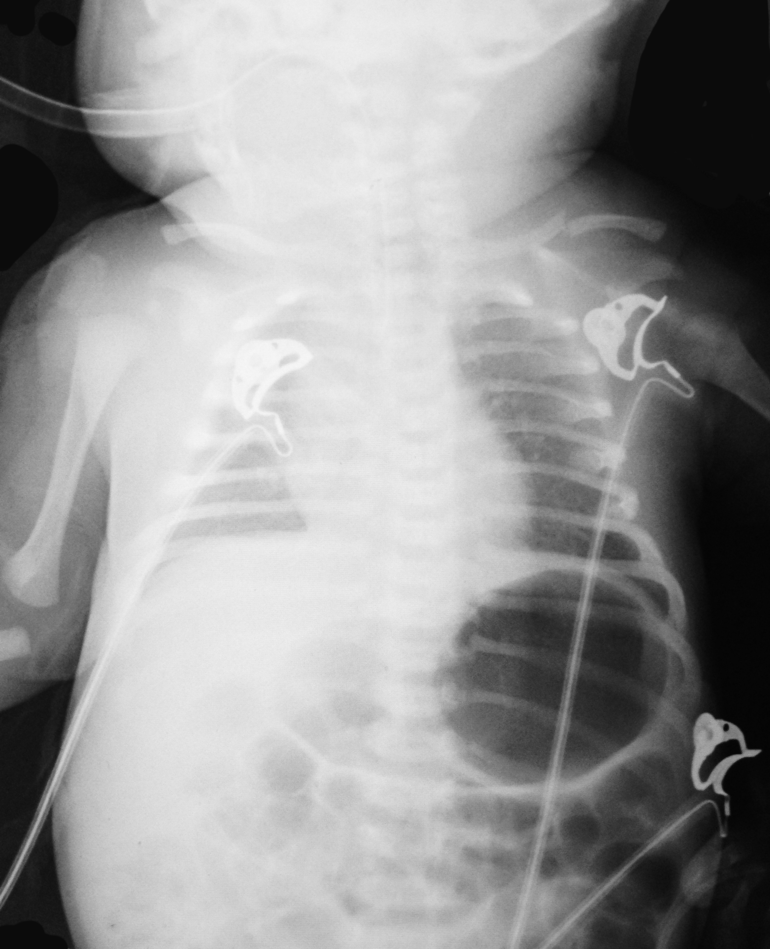
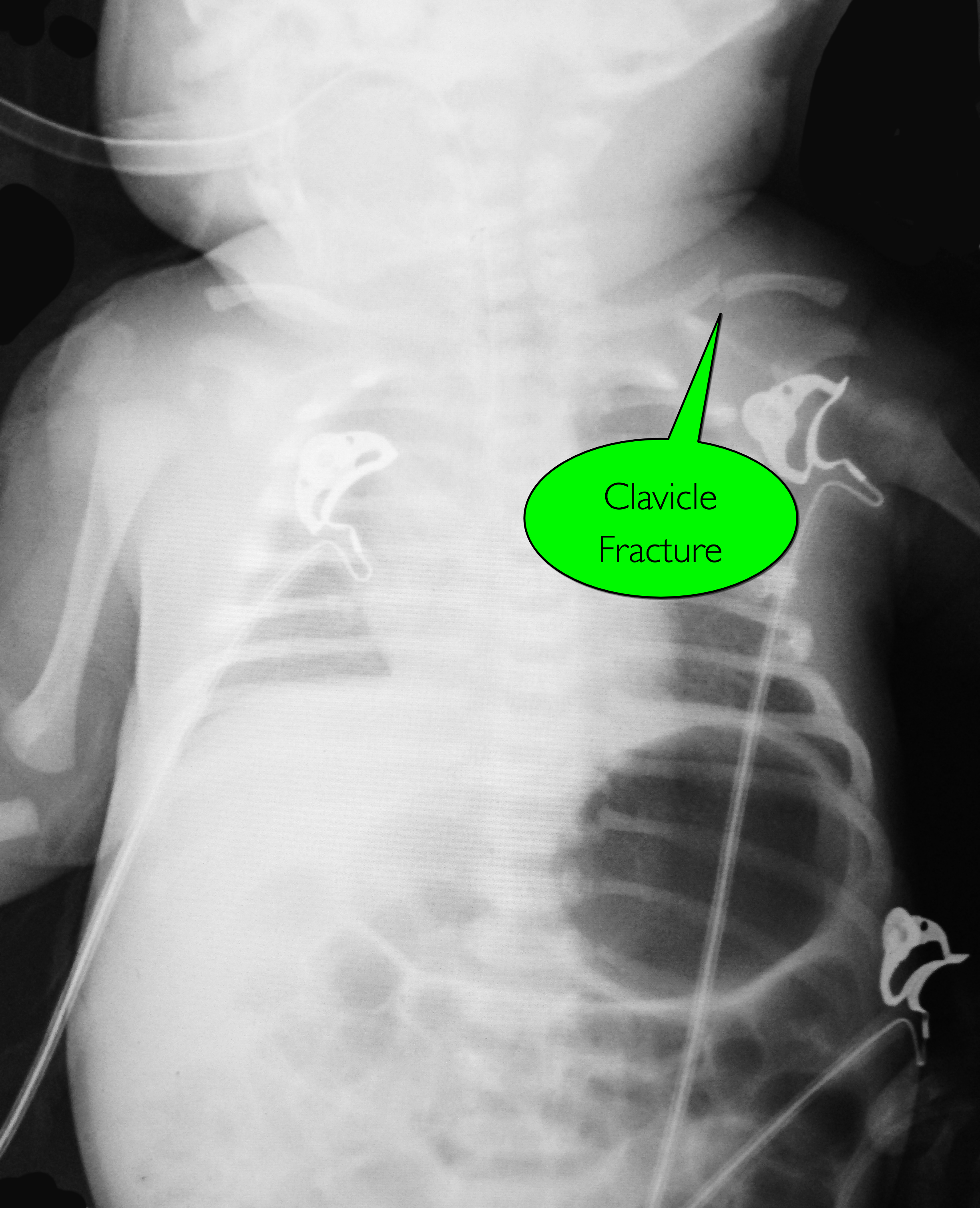
7d old child presents with difficulty feeding,vomiting one time, and now with intermittent apneic episodes. What's the diagnosis? (Careful....this one is tricky!)
Show Answer
Show References
Question
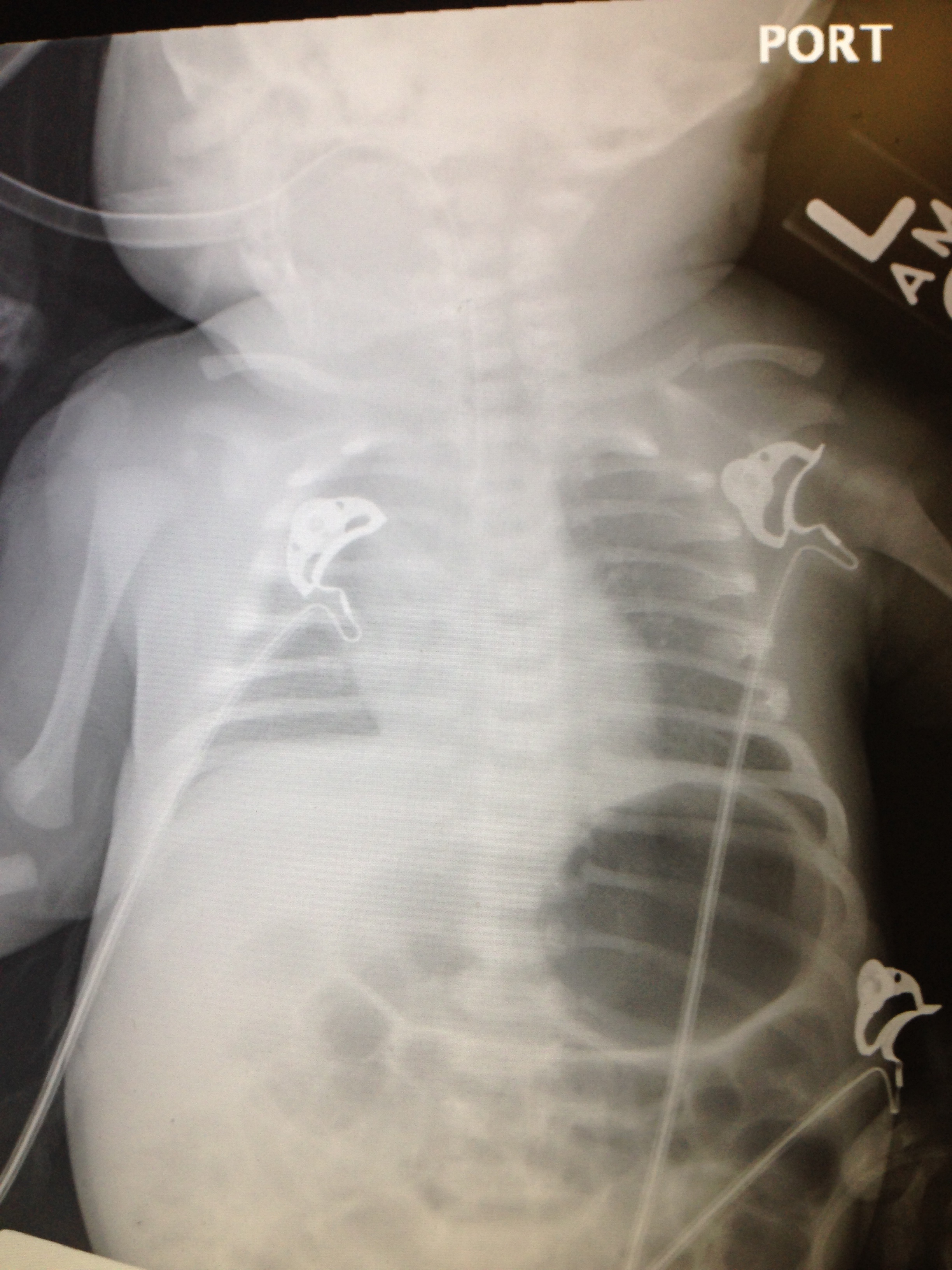
Q: What is wrong with this baby? And what Dx should you entertain?
Previously healthy 7d old presents after difficulty feeding, one episode of vomiting and now with intermittent apneic episodes.
Show Answer
Show References
Attachments
Kounis Syndrome (Part I)
- Kounis & Zavras (1991) described the syndrome of allergic angina and allergic myocardial infarction, currently known as Kounis syndrome (KS). Braunwald (1998) noted vasospastic angina can be induced by allergic reactions, with mediators such as histamine and leukotrienes acting on coronary vascular smooth muscle.
- Two subtypes have been described: type I, occurring in patients without predisposing factors for CAD often caused by coronary artery spasm and type II, occurring with angiographic evidence of coronary disease when the allergic events induce plaque erosion or rupture.
- This syndrome has been reported in association with a variety of medical conditions, environmental exposures, and medication exposures. Entities such as Takotsubo cardiomyopathy, drug-eluted stent thrombosis, and coronary allograft vasculopathy also appear to be associated with this syndrome.
- Clinical presentation includes: symptoms and signs of an allergic reaction and acute coronary syndrome: chest pain, dyspnea, faintness, nausea, vomiting, syncope, pruritus, urticaria, diaphoresis, pallor, palpitations, hypotension, and bradycardia.
Show References
Beta-lactam antimicrobials (penicillins, cephalosporins, and carbapenems) are frequently used for empiric and targeted therapy in critically ill patients. They display time-dependent killing, meaning the time the antibiotic concentration is above the minimin inhibitory concentration (MIC) is associated with improved efficacy.
Two new pharmacodynamic/pharmacokinetic studies suggest that current beta-lactam antimicrobial dosing regimens may be inadequate.
- In patients from 68 ICUs across 10 countries, use of intermittent infusions (compared to extended and continuous infusions) and increasing creatinine clearance were risk factors for MIC target non-attainment. [1]
- A second group specifically investigated the pulmonary penetration of piperacillin/tazobactam in critically ill patients and found that intrapulmonary exposure is highly variable and unrelated to plasma exposure and pulmonary permeability. [2]
Antimicrobial dosing in critically ill patients is complex. Current dosing of beta-lactams may be inadequate and needs to be studied further with relation to clinical outcomes.
Show References
INFLUENZA
What is it?
- Common disease that causes significant morbidity and mortality worldwide
- Both seasonal and pandemic influenza occurs
- Vaccination can decrease disease incidence and spread
- Treatment can decrease disease severity
- Orthomyxoviruses (RNA virus)
- 6 genera
Why do we care about influenza?
- Pandemic Influenza
- Can infect and kill young, healthy people
- 1918 H1N1 Influenza pandemic infected an estimated 500 million globally
- Approximately 20% of the world’s population
- Killed an estimated 50 million (may be as high as 100 million)
- 2%-3% of the world’s population died
- Seasonal Influenza
- Most often causes severe disease in the very young, very old, and those with chronic illnesses
- Estimated between 3-5 million cases of severe illness around the world annually
- Between 250,000 and 500,000 deaths
Bottom line
- Influenza is a potentially life threatening disease.
- Both seasonal and pandemic influenza are global concerns.
- Morbidity and mortality can be decreased through appropriate vaccination and treatment
Cardioactive steroids are among the many treatments used for CHF, and for the control of ventricular response rate in atrial tachydysrhythmias. There are many sources of cardioactive steroids:
Pharmaceutial: Digoxin, Digitoxin
Plants: Oleander, Yellow Oleander, Foxglove, Lily of the Valley, Dogbane, Red Squill
Animal: Bufo marinus toad
It is a potent Na+-K+-ATPase inhibitor and can lead to hyperkalemia in acute ingestion with associated signs and symptoms of N/V, abdominal pain, bradycardia and possibly, hypotension.
Toxicity should be suspected with bidirectional ventricular tachycardia or atrial tachycardia with high-degree AV block
Therapeutic range of digoxin of 0.5 - 2.0 ng/mL is helpful but not a sole indicator of toxicity
Indication for antidote (Digoxin-specific Antibody Fragments) include:
1) Digoxin-related life-threatening dysrhythma
2) Serum K+ > 5.0 mEq/L in acute ingestion
3) Serum digoxin concentration >15ng/mL at any time, or >10 ng/mL 6 hours postingestion
4) Ingestion of 10 mg in adult; 4 mg in pediatric
5) Poisoning by non-digoxin cardioactive steroid
Show References
The last Back to the Basics post discussed the use of vasopressors to improve hemodynamics by increasing arterial (and venous) tone. This time we’ll discuss the use of agents to increase inotropy for patients with severe systolic dysfunction / failure.
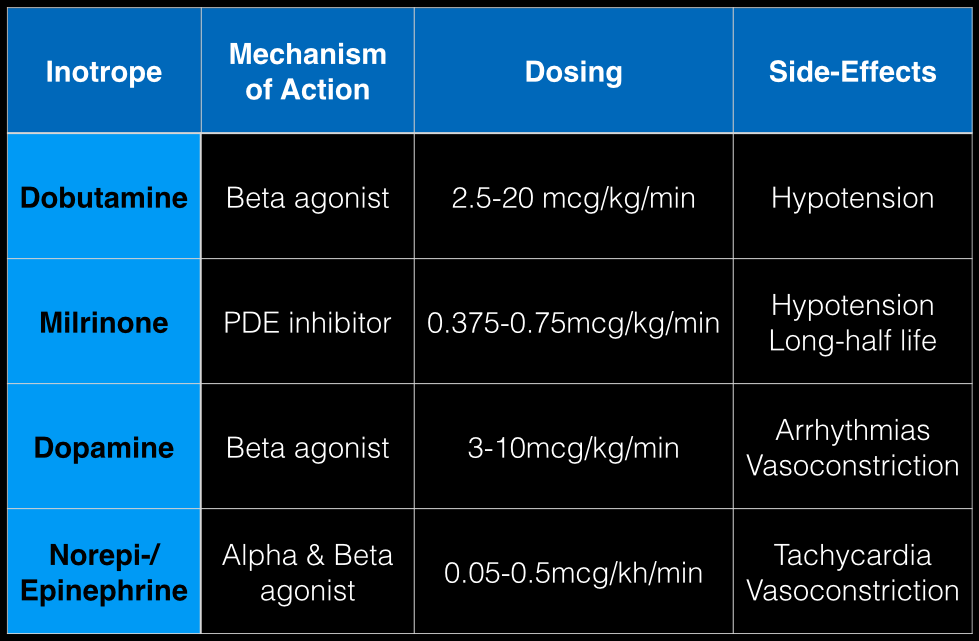
Dobutamine: a direct b1 and b2-receptors agonist. It has no peripheral vasoconstrictor properties, so if blood pressure increases it occurs secondary to increased cardiac output. Unfortunately, blood pressure may be decreased in some patients due to its peripheral vasodilatory effects; in these cases it may need to be used with a vasopressor.
Milrinone: augments contractility by increasing intracellular Ca levels via cellular phosphodiesterase inhibition. Because it does not work on beta-receptors, it might be preferred for patients taking beta-blockers requiring inotropic support. It may cause peripheral vasodilation and hypotension, but this may be a benefit if pulmonary artery pressure is elevated as reductions in pulmonary artery pressure lead to improvements in right ventricular function. It has a long-half life and should be avoided in patients with renal impairment.
Dopamine: chemical precursor to norepinephrine and technically a vasopressor. At moderate doses (3-10 mcg/kg/min) it works on beta-receptors to increase myocyte contractility. At higher doses works primarily as a vasopressor, which may reduce cardiac output due to higher afterload.
Norepinephrine/epinephrine: has alpha and beta properties that lead to increased peripheral vasoconstriction, but also increases inotropy and chronotropy (faster heart rate)
Show References
Question
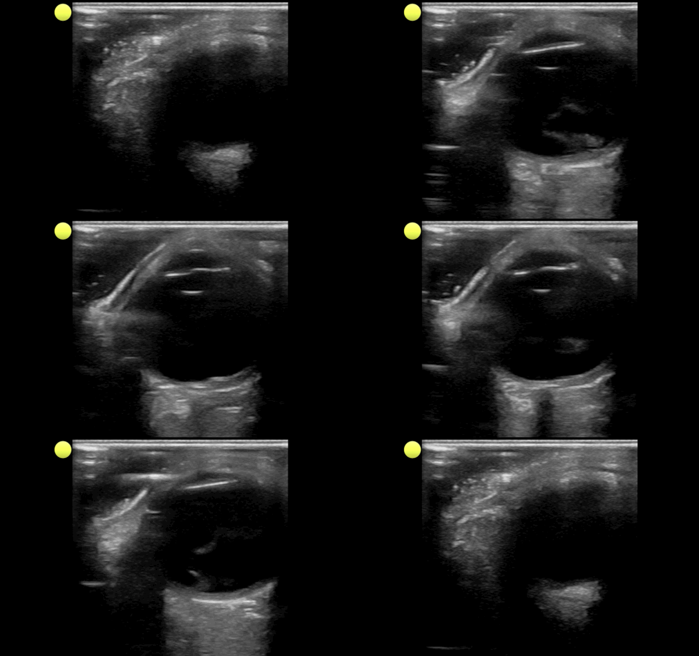
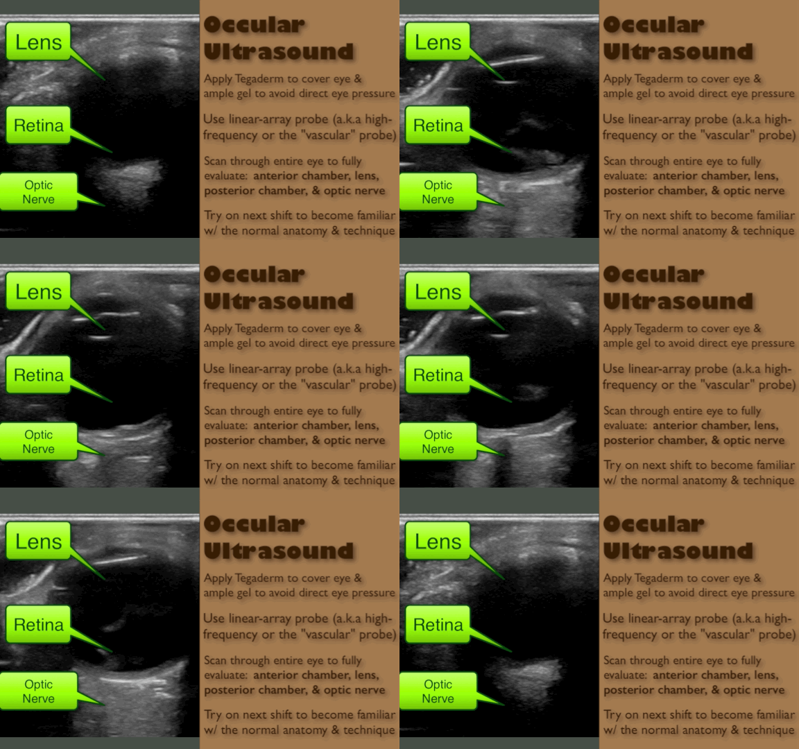
Person presents with painless vision loss after seeing flashes of light. Ultrasound is below. What's the diagnosis?
Show Answer
Show References
Ventricular Arrhythmias Originating from the Moderator Band
- Ventricular arrhythmias originating from the moderator band (MB) often have a distinct morphology
- Typically MB arrhythmias have a left bundle branch block pattern, QRS with a late precordial transition (>V4), a rapid down stroke of the QRS in the precordial leads, and a left superior frontal plane axis
- MB arrhythmias are often associated with PVC-induced ventricular fibrillation
- Catheter ablation is quite effective at termination of the arrhythmias and facilitated with intracardiac echocardiography (ICE)
Show References
Back pain with radiculopathy can be very distressing to a patient and they have heard from their medically savvy neighbor that a MRI is the way to go. Now, armed with this knowledge, they are in your ED with earplugs in hand...
A few minutes of reassurance and education can save in both cost and ED throughput.
In one study researchers performed MRIs on asymtomatic adult patients.
Almost two-thirds (64%) had abnormal discs
Just over half (52%) had bulging discs
Almost a third (31%) had disc protrusions
Further, finding a bulging disc already suggested by your history and physical examination does not change management. The majority of these patients improve with conservative treatment within four to to six weeks.
Restrict ED MRI use for the evaluation of suspected cauda equina, epidural abscess and spinal cord compression.
There are numerous different causes of pediatric hemorrhagic diarrhea. Consider a pediatric patient with bloody diarrhea as being at risk for developing hemolytic uremic syndrome. Most cases of hemolytic uremic syndrome are caused by O157:H7 strains of E Coli that release Shiga-like toxin from the gut. Systemic release of the toxin causes microvascular thromboses in the renal microvasculature. The characteristic microangiopathic hemolysis results with anemia, thrombocytopenia and peripheral schistocytes seen on laboratory studies, in addition to acute renal failure.
Antibiotics have been controversial in the treatment of pediatric hemorrhagic diarrhea due to concern that they worsen toxin release from children infected with E Coli O157:H7 and thus increase the risk of developing hemolytic uremic syndrome. Numerous previous studies have provided conflicting data regarding the true risk (1). A recent prospective study showed antibiotic treatment increases the risk (2). Most recommendations warn against using antibiotics to treat pediatric hemorrhagic diarrhea unless the patient is septic.
Bottom line: Avoid treating pediatric hemorrhagic diarrhea with antibiotics
Show References
When you prescribe certain medications, it may require some further instructions to avoid ethanol or a disulfiram like reaction (nausea, vomiting, flushing) may occur. Keep this short list in your brain:
1) Particular cephalosporins: cefotetan is a the one more likely
2) Nitrofurantoin
3) Sulfonylureas: chlorpropamide and tolbutamide
4) Metronidazole
5) Trimethoprim-sulfamethoxazole
Show References
Jolt accentuation, the exacerbation of a headache with horizontal rotation of the neck, or shaking of the stretcher in the less cooperative patient, has been promoted for the past few years as the "go-to" test to assess for meningeal irritation in patients with headache. Previous studies have quoted sensitivities as high as 97.1%. (1)
A new prospective study in AJEM challenges this belief by looking at a total of 230 patients with headaches and subsequent LPs. 197 of them had the jolt accentuation test done, which had a sensitivity of only 21% for pleocytosis (defined as greater than or equal to 5 cells/high power field in the 4th CSF tube). Kernig's and Brudzinski's signs both did even more poorly, with a sensitivity of 2% each. (2)
Show References
What is a massive transfusion?
- Can be institution dependent but usually means greater than 10 Units of blood products transfused within 24hrs.
- Most hospitals have this as a protocol that a physician can order to notify the blood bank that a large volume of blood products may be required rapidly.
When would I use this?
Indications:
-Systolic Blood pressure < 100
-Unable to obtain blood pressure
AND
-Penetrating torso trauma
-Positive FAST
-External blood loss
-Plans to go to the OR
How do I give it?
- The transfusion ratio is usually 1:1:1 or 2:1:1
- Give 1 unit PRBC, then 1 U FFP, and alternate until 6 units of each have been given and then 1 bag of apheresis platelets (6 equivalent units). Can repeat as needed.
Does this apply for just traumatic bleeding?
- Although this data was based on soldiers in the recent Iraq Wars, it has been used for medical patients as well.
- Therefore, consider using in upper GI bleeds, post-partum hemorrhage, etc.
Are there other agents I can use?
- There is some data to give tranexamic acid early (less than three hours from injury) in trauma patients who are hypotensive and are having severe bleeding.
What am I trying to do with this protocol?
- Control hemorrhage
- Use the best products possible
- Prevent hypothermia
- Prevent hemodilution
- Treat coagulopathy
Show References
Question
16 yo M with pleuritic right upper chest pain that started today. He is suffering from an asthma exacerbation currently in the setting of URI with cough. He is afebrile, tachycardic to 140-150s, respiratory rate 20, and sats 98% on room air. ECG was performed which incidentally diagnosed this patient WPW and he went for ablation as an outpatient. His chest x-ray showed:
Besides a bad day, what do we call this chest x-ray finding?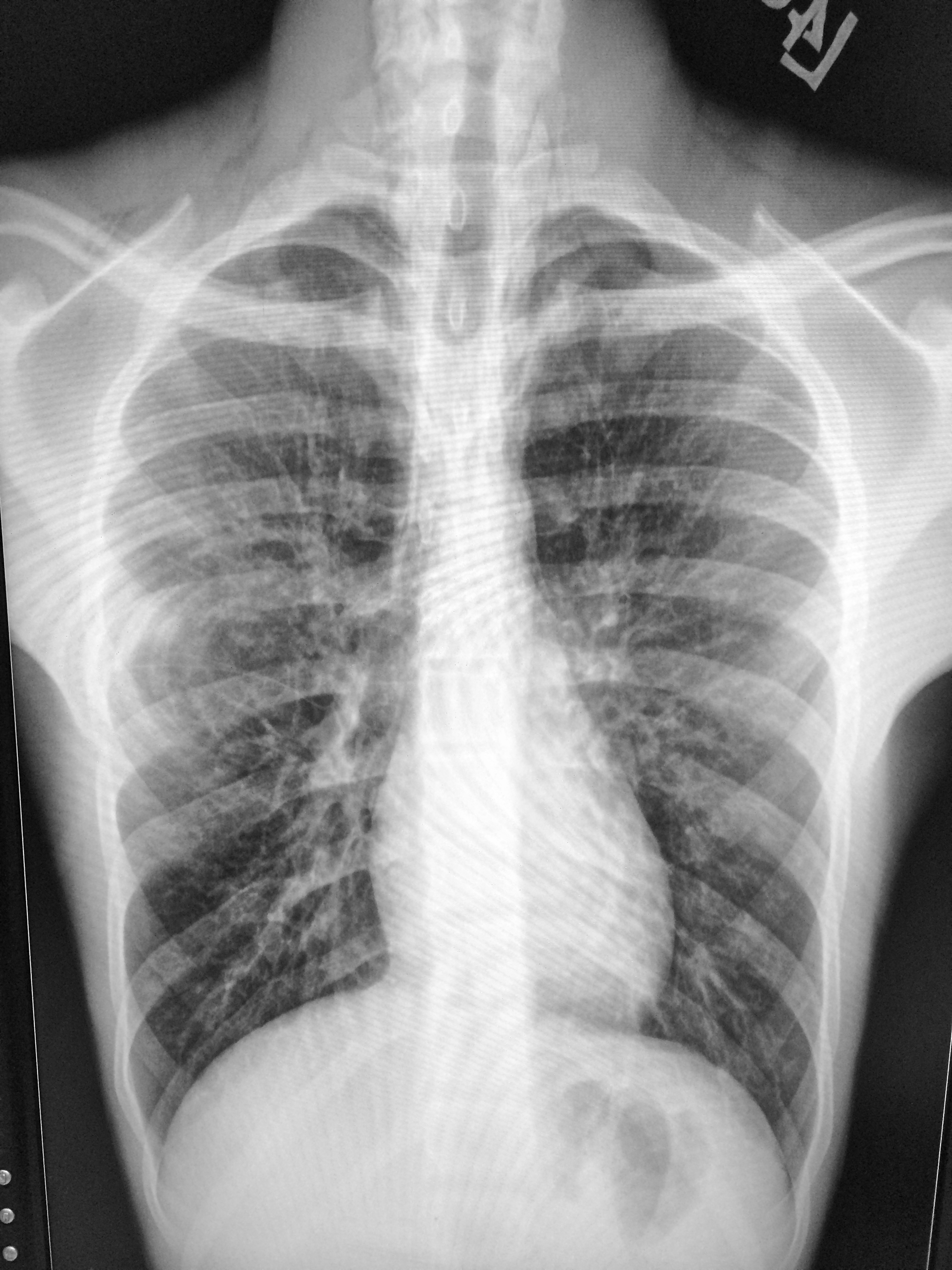
Show Answer
Show References
Attachments
Tendon Lacerations:
- Flexor tendon lacerations have historically not been repaired by emergency providers due to the extensive pulley systems involved and possibility of loss of mobility from scarring.
- However, if both ends of the tendon can be visualized in the ED it is not unreasonable to place 1 or 2 horizontal mattress sutures between the two ends to prevent retraction of the proximal portion which can make a formal repair more difficult.
- These injuries have a very high complication rate so most will defer to a hand surgeon for definitive treatment.
- Extensor tendon lacerations can be repaired by emergency providers.
- Most often these repairs are limited to 6-8. See image at http://www.boneandjoint.org.uk/content/focus/extensor-tendon-injury
- One technique is to use a running horizontal mattress suture with non-absorbable nylon sutures.
- The ultimate strength of the repair is dependent on the number and size of the sutures placed.
- Careful placement of the sutures can prevent gap formation between the ends when the tendon is stressed.
- A good discussion on tendon repairs can be found at http://www.boneandjoint.org.uk/content/focus/extensor-tendon-injury
A reasonable approach to all tendon lacerations is to loosly reapproximate the wound and splint the hand in the position of function until the patient can be seen by a hand surgeon in the next 1-3 days. These injuries do not require immediate surgical repair, and with the high rate of complications it is probably best to discuss with your hand surgeon before attempting a repair.