University of Maryland School of Medicine logo
Emergency Medicine
All Departments
UMSOM Assistant
UMEM Educational Pearls
  • Home
  • Departments
  • Emergency Medicine
  • CME
  • UMEM Educational Pearls
  • CME
  • Courses and Conferences
  • Free Open Access Medical Education
  • UMEM Educational Pearls

Search

1-20 of 321 results by Haney Mallemat

Previous |  1 |  2 |  3 |  4 |  5 |  6 |  ... |  17 |  Next

Title: End-Tidal CO2: Beyond the Waveform

Category: Critical Care

Posted: 5/31/2016 by Haney Mallemat, MD

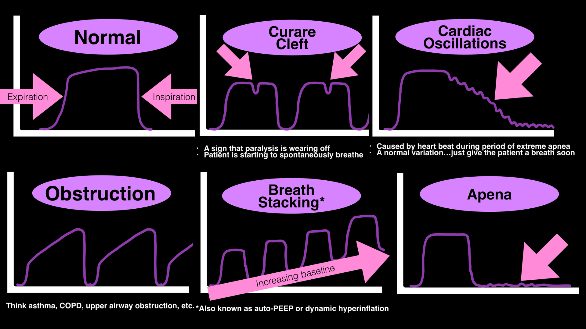
  • Many clinicians use end-tidal CO2 to monitor respirations during procedural sedation or mechanical ventilation
  • Typically either the presence (or absence) of a "normal" waveform or the quantitative value is used, however a lot more information can be gathered from the actual shape of the waveform; below are a few examples.
  • For more examples of interpreting waveforms, click HERE.
Show References

Follow me on Twitter (@criticalcarenow)a


Title: VF or pulseless VT...just give saline?

Category: Critical Care

Posted: 4/5/2016 by Haney Mallemat, MD

  • Amiodarone and lidocaine are commonly used antiarrhythmics for ventricular fibrillation (VF) or pulseless ventricular tachycardia (VT). Their efficacy towards survival to hospital discharge and neurological outcome, however, has been questioned.
  • A recently published study in the NEJM evaluated these drugs by performing a double-blind, randomized, placebo-control trial. The trial evaluated patients presenting with out of hospital cardiac arrest secondary to VF or pulseless VT that is refractory to one or more shock.
  • The trial randomized 3,026 patients to receive amiodarone (974), lidocaine (993), or normal saline (i.e., placebo) (1,059); the primary outcome was survival to hospital discharge and the secondary outcome was favorable neurological outcome at hospital discharge. Several sub-group analyses were planned a priori.
  • No statistically significant difference was found in hospital survival or neurologic outcomes between any of the groups. Patients who had a witnessed arrest and bystander CPR had higher rates of survival with either lidocaine or amiodarone compared to saline while there was no difference between the two.

Show References

Follow me on Twitter (@criticalcarenow)


Title: Should we add VTI at the bedside?

Category: Critical Care

Posted: 3/8/2016 by Haney Mallemat, MD

  • The RUSH exam is a rapid way to identify the cause of shock using ultrasound. What's the RUSH exam? Click here
  • The RUSH exam does not include an assessment of volume responsiveness (VR), but a new article by Blaivas, Aguiar, and Blanco suggests that it should be.
  • VR has classically been assessed by determining the stroke volume before and after a passive leg raise or a fluid bolus. Click here for a video on how to calculate the stroke volume (skip to 21:30 in the video)
  • The authors claim that VR can further be simplified by not measuring the left ventricular outflow tract (LVOT) and only comparing changes in the velocity-time integral (VTI). The assumption is that the LVOT is constant and doesn't change in most circumstances; a change of VTI that is greater than 15% suggests that the patient is VR
  • Further validation is required to determine the degree of benefit to adding VTI to the RUSH exam, however measuring VTI is a skill that can be done with relatively little training and is clinically helpful.

Show References

Follow me on Twitter (@criticalcarenow)

Blaivas, M. et al. Rapid Ultrasound in Shock (RUSH) Velocity-Time Integral: A Proposal to Expand the RUSH Protocol. J Ultrasound Med. 2015 Sep;34(9):1691-700


Title: What's the Diagnosis?

Category: Visual Diagnosis

Posted: 2/29/2016 by Haney Mallemat, MD

Question

19 year-old male complaining of left arm pain one week after injecting anabolic steroids into his shoulder. What's the diagnosis?
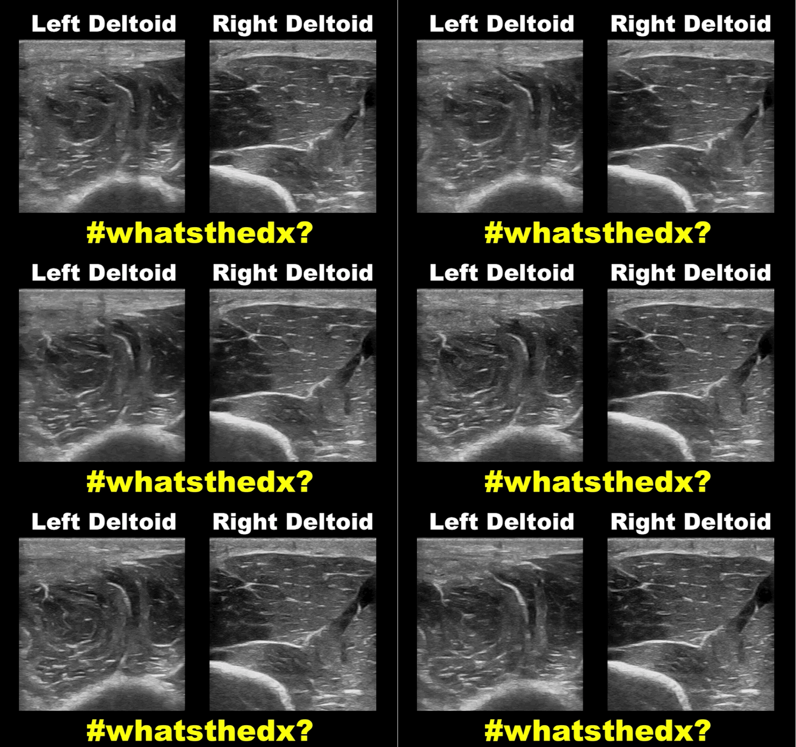
Show Answer
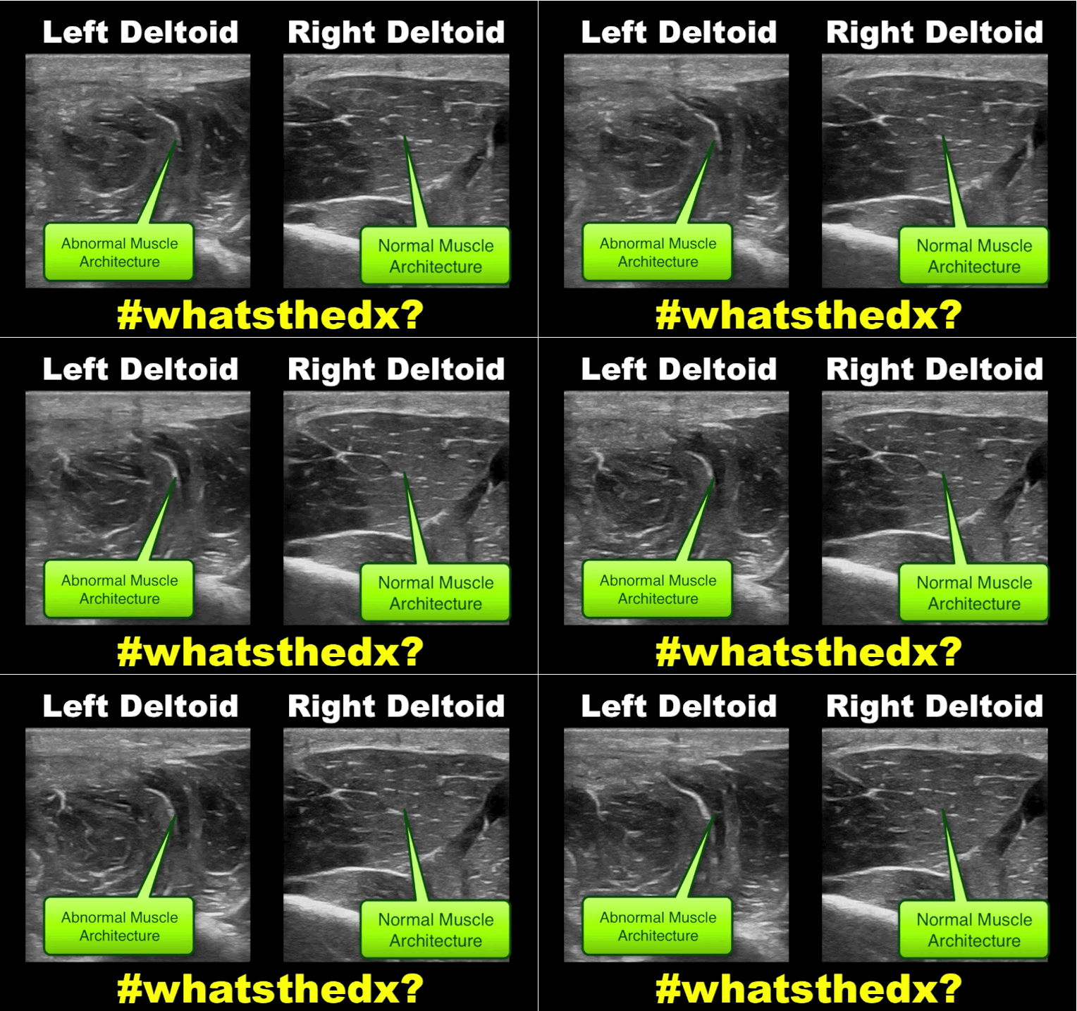
Myositis of the deltoid muscle

Show References

Follow me on Twitter (@criticalcarenow)


Title: TEE for You and Me?

Category: Critical Care

Posted: 2/9/2016 by Haney Mallemat, MD

  • Transthoracic echocardiography (TTE) is an essential tool during cardiac arrest because it identifies potentially reversible causes (e.g., tamponade, massive PE, etc.).
  • One of the limitations of TTE is that it is sometimes difficult to assess the heart in less than ten seconds (i.e., during a pulse check) and good views of the heart sometimes hard to obtain. Transesophageal echocardiography (TEE) offers the potential to overcome these obstacles.
  • TEE not only allows continuous visualization and better imaging of the heart during arrest, but it also allows the assessment of compression depth, and whether the heart is being correctly compressed during CPR.
  • Here is what a TEE probe looks like, here is an example of a TEE during arrest, and here is a podcast by @ultrasoundpodcast on the literature for using TEE during cardiac arrest.

Show References

Follow me on Twitter (@criticalcarenow)


Title: What's the Diagnosis? Case by Dr. Ali Farzad

Category: Visual Diagnosis

Posted: 1/18/2016 by Haney Mallemat, MD

Question

23 year-old female presents complaining of progressive right lower quadrant pain after doing "vigorous" exercise. CT abdomen/pelvis below. What’s the diagnosis? (Hint: it’s not appendicitis)
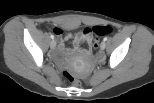


Show Answer
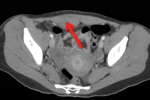
Rectus sheath hematoma

Rectus Sheath Hematoma (RSH)

Rectus muscle tear causing damage to the superior or inferior epigastric arteries with subsequent bleeding into the rectus sheath; uncommon cause of abdominal pain but mimics almost any abdominal condition.

Diagnose with CT, but try using ultrasound (thanks Dr. Joseph Minardi)

May occur spontaneously, but suspect with the following risk factors:

  • Coagulopathy (#1 cause); acquired (e.g., warfarin) or inherited disorder
  • Rectus muscle trauma
  • Vigorous or sudden contraction of rectus muscle
  • Increased intra-abdominal pressure from vigorous coughing
  • Pregnancy (gestation, labor, or post-partum)

Typically a self-limiting condition, but hypovolemic shock may result from significant hematoma expansion.

Hemodynamically stable (non-expanding hematoma): conservative treatment (rest, analgesia, and ice)

Hemodynamically unstable (expanding hematoma): treat with fluid resuscitation, reversal of coagulopathy, and transfusion of blood products.


Show References

Follow me on Twitter (@criticalcarenow)


Title: Ventilation during CPR, "Low and Slow" is the way to go

Category: Critical Care

Posted: 1/12/2016 by Haney Mallemat, MD (Updated: 1/16/2016)

There are so many variables to monitor during CPR; speed and depth of compressions, rhythm analysis, etc. But how much attention do you give to the ventilations administered?

The right ventricle (RV) fills secondary to the negative pressure created during spontaneously breathing. However, during CPR we administer positive pressure ventilation (PPV), which increase intra-thoracic pressure thus reducing venous return to the RV, decreasing cardiac output, and coronary filling. PPV also increases intracranial pressure by reducing venous return from the brain.

So our goal for ventilations during cardiac arrest should be to minimize the intra-thoracic pressure (ITP); we can do this by remembering to ventilate "low (tidal volumes) and slow (respiratory rates)"

  • Low: Use only one-hand while bagging, this will give the patient 500-600cc per breath. Using two-hands provides ~900-1,000cc per squeeze (more than we normally ventilate patients who have a pulse).
  • Slow: Ventilate patients at 8-10 breaths per minute. The less you ventilate the less time the patient spends with positive ITP. Observational studies have demonstrated that providers ventilate too fast during code so the use of a metronome or timing light provides critical feedback.


Show References

Follow me on Twitter (@criticalcarenow)


Title: What's the Diagnosis? Guest submission by #ChileEM Dr. Pedro Verdugo

Category: Visual Diagnosis

Posted: 1/11/2016 by Haney Mallemat, MD (Updated: 3/10/2016)

Question

What’s the name of this CT finding and name two potential causes?

Show Answer

Pneumobilia (air in the biliary tree). Be careful, this must be distinguished from portal venous gas.

Diagnoses to consider when pneumobilia is present:

  • Infection
    • Cholangitis
    • Liver abscess communicating with biliary tree
    • Emphysematous cholecystitis
  • Biliary instrumentation (e.g., recent ERCP)
  • Incompetent sphincter or Oddi
    • passage of a gallstone
    • sphincterotomy
    • certain medications
  • Biliary-enteric fistula (e.g., Peptic ulcer disease, gallstone ileus, trauma,, etc.)

http://radiopaedia.org/articles/pneumobilia

Show References

Follow me on Twitter (@criticalcarenow)


Title: What's the Diagnosis? Case by UMEM alumni, Dr. Bethany Radin

Category: Visual Diagnosis

Posted: 12/28/2015 by Haney Mallemat, MD

Question

79 year-old male with headaches, ataxia, falls, and difficulty urinating. What's the diagnosis?
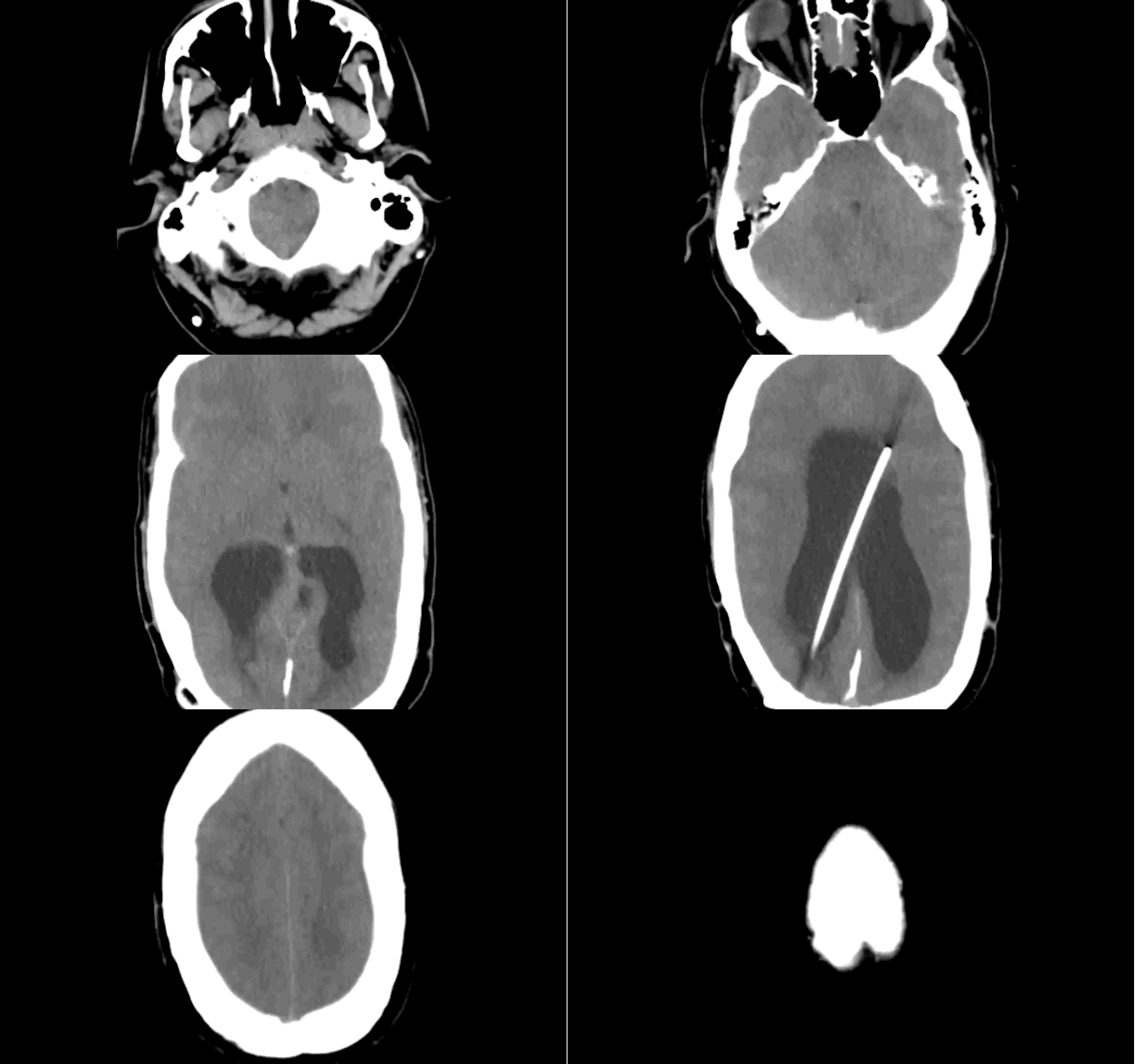
Show Answer

Diagnosis: Ventriculomegaly secondary to Normal Pressure Hydrocephalus

An approach to ventriculomegaly

Ventriculomegaly is due to cerebral atrophy (e.g., Parkinson disease) or increased cerebrospinal fluid (CSF) within the ventricles. Increased CSF is due to:

  • Increased CSF production (e.g., choroid-plexus papilloma), or
  • Impaired CSF re-absorption (e.g., subarachnoid-villi inflammation), or
  • Obstruction of CSF flow (e.g., non-communicating hydrocephalus)

Congenital causes of ventriculomegaly:

  • Neuro-tube defect (e.g, myelomeningocele)
  • Aquaductal stenosis
  • CNS malformation (e.g., Dandi-Walker syndrome)
  • Intrauterine infection (e.g., CMV, rubella, etc.)

Acquired causes of ventriculomegaly:

  • Paget's disease (obstruction of CSF flow)
  • Bacterial or viral meningitis (impaired CSF absorption)
  • Tumor (obstruction of CSF flow)
  • Post-hemorrhagic (e.g., trauma, aneurysmal rupture, AVM, coagulopathy) impaired absorption and/or obstruction of flow

Show References

Follow me on Twitter (@criticalcarenow)


Title: What's the Diagnosis? The Critical Care Edition

Category: Visual Diagnosis

Posted: 12/15/2015 by Haney Mallemat, MD

Question

A patient arrives in acute respiratory distress with left sided chest pain. Ultrasound of the left anterior chest is shown; what's the diagnosis and name one false positive?
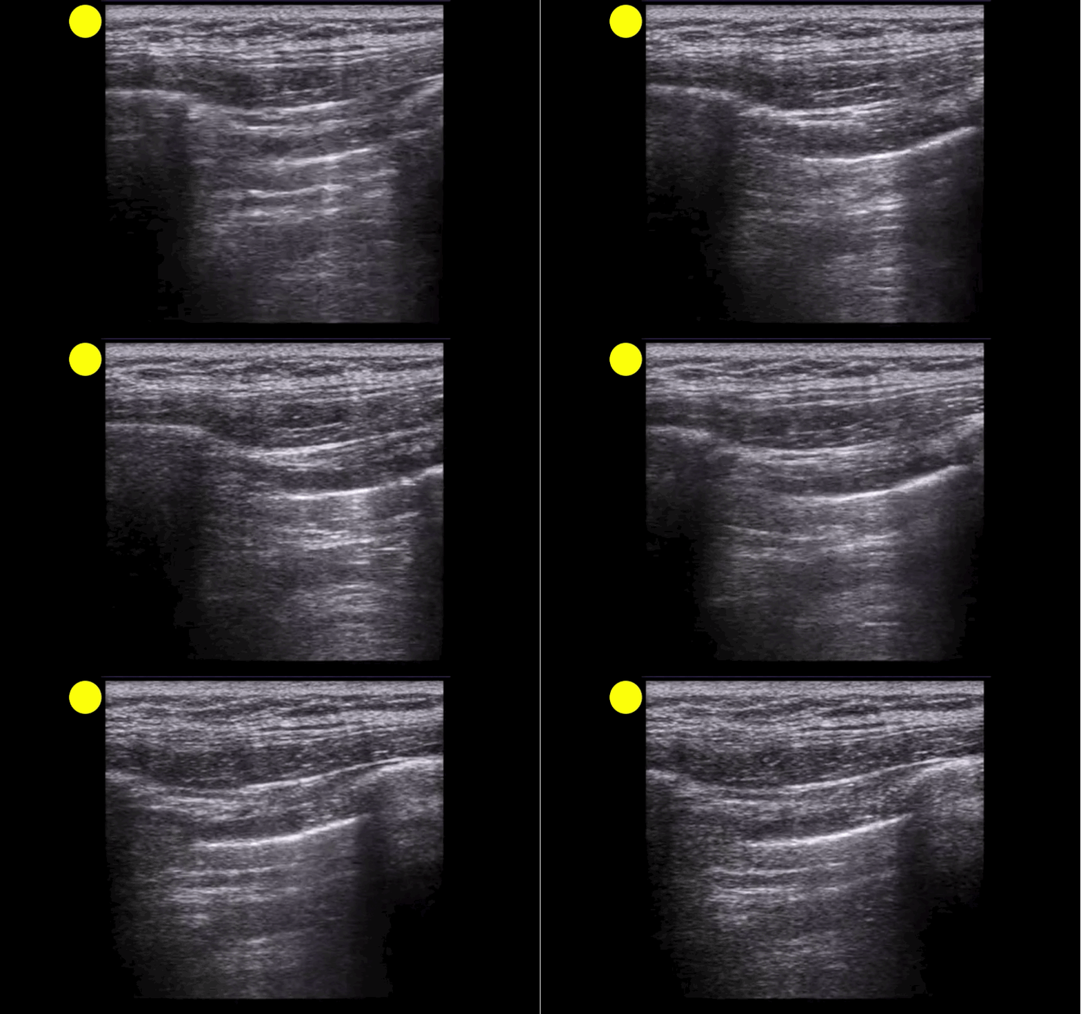
Show Answer

Lung point indicating pneumothorax (PTX)....see below for the false positives

What's the (Lung) Point

  • Separation of the visceral and parietal pleural secondary to a PTX leads to well recognized loss of lung sliding on ultrasound. 
  • Lung point (LP) on ultrasound is where the transition between pleural sliding and the loss of this sliding is demonstrated. Previous studies have reported that the LP has 100% specificity for a PTX.
  • Although this is a great sign to demonstrate PTX, be aware that there are several LP mimics:
    • lung-heart interface
    • lung-diaphragm interface (lower thorax laterally)
    • pleural effusion
    • blebs
  • Check out these great #FOAM posts by @ultrasoundjelly & @ultrasoundMD for more on the LP (and their mimics). Check them out here & here

Show References

Follow me on Twitter (@criticalcarenow)


Title: What's the Diagnosis? Case by UMEM alumni Dr. Adeleke Oni

Category: Visual Diagnosis

Posted: 12/14/2015 by Haney Mallemat, MD

Question

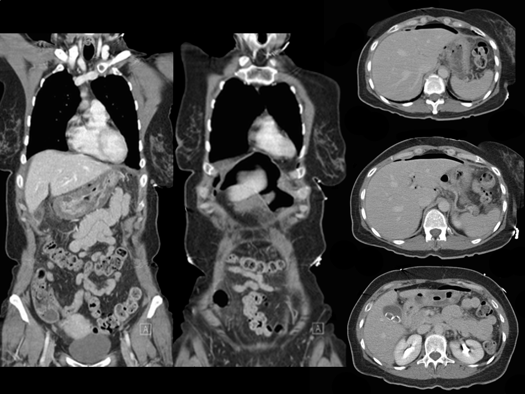
A patient presents with the sudden of onset chest and abdominal pain which woke her up at 2am. She has abdominal tenderness and rebound on exam, what's the diagnosis?

Show Answer

Ruptured gastric ulcer with pneumoperitoneum (CT scan below)

Show References

Follow me on Twitter (@criticalcarenow)


Title: What's the Diagnosis? By Dr. Samira Bhattacharya and Dr. Girish Sethuraman

Category: Visual Diagnosis

Posted: 12/8/2015 by Haney Mallemat, MD (Updated: 12/8/2015)

Question

27 year-old presents after being punched in the face. Decreased vision in left eye, what's the diagnosis?

Show Answer

Lens disclocation

Show References

Follow me on Twitter (@criticalcarenow)


Title: What's the Diagnosis? Image by Dr. Jeffrey Leiter

Category: Visual Diagnosis

Posted: 11/30/2015 by Haney Mallemat, MD

Question

Patient presents with right elbow pain after a fall. What's the diagnosis and what other injury should you look for?
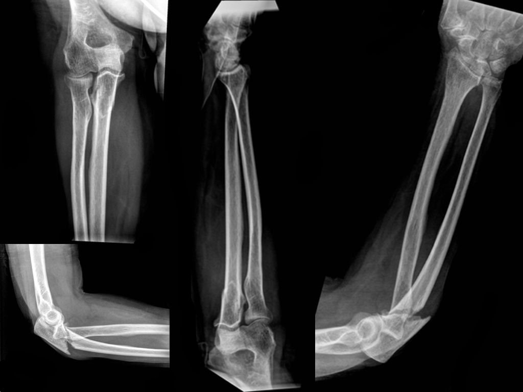
Show Answer

Displaced fracture of the proximal ulna.

Pearls

  • Always look for injury at the radial head when the proximal ulna is fractured, specifically looking for a radial head dislocation; this pattern is known as a Monteggia fracture.
  • Remember the mnemonic MUGR (MU-GR)
    • Monteggia fracture-dislocation = Ulnar fracture with a proximal radial dislocation
    • Galeazzi fracture-dislocation = Radial fracture with a distal radioulnar dislocation

Show References

Follow me on Twitter (@criticalcarenow)


Title: What's the Diagnosis? Case by Dr. Mak Moayedi

Category: Visual Diagnosis

Posted: 11/23/2015 by Haney Mallemat, MD (Updated: 12/5/2015)

Question

An elderly patient presents with a history of weight loss and chronic constipation. The abdominal Xray is shown below. What's the diagnosis?

This one is tricky so here's a hint: why is the right kidney and psoas muscle so well defined?
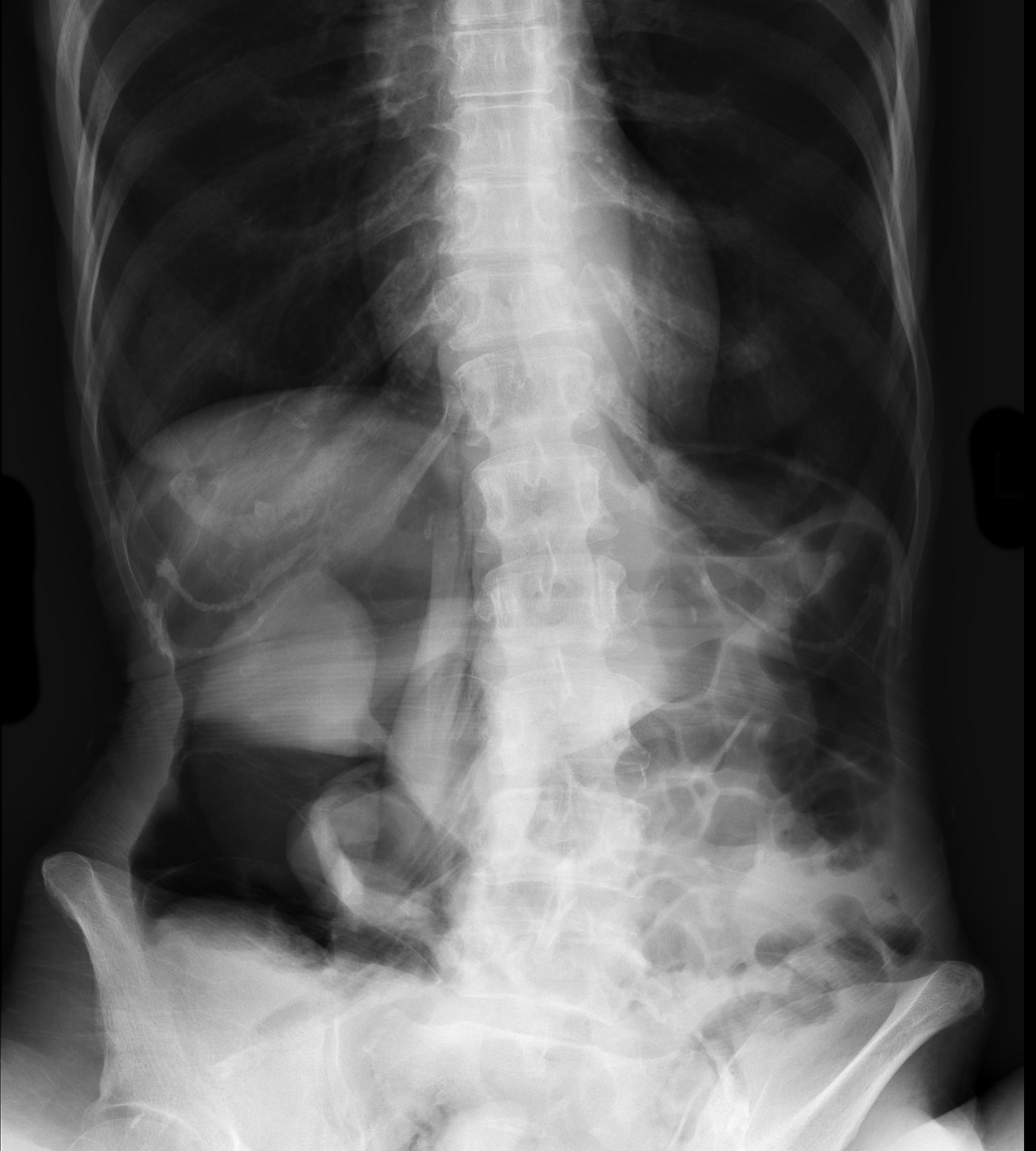
 

Show Answer
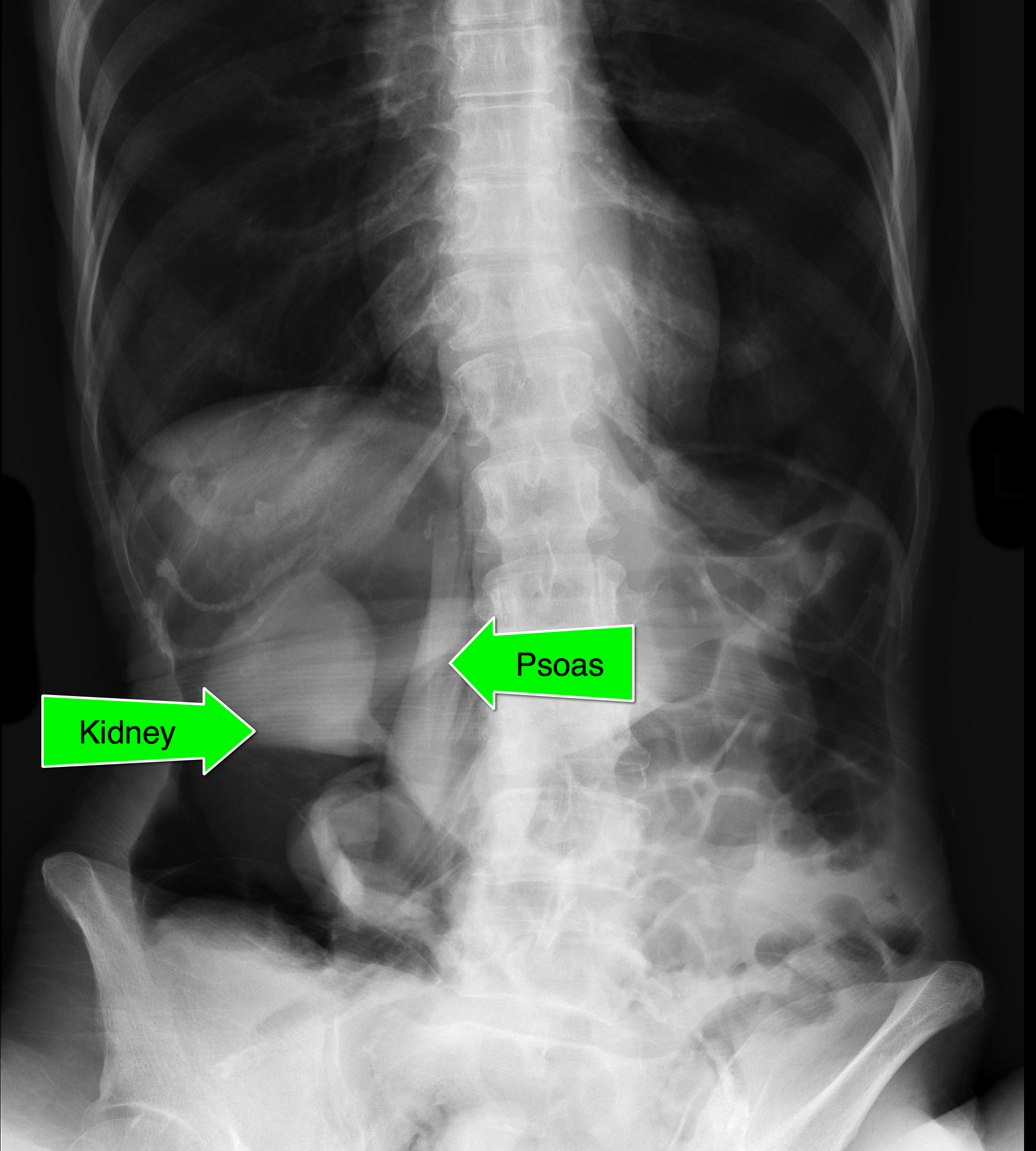
  • Massive retroperitoneal and peritoneal free-air from ascending colon rupture
  • Colon cancer was eventually diagnosed in the operating room.
  • Notice that the kidney and psoas muscle outlined by the free-air below.

Show References

Follow me on Twitter (@criticalcarenow)


Title: What's the Diagnosis?

Category: Visual Diagnosis

Posted: 11/2/2015 by Haney Mallemat, MD

Question

Patient complains of facial and neck swelling, what's the diagnosis?

Show Answer

Subcutaneous emphysema

Show References

Follow me on Twitter (@criticalcarenow)


Title: Is there a secret menu at the Blood Bank?

Category: Critical Care

Posted: 10/20/2015 by Haney Mallemat, MD

There is more than the standard preparations of plasma, platelets, and PRBCs in the blood bank. Certain patients will require these specialized preparations when a transfusion is required. Here are three to know:

  • Leukoreduced (PRBCs are run through a filter to reduce the total WBC burden)
    • Most of the blood in USA is leukoreduced
    • Should be requested for pre-transplant patients and patients who previously experienced febrile non-hemolytic reactions
  • Irradiated PRBCs (radiation incapacitates donor WBCs)
    • Irradiation prevents the fatal transfusion-associated graft versus host disease, which occurs in patients who are severely immunosuppressed or who are closely related to the blood product donors.
  • Washed RBCs/platelets (washing removes plasma, cell fragments and excess potassium)
    • Washed cells are used for neonates/pediatric patients due to sensitivity to potassium in normal products; in adults, it is used for patients with prior allergic reactions to blood products or IgA deficiency

Show References

Follow me on Twitter (@criticalcarenow)


Title: What's the Diagnosis? Case by Dr. Samuel Cordeiro

Category: Visual Diagnosis

Posted: 10/19/2015 by Haney Mallemat, MD

Question

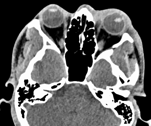
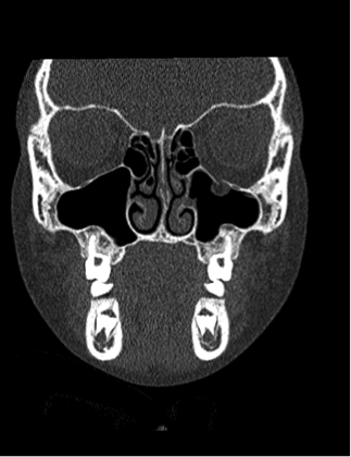
8 year-old female presents with nausea, vomiting, double-vision and inability to move her left eye upwards after being kicked in the face at school. What's the diagnosis?
Show Answer

Orbital floor fracture with entrapment of the inferior rectus muscle.

  • Orbital floor fractures are the most commonly fractured part of the pediatric orbit. Although treated conservatively in adults, pediatric patients can benefit from early repair.
  • Children are at increased risk for a “trap-door” type fracture, which can entrap the extra-ocular muscles because their orbital floor is more flexible.
  • Signs of entrapment include abnormal extra-ocular movements, diplopia, nausea, and vomiting requiring urgent subspecialty evaluation.
  • Bradycardia may occur secondary to the oculo-cardiac reflex when muscle entrapment occurs and is a helpful clue when present


Show References

Follow me on Twitter (@criticalcarenow)


Title: What's the Diagnosis? Case by Dr. Kathleen Stephanos

Category: Visual Diagnosis

Posted: 10/12/2015 by Haney Mallemat, MD

Question

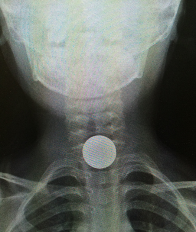
5 year-old boy who presents with sudden onset hoarse voice, and drooling without a fever. 
Show Answer

Coin lodged in the esophagus

Coin ingestions

  • More than 100,000 foreign body ingestions are reported in children each year; coins are #1 cause
  • Although not always 100% true, coins typically appear circular on an AP Xray of the neck when in the esophagus and linear when in the trachea
  • Coins usually pass without issue, but warning signs are drooling, dysphagia, hoarse voice, wheezing, or stridor
  • 10-20% require endoscopy for removal and 1% require surgery
  • Objects lodged in the middle esophagus should raise concern for underlying pathology such as strictures, masses, or webs

Show References

Follow me on Twitter (@criticalcarenow)

Waltzman ML, Baskin M, Wypij D, Mooney D, Jones D, Fleisher G. A randomized clinical trial of the management of esophageal coins in children. Pediatrics. 2005;116(3):614.

Cevik M, Gókdemir MT, Boleken ME, Sogut O, Kurkcuoglu C. The characteristics and outcomes of foreign body ingestion and aspiration in children due to lodged foreign body in the aerodigestive tract. Pediatr Emerg Care. 2013;29(1):53.


Title: What's the Diagnosis? Image by mi amigo, Dr. Pedro Verdugo.

Category: Visual Diagnosis

Posted: 10/6/2015 by Haney Mallemat, MD (Updated: 10/7/2015)

Question

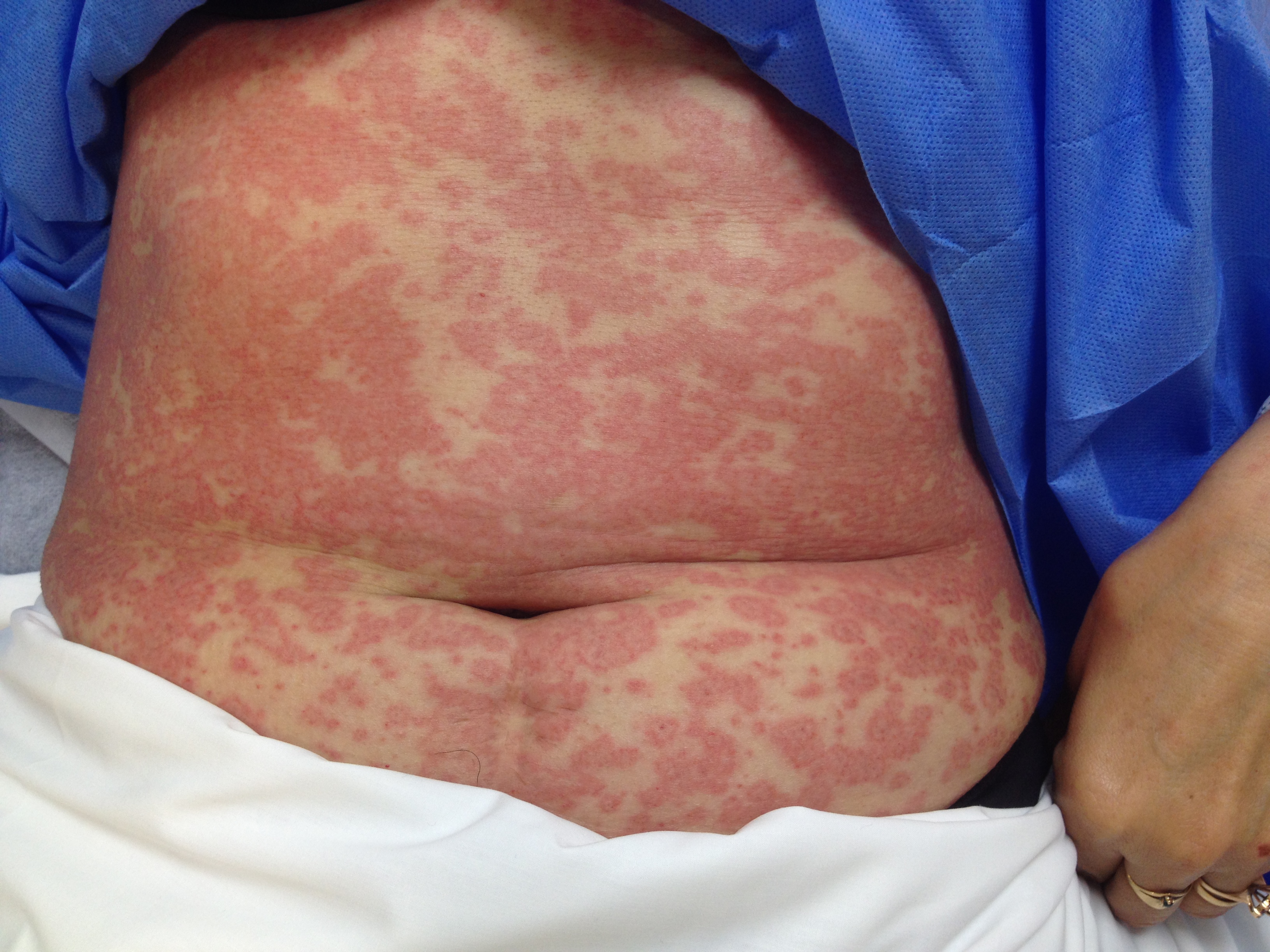
Patient presents after being started on an antibiotic for cellutlitis of lower extremity. What's the diagnosis and what are some other etiologic agents (name 3)
Show Answer

Erythema Multiforme (minor)

  • Acute, self-limited rash affecting the skin; mucus membranes are spared in the minor variant although EM major involves one mucus membrane.
  • Erythematous, macupapular, and blanching with central clearing; it is often puritic
  • Several etiologic factors that have been identified. Most commonly:
    • Infections: viral (e.g., HSV), bacterial (e.g., strep), fungal (e.g., histoplasmosis)
    • Drugs: antibiotics (e.g., sulfa drugs, PCN, etc.), NSAIDs
    • Miscellaneous: collagen vascular diseases, pregnancy, and malignancy
  • It is self-limiting and will resolve in 2-6 weeks.
  • Treat by stopping offending agent and supportive care (e.g., diphenhydramine for purtitis)

Show References

Follow me on Twitter (@criticalcarenow)


Title: What's the Diagnosis? Case by Dr. Zachary Dezman

Category: Visual Diagnosis

Posted: 9/28/2015 by Haney Mallemat, MD

Question

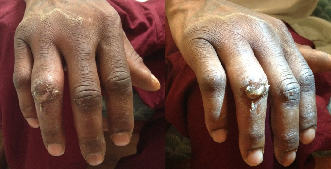
26 year-old male presents with a swollen 4th digit and pain during extension, what’s the diagnosis?
Show Answer

Answer: Infectious Flexor Tenosynovitis

Infectious Flexor Tenosynovitis

  • Closed space infection of the flexor tendon sheath; an orthopedic emergency
  • Typically an infection with skin flora secondary to penetrating trauma
  • Remember Kanavel's cardinal signs via mnemonic B.E.S.T. (the "BEST" mnemonic):
    • B: Bend finger (i.e., finger held in slight flexion)
    • E: Extension pain (i.e., pain on passive extension)
    • S: Sausage-like digit (i.e., fusiform swelling
    • T: Tenderness along tendon sheath
  • If early and not severe a course of IV antibiotics covering skin flora may be tried. Surgical intervention, however, is often necessary so early consultation with a hand surgeon is highly recommended

Show References

Follow me on Twitter (@criticalcarenow)


Previous |  1 |  2 |  3 |  4 |  5 |  6 |  ... |  17 |  Next
University of Maryland School of Medicine logo
  • Departments
  • Programs
  • Research Centers
  • Institutes
  • Graduate Education
  • CIBR Cores
  • Offices of the Dean
    • University of Maryland, Baltimore
    • University of Maryland Medical Center
    • Medical Alumni Association of the University of Maryland
    • Health Sciences and Human Services Library
University of Maryland School of Medicine
655 W. Baltimore Street
Baltimore, MD 21201
Contact Us

© University of Maryland School of Medicine

    • Webmaster
    • Web Accessibility
    • Site Index
    • UMB Hotline
    • Employment
    • Map