University of Maryland School of Medicine logo
Emergency Medicine
All Departments
UMSOM Assistant
UMEM Educational Pearls
  • Home
  • Departments
  • Emergency Medicine
  • CME
  • UMEM Educational Pearls
  • CME
  • Courses and Conferences
  • Free Open Access Medical Education
  • UMEM Educational Pearls

Search

1-4 of 4 results by Ashley Strobel


Title: Happy New Year 2015

Category: Pediatrics

Keywords: intraosseous access, pediatrics (PubMed Search)

Posted: 1/3/2015 by Ashley Strobel, MD (Updated: 7/21/2026)

Are you comfortable with Intraosseous Catheter Placement in Children during a code?  A pediatric code or child in distress is also distressing to care providers.  Your staff may not feel comfortable with IO access in children. Read on to be more comfortable with your options as IO access in children can be difficult, especially the chubby toddlers.  The basics for a patient in distress are "IV, O2, Monitor".  Access is vital to giving resuscitation medications.

Indications for IO access: Any child in whom IV access cannot readily be obtained, but is necessary.

All IOs are 15G for infusion equal to central vascular access.  

Different colors indicate different sizes:

  • Pink=15 mm
  • Blue=25 mm
  • Yellow=45 mm

Preferred sites:

  1. Proximal tibial (place a towel in popliteal fossa to bend the leg, pinch tibia and 1 finger width below the patella inferior and medial if you can’t palpate the tibial tuberosity)
  2. Distal tibia (proximal to medial malleolus by 1 finger width)—preferred in older children
  3. Proximal Humerus (internally rotate humerus and 1 finger width below surgical neck)
  4. Distal Femoral (1-2 finger widths superior to femoral epicondyles)

Kids-do NOT use the sternum or distal radius

The reference from NEJM has videos to review placement and different tools (manual, EZ IO, and autoinjector).

Show References

Joshua Nagler, M.D., and Baruch Krauss, M.D., Ed.M. Intraosseous Catheter Placement in Children.  N Engl J Med 2011; 364:e14.


Title: Emergency Physician Bedside Ultrasound for Appendicitis

Category: Pediatrics

Keywords: appendicitis, ultrasound, bedside (PubMed Search)

Posted: 11/8/2014 by Ashley Strobel, MD

Emergency Physician Bedside Ultrasound for Appendicitis

Why?

To reduce length of stay, improve patient care, and reduce radiation exposure in young patients.

How?

Start with pain medication so you get a better study. (Consider intranasal fentanyl for quicker pain relief and diagnostics in pediatrics.) Study results are also improved with a slim body habitus.

Place the patient supine

Use a high-frequency linear array transducer

Start at the point of maximal tenderness in the RLQ

Transverse and longitudinal planes "graded compression" to displace overlying bowel gas which usually has peristalsis (See Sivitz, et al article for images of "graded compression")

Appendix is usually anterior to the psoas muscle and iliac vein and artery as landmarks

Measure from outer wall to outer wall at the most inflamed portion of the appendix (usually distal end)

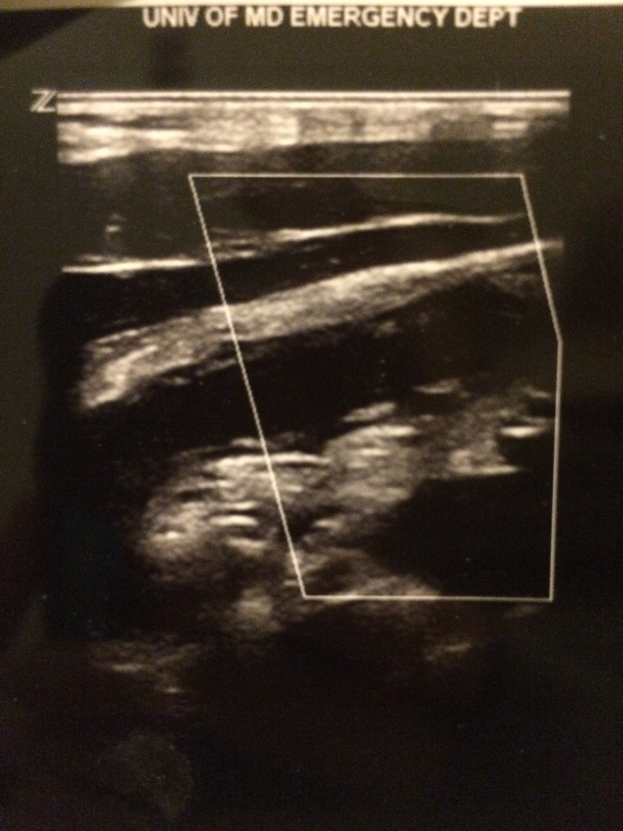
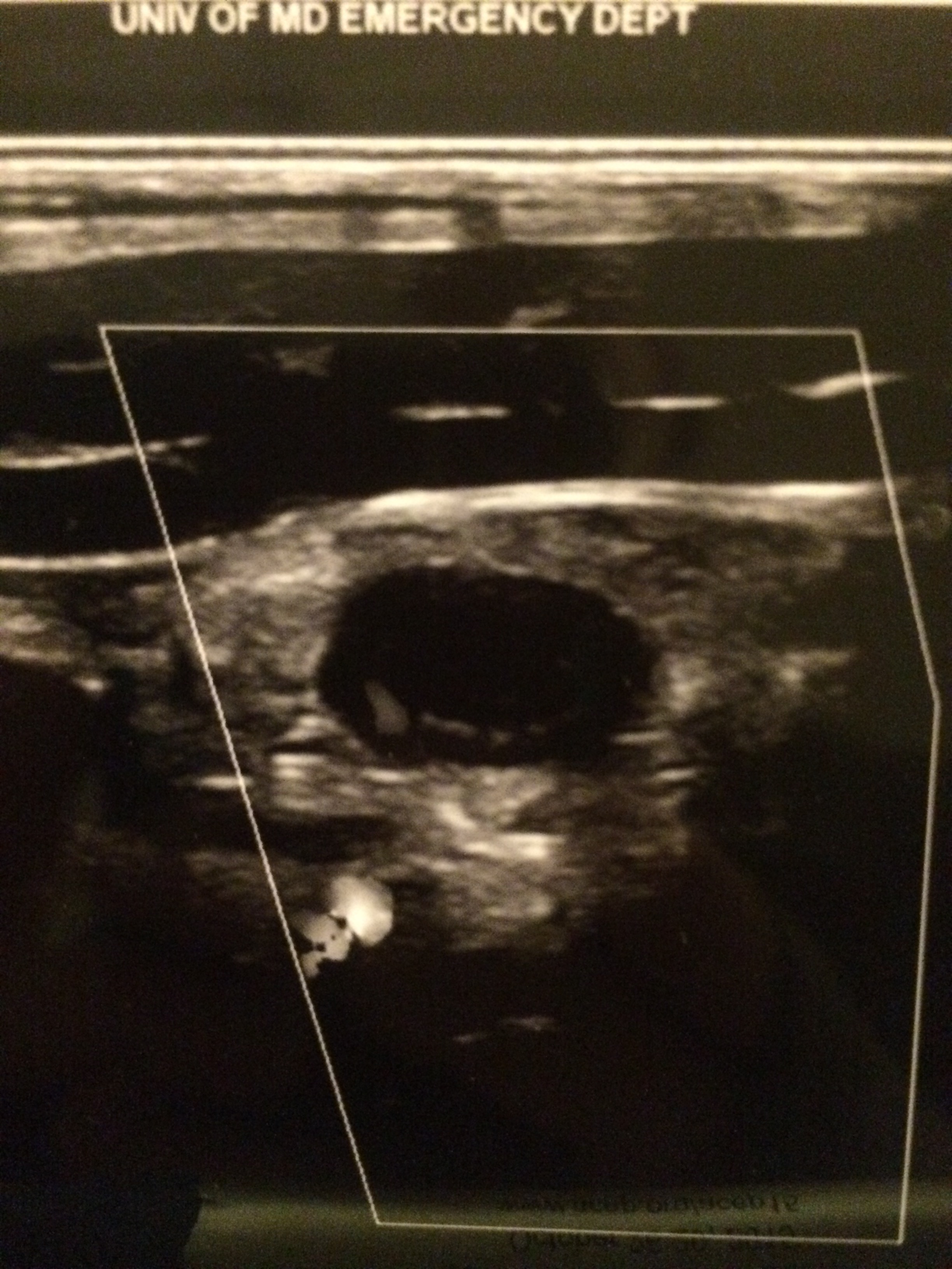
Example:
Positive study:

A non-compressible, blind-ending tubular structure in the longitudinal axis >6 mm without peristalsis (see second image above with 8.3 mm diameter measurement)

A target sign in the transverse view (see first image above)

Additional suggestive findings: appendiceal wall hyperemia with color Doppler, appendicoliths hyperechoic (white) foci with an anechoic (black) shadow, periappendiceal inflammation or free fluid

Negative study:

Non-visualization of the appendix with adequate graded compression exam in the absence of free fluid or inflammation.

Limitations for visualization and possible false negative result:

Retrocecal appendix and perforated appendix are difficult to visualize with US.

Pitfalls:

US has good specificity (93% in Sivitz et al article), but limited sensitivity (85% in Sivitz et al article), so trust your clinical judgement. You may need a MRI (pregnant/pediatrics) or CT as they have improved, but not perfect sensitivity.

Show References

Valesky, et al. Focus On: Ultrasound for Appendicitis. ACEP Now. June 2012.

Sivitz AB, Cohen SG, Tejani C. Evaluation of Acute Appendicitis by Pediatric Emergency Physician Sonography. Annals of Emerg Med. Oct 2014; 64: 358-363.

Attachments

  • 1411081818_Appendicitis_Blind_End_Pouch_US.jpg (1,372 Kb)
  • 1411081818_Target_Sign_US_Appendix.jpg (1,461 Kb)


Title: Visual Diagnosis Pediatrics: Case thanks to Ari Kestler MD (@KestlerMD) and Haney Mallemat MD (@CriticalCareNow)

Category: Pediatrics

Keywords: non-accidental trauma, clavicle fracture, neonate, pediatrics, abuse (PubMed Search)

Posted: 10/4/2014 by Ashley Strobel, MD

Question

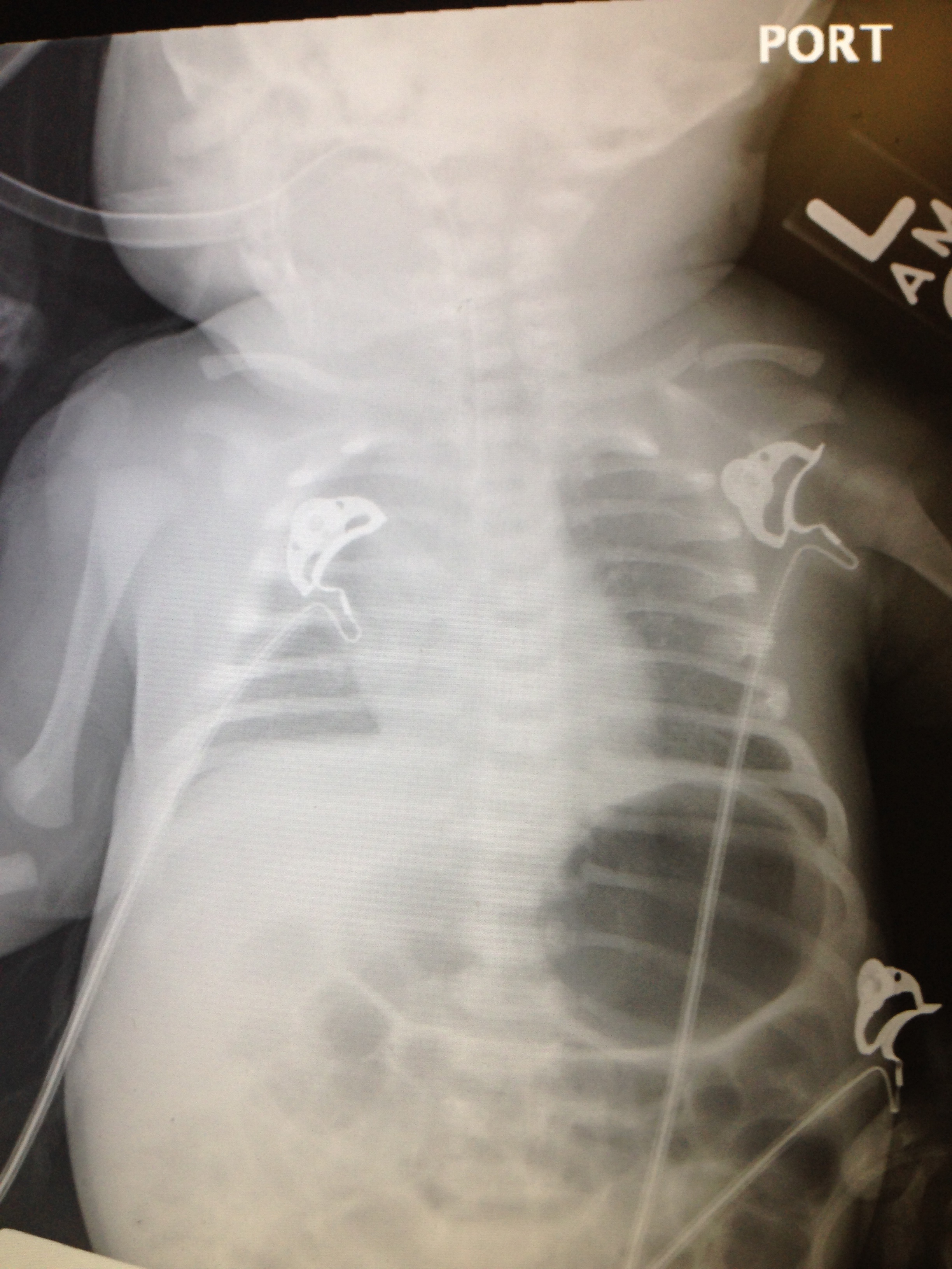
Q: What is wrong with this baby? And what Dx should you entertain?

Previously healthy 7d old presents after difficulty feeding, one episode of vomiting and now with intermittent apneic episodes.
 

Show Answer

Non-accidental trauma (NAT) is most prevalent in children 0-3 months of age.

Radiographically classic metaphyseal lesions, rib fractures, and multiple fractures in various stages of healing are most commonly described in child abuse cases.

How do we know this is not just birth trauma from a shoulder dystocia, LGA (large for gestational age), or difficult vaginal delivery?

The key is dating the fracture.  In this recent publication by Walters MM et al, prior to 8 days of life, 100% of radiographs did NOT have callus present. Callus formation is highly unlikely in fractures less than 9 days old, and typically appears by 15 days old.  Callus thickness decreases inversely with fracture age.  Additionally, subperiosteal new bone formation is highly unlikely in fractures less than 7 days old and typically appears by 10 days old.  Subperiosteal new bone formation increases in thickness inversely with fracture age.  Therefore, a clavicle fracture in a 7 day old without subperiosteal new bone formation or callus is unlikely from birth trauma and NAT should be considered.

How can you tell if subperiosteal new bone formation is present? 

Subperiosteal new bone formation appears as a hazy cortical margin or a thin layer of bone separated from the original cortex by a discrete lucent interval.  The new bone increases in thickness with time and may evolve to appear as a lamellated or multilayered linear hyperdensity parallel to the cortex of the bone.  See referenced article for great picture examples.

NAT Work-up:

CT head without contrast if ≤2 yo

Skeletal Survey if ≤ 2 yo

AST, ALT, amylase, lipase, CBC, Manual Differential, BMP, UA, Urine Toxicology

Consults: Ophthalmology, Social Work, Child Protection

OH BUTT TUBE (Dark Green Top Sodium Heparin) for further inpatient team studies

ALTE Work-up:

Guided by history, however consider the following:

Full sepsis evaluation for neonate <30 days

ECG

Possible reflux or seizure evaluation

Consider NAT or Pertussis/RSV with cyanosis

It is controversial to send these infants home from the ED.  Typically they benefit from 24 hours of monitoring, but this is a pearl for another day.

Show References

See article for further pictures of subperiosteal new bone formation:

Walters MM, Forbes PW, Buonomo C, and Kleinman PK.  Healing Patterns of Clavicular Birth Injuries as a guide to fracture dating in cases of possible infant abuse.  Pediatric Radiology.  October 2014; 44: 1224-1229.

@AstrobelMD

Attachments

  • 1410041359_Clavicle_Fracture.jpg (1,743 Kb)


Title: A young asthmatic with a bad day: Visual Diagnosis

Category: Pediatrics

Keywords: Macklin Phenomenon, asthma, pneumomediastinum (PubMed Search)

Posted: 9/22/2014 by Ashley Strobel, MD

Question

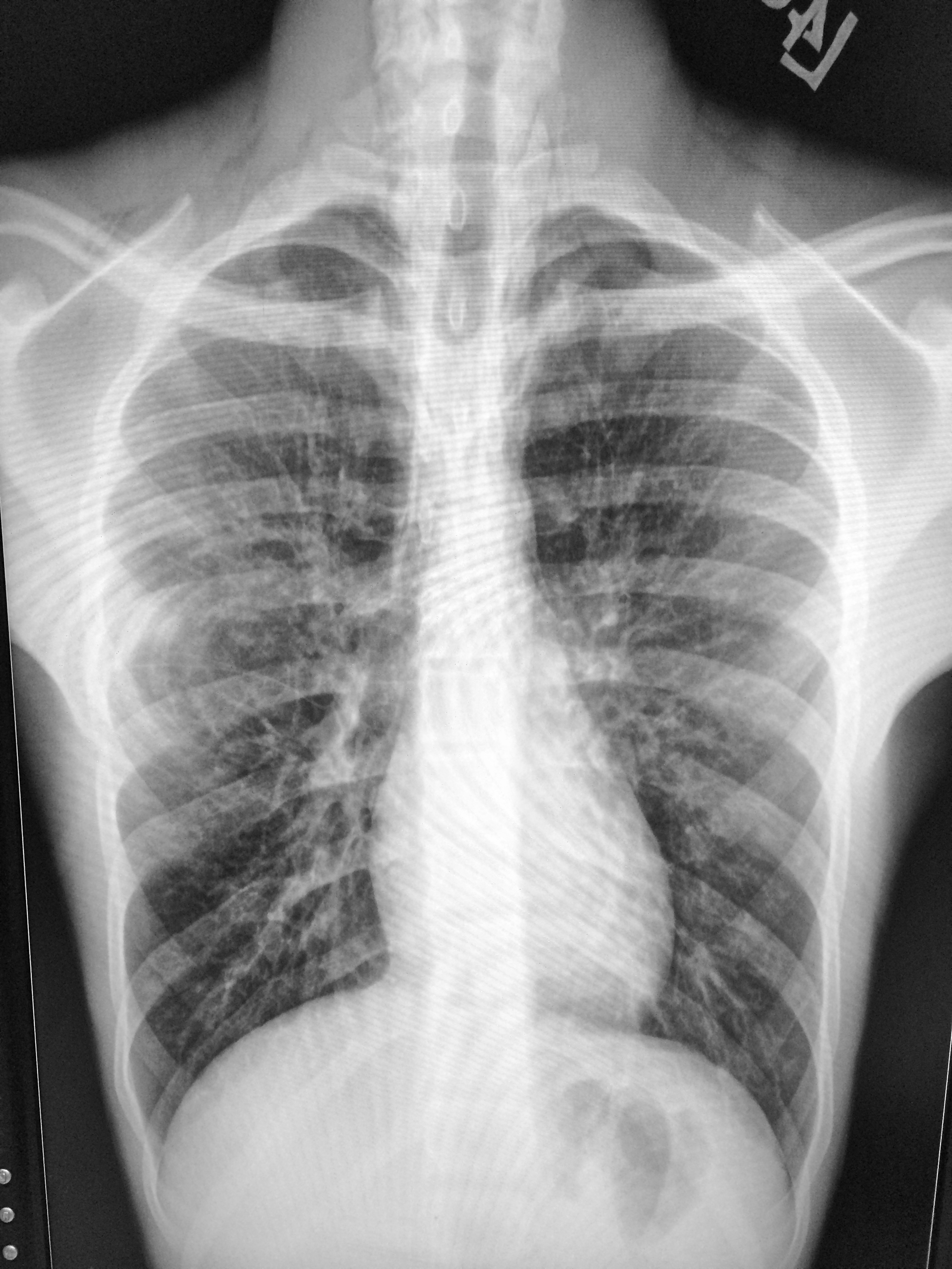
16 yo M with pleuritic right upper chest pain that started today.  He is suffering from an asthma exacerbation currently in the setting of URI with cough.  He is afebrile, tachycardic to 140-150s, respiratory rate 20, and sats 98% on room air.  ECG was performed which incidentally diagnosed this patient WPW and he went for ablation as an outpatient.  His chest x-ray showed:

Besides a bad day, what do we call this chest x-ray finding?
Show Answer

Macklin Phenomenon

-asthma exacerbation rupture of the alveoli causing pneumomediastinum

-typically a young man

-most common chief complaint is chest pain


Physical Exam: Hamman’s sign may be present (crackle with heartbeat) or subcutaneous emphysema


Etiology: Esophagus, lungs, or bronchial tree

Rupture of alveoli: asthma exacerbation (bronchial hyper-reactivity/constriction), barotrauma, valsalva maneuvers (lifting, childbirth), deep respiratory maneuvers/Valsalva (strenuous exercise or FVC breathing), drug use (crack cocaine causing bronchial constriction, marijuana), vomiting, blunt thoracic/abdominal trauma, scuba diving with rapid ascent


Aerodigestive tract injuries: bronchoscopy tracheobronchial injuries, laryngeal fx, bronchial fx, tracheal neoplasm, esophageal injuries (Boerhaave syndrome, paripartum, asthma exacerbation, esophageal neoplasm)


Extension from neck: head/neck sx, RPA/PTA, dental abscess/extractions


Extension from RP/chest wall: rupture RP hollow viscus


Management:

-self -limited

-treat underlying condition

-swallow study for all cases following emesis to rule out Boerhaave’s syndrome

-no repeat CXR, advance diet as tolerated, 23 hour observation

-Al-Mufarrei, et al suggest without trauma, pleural effusion, hemodynamic instability, pneumoperitoneum, or severe vomiting, the finding of spontaneous pneumomediastinum (with or without Meckler’s triad of esophageal rupture: vomiting, lower chest pain, and cervical subcutaneous emphysema after overindulgence) usually leads to unnecessary radiologic investigations, dietary restriction, and antibiotic administration

-surgery for decompression

Show References

Gray JM and Hanson GC.  Mediastinal emphysema: aetiology, diagnosis, and treatment.  Thorax.  1966; 21: 325-332.


Al-Mufarrej F, Badar J, Gharagozloo F, Tempesta B, Strother E, Margolis M.  Spontaneous pneumomediastinum: diagnostic and therapeutic intervnetions.  Journal of Cardiothoracic Surgery.  November 2008; 3: 59.

Attachments

  • 1409221530_IMG_5821.jpg (1,850 Kb)


University of Maryland School of Medicine logo
  • Departments
  • Programs
  • Research Centers
  • Institutes
  • Graduate Education
  • CIBR Cores
  • Offices of the Dean
    • University of Maryland, Baltimore
    • University of Maryland Medical Center
    • Medical Alumni Association of the University of Maryland
    • Health Sciences and Human Services Library
University of Maryland School of Medicine
655 W. Baltimore Street
Baltimore, MD 21201
Contact Us

© University of Maryland School of Medicine

    • Webmaster
    • Web Accessibility
    • Site Index
    • UMB Hotline
    • Employment
    • Map