
Ketamine for RSE?
- Up to 43% of patients with status epilepticus may progress to refractory status epilepticus (RSE).
- Propofol, midazolam, and barbituates are often recommended for patients with RSE.
- Importantly, all of these medications may be limited by hypotension and respiratory depression.
- Ketamine is emerging as adjuvant therapy for patients with RSE.
- The loading dose of ketamine ranges from 0.5 to 3 mg/kg, followed by a maintenance infusion of 0.3 to 4 mg/kg/h.
Show References
Show References
Ataxia is an important clinical sign of cerebellar pathology, but how is it actually described?
Stance ataxia: inability to stand with feet together for more than 30 seconds
Gait ataxia
Sensory ataxia: the first 2 elements, in addition to a positive Romberg sign
Truncal ataxia: oscillation of body while sitting or standing
Limb ataxia: functional impairment in performing actions such as writing or buttoning and improves with slowing down the movement
Dysdiadokinesia: impairment of rapidly alternating movement
Intention tremor: tested by finger-to-nose and heel-to-shin.
Dysmetria: pastpointing or undershooting on finger-chasing or shin-tap.
Dysarthria: irregular and slow speech with unnecessary hesitation
Nystagmus and other ocular disturbances, such as ocular flutter and opsoclonus.
The first 3 are present in both cerebellar pathology and loss of proprioceptive input, the rest are usually due to cerebellar pathology or ataxic syndrome.
Show References
Is it possible to have a patient present in diabetic ketoacidosis (DKA) with both negative serum and urinary ketone levels?
A case report published in American Journal of Emergency Medicine by Jehle et al provides a helpful reminder of this phenomenon (1). The degree of acidosis is directly related to the ratio of the various ketones/ketone metabolites: acetone, acetoacetate and beta-hydroxybutyrate present in the serum. The proportion of each respective substance is determined by the existing redox state in the blood. At any given time, acetoacetate and beta-hydroxybutyrate exist in an equilibrium dependent upon the ratio of NAD+ and NADH(fig.1). These substances freely convert with the assistance of the enzyme beta-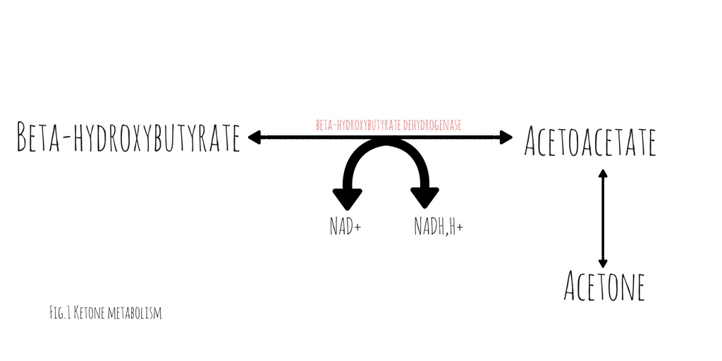 hydroxybutyrate dehydrogenase (2). This conversion requires the donation of a hydrogen atom from NADH. The balance between beta-hydroxybutyrate and acetoacetate, is determined by the ratio of NADH to NAD+. Acetoacetate will freely degrade into acetone through non-enzymatic decarboxylation. Early in DKA, acetoacetate is the most prevalent substance. As the disease progresses and the serum ratio of NADH to NAD+ increases, the proportion of beta-hydroxybutyrate rises, decreasing the quantity of acetoacetate and acetone.
Traditional serum and urinary ketone assays react strongly to acetoacetate but neither reliably react with beta-hydroxybutyrate. Patients in whom the majority of their anion gap is filled by beta-hydroxybutyrate, urinary or serum ketone levels may be negative. In such cases, serum beta-hydroxybutyrate assays would be positive but are not universally available.
It is important to note, with resuscitation and insulin therapy, the ratio of NADH/NAD+ will start to normalize causing an increase in the quantity of acetoacetate. As the patient improves and the anion gap clears, the degree of ketones detected in the serum and urine will paradoxically increase.
Show References
According to the 4th International Patellofemoral Pain Research Retreat recently published in British Journal of Sports Medicine, the core criterion required to define Patelofemoral Pain (PFP) syndrome is pain around or behind the patella, which is aggravated by at least one activity that loads the patellofemoral joint during weight bearing on a flexed knee (eg, squatting, stair ambulation, jogging/running, hopping/jumping).
Additional criteria (not essential):
- Crepitus or grinding sensation emanating from the patellofemoral joint during knee flexion movement
- Tenderness on patellar facet palpation
- Small effusion
- Pain on sitting, rising on sitting, or straightening the knee following sitting
PFP is common in young adolescents, with a prevalence of 7–28%, and incidence of 9.2%.
Stay tuned for recommendations on treatment and diagnosis.
Show References
From 2010-2014 ED visits in the US for injuries from trampoline parks (TPI) increased from 581 visits per year to 6932 visits per year. There was no change in the number of injuries related to home trampoline use. TPI were more likely to involve the lower extremity, be a dislocation and warrant admission and less likely to involve the head.
Bottom line: TPIs are increasing and have a different injury pattern compared to home trampolines.
Show References
In pediatric population, small dose or single pill ingestion can potential result in severe or lethal toxicity.
Clinicians should be mindful of potential toxicity following xenobiotic exposure (below) in pediatric population, especially under the age of 5 years old, even if the patient may initially appear asymptomatic.
- Benzocaine
- B-adrenergic antagonist (sustained release)
- Calcium Channel blockers (sustained release)
- Camphor
- Clonidine
- TCAs
- Diphenoxylate/atropine (Lomotil)
- Toxic alcohol (methanol or ethylene glycol)
- Methylsalicylate
- MAO-Is
- Opioids
- Phenothiazines
- Quinine or chloroquine
- Sulfonylureas
- Theophylline
Suspected ingestion of above medications/xenobiotics may warrent observation up to 24 hours in asymptomatic pediatric population.
July 2016 was the hottest month ever recorded. As the temperature of the planet continues to rise, changes in infectious disease patterns of transmission are likely to occur. Many formerly exotic diseases, such as Zika, West Nile, malaria and cholera are spreading to new areas. Some of these are now found in the United States. Below is a table (from M. L. Wilson) that exemplifies how environmental changes can impact infectious diseases.
Bottom Line: Remain vigilant for unusual infectious diseases, especially among travelers. However recognize that many diseases have the potential to spread to the U.S., especially considering ongoing environmental changes.

Show References
47yo M chef presents to your ED with 2 days of worsening left hand pain after sustaining a puncture wound to hand at work. The hand is red and swollen and he complains of pain. Interestingly, his index and middle digits are in an ABducted position at rest.
Collar Button Abscess
Web space infection of the palmer AND dorsal hand
The Palmer aponeurosis prevents volar extension (but promotes dorsal encroachment)
The pus spreads between the MC bones and erupts dorsally....creating a DORSAL abscess.
Loss of palmer concavity is seen.
ABduction of the adjacent digits, resulting in a "V" configuration with the apex pointing to the site of infection. This would not happen from simple pus in the dorsal space!
Can be missed if only focused on the dorsal hand. The palm will show the original injury (splinter, cut, foreign body)
Treatment is urgent surgical drainage.
http://www.eplasty.com/article_images/eplasty16ic06_fig1.gif
Show References
Question
Show Answer
Show References
Attachments
Zika Virus-associated GBS
- Zika virus has been shown to trigger Guillain-Barre Syndrome (GBS) at a rate similar to Campylobacter jejuni infections.
- In patients with Zika virus-associated GBS, neurologic deterioration has been rapid, with approximately 33% of patients developing respiratory distress.
- For patients who have required intubation, the duration of mechanical ventilation and length of ICU stay has been very long.
- Consider Zika virus-associated GBS in patients with muscle weakness, facial palsy, or paresthesias in the setting of a travel or exposure history to the virus.
Show References
Amiodarone 150 mg IV over 10 minutes and procainamide IV 20-50 mg/min (up to 17 mg/kg) are two antiarrhythmic medications recommended in the American Heart Association (AHA) Guidelines for Cardiopulmonary Resuscitation and Emergency Cardiovascular Care for stable wide QRS complex tachycardia. [1]
What they did:
Multi-center, prospective, randomized, open-label trial comparing the incidence of major cardiac events in the acute treatment of hemodynamically stable patients with wide QRS monomorphic tachycardia (presumed to be VT) using amiodarone 5 mg/kg IV infused over 20 minutes versus procainamide 10 mg/kg IV infused over 20 minutes. [2] The study period was 40 minutes, starting from the beginning of the infusion.
What they found:
- Analysis included 62 (n=33 procainamide, n=29 amiodarone) patients from 16 hospitals
- Fewer patients treated with procainamide experienced major cardiac events during the study period compared to those who received amiodarone (9% vs 41%; OR =0.1, 95% CI 0.03-0.6; P=0.006). The most frequent adverse cardiac event was severe hypotension requiring electrical cardioversion.
- Termination of VT occurred more frequently in patients treated with procainamide (67% vs 38%; OR =3.3, 95% CI 1.2-9.3; P=0.026).
Application to clinical practice:
- Medication doses and patient weights are not reported in the results. A comparison of the doses used in the PROCAMIO study to those recommended in the AHA guidelines for a 70 kg and 100 kg patient are as follows:
- Procainamide:
- 70 kg patient would receive procainamide 35 mg/min for 20 minutes. This is in the middle of the dose range recommended by the AHA.
- 100 kg patient would receive procainamide 50 mg/min for 20 minutes. This is the upper limit of the dose range recommended by the AHA.
- Amiodarone:
- 70 kg patient would receive amiodarone 350 mg over 20 minutes. This is approximately equal to administering 2 doses of 150 mg at the infusion rate recommended in the AHA guidelines.
- 100 kg patient would receive amiodarone 500 mg over 20 minutes. This is approximately equal to administering 3 doses of 150 mg at an infusion rate over 1.5 times higher than that recommended in the AHA guidelines.
- Procainamide:
- The study size was small, and depending on patient weights, it is possible that amiodarone dosing was more aggressive compared to doses commonly used in the US. However, the results suggest that procainamide could offer improved safety and efficacy over amiodarone for stable wide QRS tachycardia.
Show References
Drug-induced hypoglycemia is an often severe and symptomatic. It is a potentially preventable cause of significant morbidity. In one large study, it accounted for 23% for hospital admissions due to adverse drug events and 4.4% of overall admissions. The majority of hypoglycemic events occur with insulin and sulfonylureas. However, multiple drugs can affect glucose homeostasis and have been cited to cause hypoglycemia in therapeutic dose alone or in combination with other medications or illness. Factors that predispose to low blood sugar include reduced food intake, age, hepatic and renal disease, and severe infection. Beware of the possibility of inducing hypoglycemia in patients taking the following:
- Ethanol
- Insulin
- Pentamidine
- Quinine
- Quinolones (Gatifloxin others rare)
- Sulfonylureas
Agents with lesser quality evidence as predisposing medications or illnesses were present:
- Ace Inhibitors (with diabetic agents)
- Propanolol ( less likely in other beta blockers)
- Trimethoprim/sulfamethoxazole (in renal compromise)
- Salicylates (high dose or intoxication)
Drugs induced hypoglycemia should always be considered in the differential diagnosis of every patient presenting with low blood glucose. Octreotide antagonizes pancreatic insulin secretion and should be considered for first-line therapy in the treatment of sulfonylurea-induced hypoglycemia particularly when glucose levels cannot be maintained by dextrose infusions. Octreotide is administered 50 mcg subcutaneously (1-10 mcg in children) every 12 hours.
Show References
Recently, there have been several news reports regarding the emergence of synthetic opioids in the U.S. and Canada. There are multiple synthetic opioids that have been identified as potential agents of abuse including W-18, U-47700, fentanyl derivatives, AH-7921 and MT-45. These compounds share a similar story with synthetic cannabinoid where they were synthesized for research purpose or by pharmaceutical companies but were not marketed. They are often sold as “research chemicals” over the internet.
In July 2016, three case reports have been published regarding several cases of U-47700 intoxication in San Diego, CA and Dallas, TX.
- Dallas, TX: A couple in their 20’s purchased U-47700 on the internet believing it to be “synthetic cocaine.” They both suffered CNS and respiratory depression after insufflation. Naloxone was not administered in both cases. The man was intubated while the woman was awake at time of presentation to the ED. U-47700 exposure was confirmed by liquid chromatography/tandem mass spectrometry.
- San Diego, CA: a 22 year old man with history of heroin abuse was found unresponsive and apneic (4 breaths per minute and pulse oximetry of 60%). He received naloxone 2 mg IV which completely reversed his CNS and respiratory depression. He admitted to purchasing U-47700 on the internet and its use prior to being found unresponsive. U-47700 exposure was confirmed using liquid chromatography/mass spectrometry.
- Central CA: 41 year old woman presented with CNS depression and pinpoint pupils after ingesting 3 tablets of “Norco” purchased from the street. Her intoxication was completely reversed with naloxone 0.4 mg IV and discharged after 4 hour observation. Fentanyl and U-47700 was detected in serum blood test.
It is unknown if currently available heroin is cut with above mentioned synthetic opioids. Like other opioid receptor agonists, administration of naloxone will likely reverse the opioid toxidrome. But clinical experience in reversing synthetic opioids intoxication with naloxone is limited.
Bottom line:
Irrespective of whether an ED patient is exposed to synthetic opioids or "traditional" opioids of abuse (prescription opioid pain medication or heroin), the management of opioid intoxication management remains unchanged for respiratory depression.
- Airway management: bag-valve assisted ventilation if needed
- Naloxone administration (initial dose: 0.04 to 0.4 mg IV) with titration as needed.
- naloxone's clinical duration of effect ranges from 30 to 90 minutes.
Show References
An Arbovirus is a virus transmitted primarily by an arthropod
- Comes from ARthropod-BOrne virus
- Arthropods include: mosquitos, ticks, sandflies
- Can have rare person-to-person transmission
- Blood borne
- Transfusion, sharing needles
- Organ transplantation
- Breast feeding
- Intrauterine
- Sexual transmission
- Blood borne
There are a number of major arbovirus families:
- Family Bunyaviridae
- Rift Valley, Crimean–Congo hemorrhagic
- Family Flaviviridae
- Dengue, West Nile, Zika, Yellow Fever
- Family Reoviridae
- Equine encephalosis
- Family Togaviridae
- Chikungunya, Western equine encephalitis
The main viral family that causes substantial human disease is the Flaviviridae family.
- Protection against mosquito bites is the best way to minimize risk for Flaviviridae infections if you are traveling in an area with these diseases.
Despite a lack of prospective data, end-tidal CO2 (ETCO2) is often proposed as a viable replacement for the traditional pulse check to identify return of spontaneous circulation (ROSC) in patients presenting to the Emergency Department in Cardiac Arrest. A recent study by Tat et al examined this very question. The authors prospectively enrolled 178 patients suffering out-of-hospital cardiac arrest (OHCA) and examined the accuracy of a rise in ETCO2 at predicting ROSC. The authors examined both a rise of 10 and 20 mm Hg in ETCO2. Of the 178 patients included in this cohort, 60 (34%) experienced ROSC. The sensitivity and specificity of ETCO2 to predict ROSC at a threshold of 10 mm Hg was 33% and 97% respectively. At a threshold of 20 mm Hg ETCO2 performed no better with a sensitivity and specificity of 20% and 99% respectively.
What this data suggests is while a rise of ETCO2 of greater than 10 is highly suggestive of ROSC, the contrary cannot be said. The absence of a spike in ETCO2 does not rule out ROSC, as the large majority of patients experiencing ROSC in this cohort did so without demonstrating a significant rise in ETCO2. This evidence suggests that ETCO2 is a poor surrogate for a pulse check.
Show References
The pediatric epiglottis is more "U" shaped, often overlies the glottic opening, and is "less in line with the trachea."1 Because of this, it has traditionally been taught that a Miller blade is the ideal laryngoscope.
Varghese et al compared the efficacy of the Macintosh blade and the Miller blade when placed in the vallecula of children between the ages of 1 and 24 months. The blades provided similar views and suffered similar failure rates. When the opposite blade was used as a backup, it had a similar success rate as the opposing blade.2 Passi et al also compared these two blades, this time placing the Miller blade over the epiglottis. Again, similar views were achieved.3
Show References
Show References
Predicting Fluid Responsiveness with ETCO2
- It is well known that almost 50% of critically ill patients do not respond to fluid resuscitaiton. For those that do not respond, indiscriminate fluid administration may be harmful.
- There is increasing emphasis on the use of dynamic markers of fluid responsiveness, namely passive leg raise (PLR), pulse pressure variation, respirophasic changes in the IVC, and many others.
- ETCO2 can also be used to assess fluid responsiveness in mechanically ventilated patients with no spontaneous respiratory effort.
- An increase in ETCO2 of at least 5% with a PLR has been shown to outperform arterial pulse pressure as a measure of fluid responsiveness.
Show References
30yo male weight lifter who 10 days ago had a painful left shoulder injury after bench press. The next morning his left anterior chest wall and left upper arm were bruised and swollen. He went to see his PCP who diagnosed him with a muscle strain. 8 days later the bruising and swelling have resolved but he still cant move his shoulder and comes to the ED.
http://321gomd.com/wp-content/uploads/2015/01/pec-major-tears.jpg
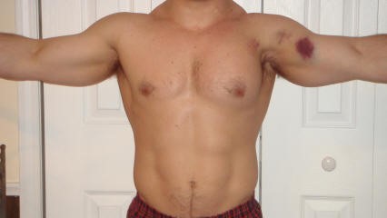
The pec major attaches to the humerus and originates from the sternum and clavicle
Injury is usually due to tendon rupture off the humerus but can also occur at the muscle tendon junction or within the muscle belly itself.
Injury is becoming increasingly common due to the popularity in power lifting sports.
Mechanism: excessive tension on a maximally eccentrically contracted muscle.
Patients will complain of pain and weakness of the shoulder.
PE: Swelling and bruising to anterior medial arm. Palpable defect and deformity or anterior axially fold (may be hidden by swelling).
Weakness and pain with adduction and internal rotation and forward flexion
Chronic presentations can be challenging to diagnose. Consider ultrasound
Non operative treatment may be indicated for partial tears (sling, ice, NSAIDs)
Operative repair of tendon avulsions is very successful. Patients age, occupation/activity level and location of injury and condition of tear are considered.