
N-acetylcysteine (NAC) is well known as the accepted antidote for acute acetaminophen (tylenol/paracetamol) overdose and is well studied for this indication. While the literature base is not nearly as strong in other causes of acute liver failure, NAC is increasingly used in these scenarios as well. In the emergency department in particular, the cause of fulminant hepatic failure is often not known. NAC may have some protective benefit in non-acetaminophen acute liver failure. Existing data do not show a mortality benefit to NAC in non-acetaminophen acute liver failure, but do show improvement in transplant-free survival. The AASLD guidelines (last revised in 2011) do not comment on NAC in non-acetaminophen acute liver failure. A common practice is to continue NAC until the INR is < 2 and AST/ALT have decreased at least 25% from their peak values.
Patients in fulminant liver failure should also be strongly considered for transfer to a center that does liver transplant, if presenting to a non-transplant center. The King's College criteria is the most commonly used prognostic score for determining need of transfer to a transplant center, but in addition to calculating a King's College score providers should generally consider consultation with a transplant hepatologist for any fulminant liver failure patient to discuss the risks/benefits of transfer for transplant evaluation.
Bottom Line: While not as strongly indicated as it is in acute acetaminophen induced liver failure, NAC should be considered in both non-acetaminophen liver failure and liver failure of unknown etiology. In addition, strongly consider consultation with a transplant hepatologist in any case of fulminant hepatic failure.
Show References
A labial adhesion is defined as a thin avascular clear plane, a raphe, between the labia minora. These adhesions which can be caused by minor trauma or infection in the absence of estrogen can cause varying degrees of obstruction.
Show References
Dihydropyridine (calcium channel blocker) overdose is one of the leading causes of death from cardiovascular drug poisoning. In contrast, angiotensin-II receptors blockers (ARBs) and angiotensin converting enzyme inhibitor (ACEIs) causes minimal toxicity in overdose. Frequently, these medications are co-ingested with dihydropridines.
Recently, a retrospective study was conducted to evaluate the hemodynamic impact of dihydropyridines with ARBs/ACEIs co-ingestion.
Results
Cohort
- 68 mixed overdoses of dihydropyridines with ARBs/ACEIs
- 21 single agent overdose (dihydropyridines)
Mixed overdose group had:
- Lower median nadir mean arterial pressure: 62 vs. 75 mmHg (p<0.001)
- Higher OR for hypotension: OR 4.5, (95% CI: 1.7 – 11.9)
- Higher OR for bradycardia: OR 8.8 (95% CI: 1.1 – 70)
- Lower minimum systolic blood pressure by 11.5 mmHg (95% CI: 4.9 – 18.1)
Higher proportion of the mixed overdose group received:
- IV fluids: OR 5.7, (95% CI: 1.8-18.6)
- Antidotes and/or vasopressor: OR 2.9 (95% CI: 1.004 – 8.6)
Conclusion
Combined overdose of dihydropyridines with ARBs/ACEIs can result in more significant hypotension.
Show References
- When dealing with weakness and sensory complaints in the ED, the time course of symptoms and physical exam findings help identify emergent conditions.
- We often talk about upper motor neuron vs. lower motor neuron signs that distinguish whether a lesion is in the central or peripheral nervous system.
- Characteristics that differentiate between central vs. peripheral nervous system pathology include:
| Central Nervous System | Peripheral Nervous System | |
| Pattern of Symptoms | • Hemibody involvement • Weakness of UE extensors • Weakness of LE flexors | • Distal involvement in polyneuropathy • Distal and proximal involvement in polyradiculoneuropathy • Proximal involvement in polyradiculopathy • Sensory often precedes motor symptoms • Pure proximal>distal weakness may be due to myopathy or NMJ disorder |
| Sensory Symptoms | • Central poststroke pain (hyperalgesia, allodynia) • Sensory level in spinal cord pathology • Proprioception involved early in dorsal column disorders | • Neuropathic pain (burning, tingling, shock-like) • Ascending sensory loss involving distal BLE>BUE in polyneuropathy • Proprioception involved late in polyneuropathy |
| Reflexes | • Hyperreflexia in affected limb(s) after acute period • Positive Babinski’s sign | • Hyporeflexia in affected limb(s) |
| Tone | • Increased after acute period | • Decreased |
Show References
Blood Pressure Management in Acute Ischemic Stroke
- Blood pressure (BP) is elevated in many patients who present to the ED with an acute ischemic stroke (AIS).
- Severe elevations in BP are associated with hemorrhagic transformation, as well as cardiac and renal complications.
- As such, it is important to know the various BP goals for patients with an AIS.
- Permissive hypertension with a BP less than or equal 220/120 mm Hg is recommended for patients not receiving IV-tPA or endovascular therapy.
- BP should be lowered to less than or equal to 180/105 mm Hg for patients who have received IV-tPA.
- BP goals for patients who have received endovascular therapy remain controversial and should be individualized based on the degree of recanalization.
Show References
Carpal Tunnel Syndrome (CTS)
The hallmark of classic CTS: pain or paresthesia (numbness and tingling) in a distribution that includes the median nerve territory, with involvement of the first three digits and the radial half of the fourth digit.
The symptoms of CTS are typically worse at night and often awaken patients from sleep.
Fixed sensory loss is usually a late finding
Involves the median-innervated fingers BUT spares the thenar eminence.
This pattern occurs because the palmar sensory cutaneous nerve arises proximal to the wrist and passes over, rather than through, the carpal tunnel.
Consider a more proximal lesion in cases involving sensory loss in the thenar eminence
Example: pronator syndrome
Nitroglycerin is a potent vasodilator used most commonly for the treatment of angina and ACS. It can also be administered as a continuous infusion for acute management of a hypertensive emergency or sympathetic crashing acute pulmonary edema.
Most are aware of asking men for history of medications for erectile dysfunction (PDE5 inhibitors: sildenafil, tadalafil, vardenafil) but many overlook the fact that men and women may be on these medications chronically for pulmonary hypertension. Men can also be on these medications for the treatment of BPH. Be broad in your history taking and do not limit the discussion to erectile dysfunction or a specific gender.
Drug interaction:
-PDE5 inhibitors prevent the breakdown of cGMP
-Nitrates are nitric oxide donors that increase the production of cGMP
-The combination can lead to excessive vasodilation
If accidentally co-administered:
There is no antidote for this medication error. Support the patient with Trendelenburg positioning, fluid administration, and if needed, vasopressors such as norepinephrine until blood pressure stabilizes.
How long should you wait to administer nitrates after a patient takes a PDE5 Inhibitor?
Sildenafil and vardenafil: 24 h after last dose*
Tadalafil > 48 h after last dose*
*Even if acute ACS event
Show References
Acute appendicitis is the most common etiology requiring urgent abdominal surgery in children in the United States. Peak incidence occurs in the second decade of life, with male patients being more commonly affected than female patients. Classic manifestations of appendicitis occur in school-aged children and adolescents, but are often absent in younger children. Infants and young children <5 years are more likely to present with nonspecific or atypical findings, resulting in delays in diagnosis and higher rates of perforation.
Diagnosis is aided by clinical factors, lab findings, and ultrasound (+/- CT or MRI if ultrasound is equivocal).
Historically, the standard of care for acute appendicitis has been urgent operative management. However, in the past several years, there has been increasing literature supporting nonoperative management (antibiotics only) in adult patients with acute uncomplicated appendicitis. Additionally, there is a growing body of evidence demonstrating the safety and efficacy of nonoperative management for uncomplicated appendicitis in children.
Hartford and Woodward provide a review of the current literature on the nonoperative management of uncomplicated appendicitis in children. They conclude:
- The majority of recent prospective studies demonstrate early treatment success (0-30 days) of approximately 90% in pediatric patients undergoing nonoperative management.
- Factors associated with failure of nonoperative management in pediatric appendicitis: longer duration of symptoms (>48 hours), younger age (<5 years), and presence of appendicolith.
- Nonoperative management has been associated with
o Lower healthcare costs at 1 year
o Fewer disability days at 1 year
o No significantly different rate of complicated appendicitis
- Most trials to date involve a 24-48 hour initial course of broad spectrum IV antibiotics followed by oral antibiotics for a total of >/= 7 days as nonoperative management. Currently, there is no consensus on antibiotic regimen.
Bottom Line: Given the current evidence, nonoperative management may be a viable treatment option for low risk pediatric patients with uncomplicated appendicitis. The literature is not conclusive, thus we as medical providers in conjunction with our surgical colleagues, should consider numerous factors when discussing treatment options for acute appendicitis with patients and their families.
Show References
Historically, there has been debate on transporting outside hospital cardiac arrests, as well a trauma, with the question of whether to "scoop and run" or "stay and play".
Could hasty transportation of cardiac arrest patients put a damper on resuscitation quality?
A recent propensity-matched study in JAMA analyzed 192 EMS agencies across 10 N American sites.
Methods:
-Resuscitation Outcomes Consortium Cardiac Epidemiologic Registry, which counted 43,969 consecutive cases of nontraumatic adult EMS-treated OHCA (median age 67, 37% of whom were women) in 2011-2015.
-25% of these patients were transported to the hospital
-Matched 1:1 with patients in refractory arrest who were resuscitated on scene
-Primary outcome was survival to hospital discharge, secondary outcome survival to hospital discharge with a favorable neurological status
Results:
-Duration of out-of-hospital resuscitation was only 6 minutes longer in the intra-arrest transport group (29.1 and 22.9 minutes; not a statistically significant difference)
-Survival to hospital discharge was 3.8% for patients who underwent intra-arrest transport and 12.6% for those who received on-scene resuscitation
-In the propensity-matched cohort, which included 27,705 patients, survival to hospital discharge occurred in 4.0% of patients who underwent intra-arrest transport vs 8.5% who received on-scene resuscitation (risk difference, 4.6% [95% CI, 4.0- 5.1])
-Favorable neurological outcome occurred in 2.9% of patients who underwent intra-arrest transport vs 7.1% who received on-scene resuscitation (risk difference, 4.2% [95% CI, 3.5%-4.9%])
-Intra-arrest transport during resuscitation was associated with worse odds of survival to hospital discharge compared to on-scene resuscitation (4% vs 8.5%, RR 0.48, CI 0.43-0.54)
-Findings persisted across subgroups of initial shockable rhythm vs. non-shockable rhythms (most common initial rhythm was aystole), as well as EMS witness arrests vs. unwitnessed arrests
Conclusion:
-This study does not support the routine transportation of patients in cardiac arrest during rescuscitation.
-The neurologically intact survival benefit associated with on-scene resuscitation is both impressive and intriguing.
-However, what implications could this have on ECPR?
Limitations:
-Potential bias due to observational nature of study
-Duration of resuscitations very similar, unknown exactly how long transport times were or if this was in urban or rural populations
-External validity not generalizable due to heterogeneity of patient populations and EMS systems
-Further randomized clinical trials are required
Show Additional Information
Show References
Physical injury patterns associated with physical elder abuse
Elder abuse is both common and underrecognized
Between 5 and 10% of US older adults are victims of elder abuse annually
For many older adults, contact with a health care provider may represent their only contact outside the home
Differentiating physical elder abuse from unintentional trauma can be very difficult
A recent study compared these two groups with a case-control design
Study cases: 100 successfully prosecuted physical elder abuse cases from a single urban ED
Physical abuse victims were more likely to have:
Bruising (78% vs. 54%)
Injuries to maxillofacial, dental or neck region (67% vs. 28%)
Particularly the LEFT side
Neck injuries 6x more common is assault
Ear injuries occurred in assault but not in falls
Absence of fracture (8% vs. 22%)
Less likely to have lower extremity injuries (9% vs. 41%)
22% of victims had no visible injuries
Most common mechanism assault with hands or fists and pushing or shoving causing a fall
Take home: Consider elder abuse especially in cases of the above red flags
Show References
- We've talked about the rising incidence of cerebral venous thrombosis (CVT) and choice of neuroimaging studies before, now let’s talk about presentation and treatment.
- Symptoms range from headache to coma with cerebral edema and intracranial hypertension depending on the veins and sinuses involved.
- Superior sagittal sinus is most frequently affected (62%) and can cause headache, hemiparesis, hemisensory loss, hemianopia, and seizures.
- Transverse sinus is also commonly involved (45%) and can cause headache, aphasia, and seizures.
- Thrombosis of the deep veins is seen in 18% of cases and can cause altered mental status, coma, and gaze palsy.
- Management includes anticoagulation, treatment of underlying cause, seizures, and intracranial hypertension.
- LMWH is preferred unless in patients with renal dysfunction or need for rapid reversal of anticoagulation.
- Endovascular intervention may be considered in severe cases that do not improve or deteriorate despite anticoagulation.
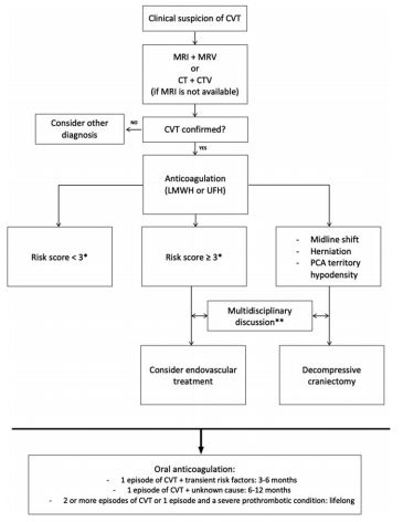
- Poor prognostic factors are:
- 2 points each - malignancy, coma, deep venous thrombosis
- 1 point each - mental status disturbances, male, intracranial hemorrhage
- Score ≥3 suggests high risk of poor outcome
Bottom Line: Severity of CVT presentation depends on the location and clot burden. Anticoagulation is key, though consider endovascular intervention if patient does not improve or deteriorates despite anticoagulation.
Show References
A retrospective study analyzed data from 757 patients with spontaneous intraparenchymal hemorrhage.
Within the first 6 hours of admission, patients who had systolic blood pressure reduction between 40 – 60 mm Hg (OR 1.9, 95% CI 1.1-3.5) or reduction ≥ 60 mm Hg (OR 1.9, 95%CI 1.01-3.8) were associated with almost double likelihood of poor discharge functional outcome (defined as modified Rankin Scale 3-6).
Additionally, large systolic blood pressure reduction ≥ 60 mm Hg in patients with large hematoma (≥ 30.47 ml) was associated with higher likelihood of very poor functional outcome (mRS 5-6).
Take home points: while more studies are still needed to confirm these observations, perhaps we may not want to drop blood pressure in patients with spontaneous intraparenchymal hemorrhage too much and too fast.
Show References
Systematic review and meta-analysis of 5 studies with a total of 929 patients comparing early vs. late initiation of norepinephrine in patients with septic shock
- all were single-center studies
- included RCTs, prospective and retrospective cohort studies
Primary outcome:
- short-term mortality of the early group was lower than that of the late group ([OR] = 0.45; 95% CI, 0.34 to 0.61)
Secondary outcome:
- no difference in ICU LOS
- time to achieved target MAP of the early group was shorter than that of the late group (mean difference = − 1.39; 95% CI, −1.81 to −0.96)
- in the three studies that assessed the volume of intravenous fluids within 6 h, the volume of intravenous fluids within 6 h of the early group was less than that of the late group (mean difference = − 0.50L; 95% CI, −0.68 to −0.3)
Caveat:
- no clear definition of “early” initiation (ranged from within 1 to 6 hrs)
Take home point:
Early norepinephrine usage may improve mortality in septic shock
Show References
- Urethral prolapse will appear as a protrusion of the distal urethra through the urinary meatus causing a “doughnut” sign.
- Risk factors include trauma, UTI, anatomical differences, and increased intraabdoiminal pressure from cough or constipation. There is a higher incidence in people of African descent.
- The chief complaint may include urethral mass and vaginal bleeding.
- There is a bimodal age distribution (prepuberty and postmetapause) due to a relative estrogen deficiency.
-Treatment is with estrogen cream and sitz baths for 4- 6 weeks.
Show References
A recent study looked at thoracic spinal fractures in the era of the trauma panscan
NEXUS Chest CT Study from 2011 to 2014 at 9 Level I trauma centers.
Goal: To describe the identification rate and types of thoracic spine fractures.
Inclusion: age over 14 years, blunt trauma occurring within 6 hours of ED presentation, and chest CT imaging during ED evaluation.
11,477 subjects, 217 (1.9%) had a thoracic spine fracture
The majority of spine fractures in patients who had both chest x-ray and CT were observed on CT only (91%). 50% had more than 1 thoracic spinal level involved (mean 2.1). 22% had associated cervical fractures and 25% had associated lumbar fractures.
64% had vertebral body fractures
45% had posterior column fractures
28% had compression fractures
6% had burst fractures
Many patients (62%) had associated thoracic injuries such as
Rib fractures (45%)
PTX (36%)
Clavicle fracture (18%)
Scapular fracture (17%)
Hemothorax (15%)
100 patients had clinically significant thoracic spine fractures.
Thoracic spine fractures are relatively uncommon in adult patients with blunt trauma.
If thoracic spine fracture is suspected clinically, radiography is not an effective screen and clinician should consider CT. If not suspected, guidelines discourage ordering CT to screen for this injury because of effective screening instruments, the diagnosis of clinically insignificant injuries and radiation exposure.
All clinically significant thoracic spine fractures would have been detected by the NEXUS Chest CT decision instrument.
https://www.mdcalc.com/nexus-chest-ct-decision-instrument-ct-imaging
Show References
Extracorporeal membrane oxygenation use is increasing in the US for acute poisoning.
A retrospective study of the National Poison Data System from 2000 to 2018 identified 407 ECMO cases (332 adults – age > 12 years, 75 pediatric – age < 12 years). Increase in ECMO use were more notable in adult population.
Characteristics
- Median age: 27 years (IQR: 15-39)
- Male: 52.6%
- Single substance exposure: 51.5%
- Median number of exposures: 3 (IQR: 2-4)
- Overall survival: 70%
Intentional exposure
- Age > 12 years: 72.6%
- Age < 12 years: 9.3%
Most common class of drug/poison exposure in adults
- Sedative/hypnotic: 26%
- Antidepressants: 25%
- Calcium channel blockers: 19%
- Opioids: 17%
Most common class of drug/poison exposure in children
- Hydrocarbons: 37%
- Antiarrhythmics: 15%
- Antihistamine: 8%
- Unknown: 8%
Most common states that used ECMO for poisoning
- Pennsylvania: 45
- Texas: 27
- Minnesota: 24
- Maryland: 22
- Michigan: 20
- New York: 20
Conclusion
- Increase in EMCO use was most notable in patients with age > 12 years
- There was no significant trend in mortality during the study period
- ECMO cases were mostly reported from urban areas
- Cerebral venous thrombosis (CVT) is thought to predominantly affect young and middle-aged females.
- Known risk factors include prothrombotic states such as malignancy and oral contraceptive use, as well as local infections and head trauma.
- The incidence of stroke in young adults is rising worldwide.
- A recent study by Otite et al. examined the incidence of CVT during 2006-2016 in New York and Florida utilizing the State Inpatient Database.
- CVT remains an uncommon condition though number of admissions increased 70%.
- Mean age of patients increased with number of hospitalizations in the elderly doubled.
- Incidence was highest in Blacks, followed by non-Hispanic Whites and Hispanics.
- This rise in incidence may be related to increased recognition, improved diagnostic studies, increased neuroimaging utilization, emerging or unknown risk factors.
Bottom Line: The incidence of CVT is increasing with rate of increase higher in males and older females. Consider CVT beyond traditional risk factors.
Show References
While the invasive monitoring of central venous pressure (CVP) in the critically ill septic patient has gone the way of also transfusing them to a hemoglobin of 10 mg/dL, it remains that an elevated CVP is associated with higher mortality1,2 and renal failure.2,3
Extrapolating from existing data looking at hepatic vein, portal vein, and renal vein pulsatility as measures of systemic venous hypertension and congestion,4,5,6 Beaubien-Souligny et al. developed the venous excess ultrasound (VExUS) grading system incorporating assessment of all 3, plus the IVC, using US to stage severity of venous congestion in post-cardiac surgery patients.7 They evaluated several variations, determining that the VExUS-C grading system was most predictive of subsequent renal dysfunction.
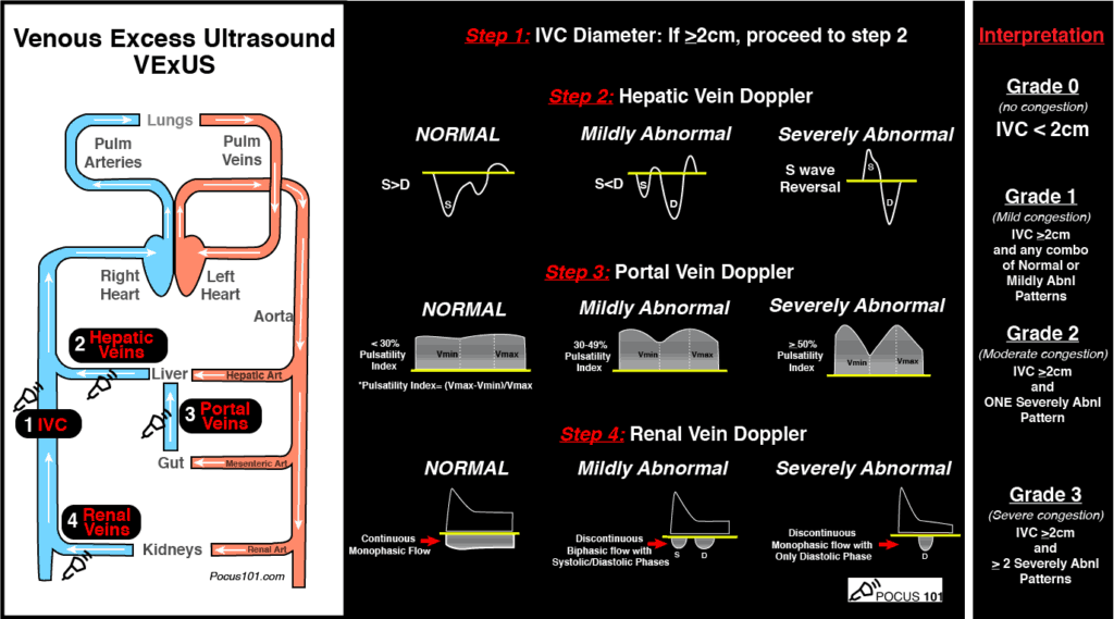
(Image from www.pocus101.com)
High Points
VExUS Grade 3 (severe) venous congestion:
- Correlated with higher CVP & NTproBNP levels, as well as overall fluid balance
- Had a 96% specificity for development of subsequent AKI
Caveats
- Evaluating all parameters yields the most benefit to avoid false positives
- Can be difficult to obtain all views (>25% of subjects excluded due to poor US image quality)
- Studied in a limited population, notably not primarily RV failure patients
Clinical Uses
- To limit harmful fluid administration in shock
- To help answer the prerenal vs cardiorenal AKI question in CHF
- To indicate when volume removal (diuresis) should be the strategy, even in patients with vasopressor-dependent shock
A great how-to can be found here:
https://www.pocus101.com/vexus-ultrasound-score-fluid-overload-and-venous-congestion-assessment/
Show References
| | Beta-blockade N=22 | Control N= 44 | OR/CI |
| Temporary ROSC, n (%) | 19 (86.4) | 14 (31.8) | OR 14.46, 95% CI 3.63-57.57 |
| Sustained ROSC, n (%) | 13 (59.1) | 10 (22.7) | OR 5.76, 95% CI 1.79-18.52 |
| Survival with neurological function, n (%) | 6 (27.3) | 4 (9.1) | OR 4.42; 95% CI 1.05-18.56 |