Search
Diphenhydramine is commonly involved in overdose or misused. Although it is primarily used for its anti-histamine property, it also has significant antimuscarinic effect.
A recent retrospective study investigated the clinical characteristics associated with severe outcomes in diphenhydramine overdose using the multi-center Toxicology Investigators Consortium (ToxIC) Registry.
Severe outcomes were defined as any of the following:
- Seizure
- Ventricular dysrhythmia
- Intubation
Results
863 cases of isolated diphenhydramine ingestion were identified between Jan 1, 2010 to Dec 31, 2016
- Females: 59.1%
- Age < 18 years: 51.3%
- Intentional ingestion: 86.0%
- Self-harm: 37.5%
- Abuse/misuse: 11.5%
Most common symptoms:
- Delirium/toxic psychosis: 40.1% (n=346)
- Agitation: 33.1% (n=286)
- Severe outcome: 15.6% (n=135)
Factors associated with severe outcome
- Intubation: self-harm ingestion and male
- Acidemia: pH <7.2
- QRS prolongation: QRS > 120 msec
- Elevated anion gap: AG >20
Conclusion
- Acidemia, QRS prolongation and elevated anion gap was associated with severe outcome in diphenhydramine toxicity
Show References
Even though acute myocardial ischemia (AMI) does not present as commonly in the pediatric patient as in the adult and the literature is limited, it is reasonable to obtain a troponin when acute cardiac ischemia is suspected based on the history and physical exam.
Recreational drugs including cocaine, amphetamine, cannabis, Spice, and K2 (cannabis derivatives) have been shown to result in myocardial injury including AMI. Coronary vasospasm secondary to drug use is well documented in the pediatric population. While cocaine use is a known risk factor for coronary vasospasm, the same condition has been reported in pediatric patients after marijuana use.
In a study of pediatric patients with blunt chest trauma, 3 of 4 patients with electrocardiographic or echocardiographic evidence of cardiac injury had elevations in troponin I above 2.0 ng/mL. Cardiac troponins are an accurate tool for screening for cardiac contusion after blunt chest trauma in pediatric patients even with limited data.
Cardiac troponins are also useful in the evaluation for myocarditis. In one study, myocarditis was the most common diagnosis (27%) in pediatric ED patients presenting with chest pain and an increased troponin. Eisenberg et al showed a 100% sensitivity and an 85% specificity for myocarditis using a troponin of 0.01 ng/mL or greater as a cut off. A normal troponin using this cutoff can be used to exclude myocarditis. Abnormal troponin in the first 72 hours of hospitalization in pediatric patients with viral myocarditis is associated with subsequent need for extracorporeal membrane oxygenation and IVIg.
Bottom line: Troponin can be used in pediatric patients with clinical concern for cardiac ischemia, cardiac contusion and myocarditis
Show References
There are different occupational hazards depending on the nature of one’s trade/skill/employment. Although healthcare providers may not always inquire about patient’s occupation, knowledge of a patient’s occupation may provide insightful information when caring for patients with acute poisoning.
From a recent retrospective study of National Poison Data System, the top 10 occupational toxicants were:
- Caustics (acids & alkalis)
- Chlorines/hypochlorites
- Carbon monoxide
- Hydrocarbons
- Cleansers/detergents
- Ammonia
- Cement
- Hydrofluoric acid
- Disinfectants
- Hydrogen sulfide
Top 10 occupational toxicants associated with fatalities were:
- Hydrogen sulfide
- Ammonia
- Carbon Monoxide
- Simple asphyxiants
- Chlorine/hypochlorites
- Alkalis
- Pyrethrins/pyrethroids
- Toluene/xylene
- Methane
- Methylene chloride
Show References
The PARAMEDIC2 trial in NEJM 2018 studied the outcomes of the use of epinephrine in outside hospital cardiac arrest (OHCA) on survival and neurological outcome.
Methods: Conducted in Britain, randomized 8007 patients to receive either epinepherine 1mg (n=4012) or placebo (n=3995) as part of standard CPR for out-of-hosptial arrest. Their primary outcome was survival at 30 days and their secondary outcomes included length of stay as well as neurological outcomes at 30 days and 3 months.
Results: The epinepherine group had improved survival to hospital admission (23% vs. 8%), at 30 days (3.2% vs. 2.4%) or at 3 months (3% vs. 2.2%). Favourable neurological outcomes, however, had no statistical difference at both hospital discharge and at 3 months.
Bottom line: Epinephrine improves ROSC, though with poor neurological outcomes.
Important facts:
- Demographics: Mean age 69 years, 35% female
- Initial cardiac rhythm: shockable 19%, Non-shockable 78%, Undetermined 2%
- Cause of Cardiac Arrest: Medical 91% Traumatic 2%, Drowning 0.2%, substance overdose 2%, Asphyxia 3%, missing data 2%
- Witness of cardiac arrest: none 37%, Paramedic 11%, Bystander 50%, missing data 1%
- CPR Performed by: Paramedic 11%, bystander 59%, missing data 2%
- Time from: emergency call to ambulance arrival 6min, emergency call to administration of drug 22 min, arrival to ambulance departure 50 min
Recently, a follow up of the PARAMEDIC2 trial was completed in Resuscitation.
They reported long-term survival, quality of life, functional and cognitive outcomes at 3, 6 and 12-months.
Results: At 6 months, 78 (2.0%) of the patients in the adrenaline group and 58 (1.5%) of patients in the placebo group had a favourable neurological outcome (adjusted odds ratio 1.35 [95% confidence interval: 0.93, 1.97]). 117 (2.9%) patients were alive at 6-months in the adrenaline group compared with 86 (2.2%) in the placebo group (1.43 [1.05, 1.96], reducing to 107 (2.7%) and 80 (2.0%) respectively at 12-months (1.38 [1.00, 1.92]). Measures of 3 and 6-month cognitive, functional and quality of life outcomes were reduced, but there was no strong evidence of differences between groups.
Bottom line: Epinephrine improves survival at 12 months, but poor neurological outcomes remain.
Show Additional Information
Show References
Bottom Line: In a recent meta-analysis the risk factors for patellofemoral syndrome are weak hip abduction strength, quadricep weakness in military recruits, and increased hip strength in adolescence.
PatelloFemoral Syndrome: Patellofemoral pain is not clearly understood and is believed to be multi-factorial. Numerous factors have been proposed including muscle weakness, damage to cartilage, patella maltracking, as well as others. Patient often complain of anterior knee that is aggravated by walking up and down stairs or squatting. Patellofemoral pain is extremely common. In the general population the annual prevalence for patellofemoral pain is approximately 22.7%, and in adolescents it is 28.9%.
Though commonly taught, the following have no evidence to support that they are a risk factor for patellofemoral syndrome: Age, Height, Weight, BMI, Body Fat or Q Angle of patella
Show References
- Idiopathic intracranial hypertension (IIH) predominantly affects women of childbearing age and is strongly associated with obesity.
- Patients can have high rates of healthcare utilization due to their chronic disabling headaches, multiple diagnostic tests and specialist consultations, and potential complications related to treatments.
- A recent study looked at trends in the incidence, prevalence, and healthcare outcomes of IIH in the 3.1 million Welsh population over a 14-year period.
- Incidence of IIH increased from 2.3/100,000 to 7.8/100,000 (p<0.001).
- Prevalence of IIH increased from 12/100,000 to 76/100,000 (p<0.001).
- Obesity (BMI >30 kg/m2) also increased from 29% to 40% of the population (p<0.001), with IIH incidence and prevalence strongly associated with BMI.
- Outcomes included blindness (0.78%), moderate visual impairment (1.9%), CSF diversion (9%) with 44% of these patients requiring at least 1 revision surgery.
- IIH patients also had 5.28 times higher rate of unscheduled hospital visits during this time period.
Bottom Line: The incidence and prevalence of IIH is increasing, likely related to rising rate of obesity. This has also been associated with more healthcare utilization compared to the general population.
Show References
Background:
The association between time intervals of ED antibiotic administration and outcome has been controversial. While single studies showed there was increased mortality associated with delayed antibiotic administration (1-3). A meta-analysis of 13 studies and 33000 patients showed that there was no mortality difference between septic patients receiving immediate Abx (< 1 hour) vs. those receiving early abx (1-3 hours) (4).
Since delay in recognition of sepsis (defined as ED triage to Abx order) and delay in antibiotics delivery (Abx order to administration) contribute to total delay of Abx administration, a new retrospective study (3) attempted to investigate the contributions of either factor to hospital mortality.
Results:
The study used generalized linear mixed models and involved 24000 patients.
For All patients and outcome of hospital mortality:
Recognition delay (ED triage to Abx order): OR 2.7 (95% CI 1.5-4.7)*
Administration delay at 2-2.5 hours (Abx order to administration): OR 1.5 (1.1-2.0)
These results was associated with non-statistical significance in patients with septic shocks.
Conclusion:
Delayed recognition of sepsis was associated with higher hospital mortality. Longer delay of abx administration was also associated with increased risk of hospital mortality.
Show References
Tranexamic acid (TXA) is an antifibrinolytic medication that has been trialed in previous small studies to treat epistaxis. The data to this point has not reliably shown a reduction in bleeding at 30 minutes, but has demonstrated an increased rate of discharge at 2 hours and a reduction in re-bleeding events.
The NoPAC study was the largest study to date on TXA for epistaxis. It was a double-blind, placebo-controlled, randomized study of TXA in adult patients with persistent atraumatic epistaxis to determine if TXA use decreased the rate of anterior nasal packing. Patients were excluded if they had trauma, out of hospital nasal packing, allergy to TXA, nasopharyngeal malignancy, hemophilia, pregnancy, or if they were referred to ENT.
Eligible patients completed 10 minutes of first aid measures followed by 10 minutes of topical vasoconstrictor application prior to randomization to either placebo of 200mg TXA soaked dental rolls inserted in the nare.
496 patients were enrolled. The average patient was 70 years old with stable vitals 150/80mmHg, HR 80 bpm with >60% on oral anticoagulants.
TXA did not reduce the need for anterior nasal packing: 100 (41.3% placebo) vs 111 (43.7% TXA) OR 1.11 (0.77-1.59). There were no differences in the rates of adverse events.
Bottom Line: TXA does not improve rates of anterior nasal packing for patients with persistent epistaxis.
Show References
Recently, there has been questions if standard n-acetylcysteine (NAC) dose is adequate for massive acetaminophen (APAP) overdose (ingestion of > 32 gm or APAP >300 mcg/mL).
A retrospective study from a single poison center (1/1/2010 to 12/31/2019) investigated the clinical outcome of massive APAP overdose (APAP > 300 mcg/mL at 4 hour post ingestion) treated with standard dosing of NAC.
Results
1425 cases of APAP overdose identified; 104 met the criteria of massive APAP overdose.
- 300-449 mcg/mL: 59.6% (n=62)
- 450-599 mcg/mL: 14.4% (n=15)
- >600 mcg/mL: 25.9% (n=27)
- No acute liver injury/hepatotoxicity: 76% (n=79)
- Hepatotoxicity: 24% (n=25)
Among cases that received NAC within 8 hours post ingestion (n=44)
- Only 9% (n=4) cases developed hepatotoxicity
Among cases that received NAC > 8 hours post ingestion (n=60)
- 35% (n=21) developed hepatotoxicity
Odds of hepatotoxicity
- 5.5 If NAC initiated > 8 hours post ingestion
- 3.8 if 4 h post ingestion APAP level >600 mcg/mL
Conclusion
- Cohort: no acute liver injury/hepatotoxicity in 76% (n=79)
- Standard NAC dosing initiated within 8 hours prevented hepatotoxicity in 91% (n=40/44)
Clinical Question:
- Does dexmedetomidine lead to better short-term and long-term outcomes than propofol in mechanically ventilated adults with sepsis?
Methods:
- Multicenter (13 US hospitals), double-blinded, 422 mechanically ventilated patients with sepsis
- Notable exclusion criteria: pregnant, 2nd- or 3rd-degree heart block or persistent bradycardia requiring intervention, indication for bzds, expected to have NMB > 48 hrs, already had received mechanical ventilation >96 hrs
- Pain was treated with opioid pushes or fentanyl gtt
- Primary end point: number of calendar days alive without delirium or coma during the 14-day intervention period
- Secondary efficacy end points included ventilator-free days at 28 days, death at 90 days, and global cognition at 6 months
Results:
- No sig difference in adjusted number of days alive without delirium or coma over the 14-day intervention period (dexmedetomidine: 10.7 days vs. propofol: 10.8 days; OR, 0.96; 95% CI, 0.74 to 1.26; P = 0.79
- No sig differences in the number of ventilator-free days at 28 days, in death at 90 days, or global cognitiion at 6 months
- Other notable findings:
- Fewer patients in the dexmedetomidine group had ARDS or signs of trial drug withdrawal
- Fewer patients in the propofol group extubated themselves
- Open-label propofol received by 13% in the dexmedetomidine group and 8% in the propofol group) and dexmedetomidine (4% in the dexmedetomidine group and 3% in the propofol group)
- Rescue midazolam was used in about half the patients, most often for procedural sedation or during NMB, 42% received antipsychotics
- Similar proportions of patients had organ dysfunction, hypotension, or severe lactic acidosis
- Symptomatic bradycardia requiring discontinuation of the trial drug was similar in the two groups
Take-home points:
- Dexmedetomidine or propofol are reasonable options for septic patients requiring mechanical ventilation without notable differences in delirium or mortality
Show References
Home management versus PCP follow-up of patients with distal radius buckle (torus) fractures
A recent study investigated outcomes of patients with distal radius buckle fractures who were randomized to
- Home removal of splint and physician follow-up as needed (home management)
Versus
- Prescribed PCP follow-up in 1-2 weeks
Noninferior study
Torus/buckle fractures of the distal radius are the most common fractures in childhood occurring on average in 1 in 25 children
This is a stable fracture typically treated with removable wrist splint and very rarely require orthopedic intervention
Outcome: functional recovery at 3 weeks
Randomized controlled trial at a tertiary care children’s hospital
All radiographs reviewed by pediatric radiologist with MSK specialization
149 patients. Mean age 9.5 years. 54.4% male
Telephone follow-up at 3 and 6 weeks following ED discharge by blinded interviewer
Primary outcome was comparison of Activities Scale for Kids-performance scores between groups at 3 weeks
Outcomes: Home management performance score was 95.4% and PCP follow-up group was 95.9%. Mean cost savings were $100.10.
Conclusion: Home management is at least as good as PCP follow-up with respect to functional recovery in ED patients with distal radius buckle fractures.
Show References
- Bell’s palsy can be caused by herpes simplex virus-1 (HIV-1) and Lyme borreliosis.
- Prior studies have found higher incidence of Bell’s palsy in colder months possibly related to reactivation of latent HSV-1.
- A recent study looked at the incidence and seasonal variation of positive Lyme tests in Bell’s palsy patients in a Lyme-endemic area (New Jersey).
- Over a 5-year period, 81% of 442 patients with Bell’s palsy were tested for Lyme and 16% tested positive.
- The months May through October had a 7.2 times higher incidence of positive Lyme tests in Bell’s palsy patients, with the peak in July.
- May through October also had a 1.3-fold increased ED visits for Bell’s palsy, also peaking in July.
Bottom Line: In a Lyme-endemic area, the incidence of positive Lyme tests and Bell’s palsy are highest in the Lyme months. This seasonal variation may help guide the management of patients with Bell’s palsy.
Show References
Background: In respiratory failure due to COPD and cardiogenic pulmonary edema, noninvasive positive pressure ventilation decreases need for intubation and improves mortality,1 while its utility in other scenarios such as ARDS and pneumonia has yet to be proven.1,2 We know that patients on NIV with delays to needed intubation have a higher mortality,1,3 but intubation and mechanical ventilation come with risks that it is preferable to avoid if possible.
So how and when can we determine that NIV is not working?
The HACOR (Heart rate, Acidosis, Consciousness, Oxygenation, Respiratory rate) score at 1 hour after NIV initiation has been demonstrated to be highly predictive of NIV failure requiring intubation.4,5
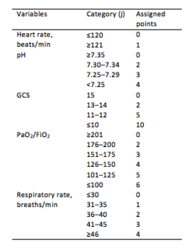
Initial development/validation: Score > 5 after 1 hour of NIV corresponds to >80% risk of NIV failure4
-
Earlier intubation (before 12 hours) in these patients = better survival
External validation: Score > 8 after 1 hour of NIV most predictive of eventual NIV failure 5
-
Average score @ 1-hour of patients with NIV success = 3.8
-
Score remained predictive at 6, 12, 24, 48 hours as well & mortality worsened as delay to intubation time increased
-
Baseline, pre-NIV score not predictive
-
Better predictive agreement in pneumonia and ARDS
Bottom Line:
-
Patients on NIV require close reassessment to prevent worsened survival due to intubation delay should invasive mechanical ventilation be indicated.
-
A HACOR score >8 after 1 hour of NIV should prompt intubation in most instances, with strong consideration given to a score >5.
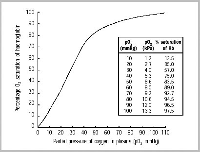
*Note: ABGs were obtained for PaO2 assessment in the above studies -- the use of SpO2 was not evaluated -- but we are often not obtaining ABGs in our ED patients with acute respiratory failure. The following chart provides an estimated SpO2 to PaO2 conversion.
WHO 2001
Caveats:
- Pulse oximetry may be inaccurate in darker skin tones (overestimated by ~2%)6 and in certain disease processes (e.g. CO poisoning, profound shock states, etc.)
- The oxyhemoglobin dissociation curve shifts right with increasing pCO2/decreasing pH (lower saturation for a given PaO2).
Show References
In children with known congenital heart disease, BNP measurements are higher in those patients with heart failure compared to those without heart failure.
The utility of BNP in differentiating a cardiac from pulmonary pathology in patients with respiratory distress has been studied in pediatrics. In one study involving 49 infants with respiratory distress, the patients with a final diagnosis of heart failure had a higher mean BNP concentration than those patients with other causes. Also, there is a suggestion that the relative change in NT proBNP levels may be useful in patients with underlying pulmonary hypertension. However, currently there is not enough literature to support the routine use of BNP or NT proBNP in acute management.
Bottom line: BNP can be useful in your patient with congenital heart disease who is decompensating and may be used in a patient where there is difficulty in differentiating a primary respiratory from cardiac etiology.
Show References
Patients with cannabis hyperemesis syndrome experience recurrent/protracted nausea/vomiting. Cases of cannabis hyperemesis syndrome may increase as cannabis use becomes more common in the United States.
A randomized control trial (triple-blind) was conducted to compare haloperidol (0.05 or 0.1 mg/kg) IV or ondansetron 8 mg IV. Primary outcome was reduction of abdominal pain and nausea from baseline (on a 10 cm visual analog scale) 2 hours after treatment.
Results
- 33 subjected were randomized to haloperidol (n=13) and ondansetron (n=17)
- 30 used 1.5 gm/day since 19 years of age.
- Haloperidol was superior to ondansetron
- 2.3 cm difference in pain and nausea
- Less use of rescue antiemetics (31% vs. 59%)
- Shorter time to ED departure (3.1 hours vs. 5.6 hours)
Conclusion
- In this small trial, haloperidol (0.05 or 0.1 mg/kg IV) was superior to ondansetron (8 mg IV) in the treatment of acute cannabis associated hyperemesis
Show References
What time of day is best for exercise to achieve weight loss goals?
Working out in the morning has traditionally held the edge, especially if done on an empty stomach.
Upon walking, elevated levels of cortisol and GH will aid in fat metabolism.
Switching to a morning workout may also decrease appetite throughout the day.
Morning exercise may also induce significant circadian phase?shifting effects. Patients report feeling more alert in the morning and get more tired at night. This may “force” people to get increased rest as poor sleep quality and duration has been associated with weight gain.
Moderate intensity aerobic exercise has been shown to cause immediate mood improvement and mental productivity. These effects can last up to 12 hours and may be a simple aid to combat job stress.
However, a recent small study looked at this question with a group of men at high risk for Type 2 diabetes.
Those that exercised in the morning had better blood sugar control and lost more abdominal fat than those who exercised in the morning.
Study: 32 adult males (58 ± 7 years) at risk for or diagnosed with type 2 diabetes performed 12 weeks of supervised exercise training either:
In the morning (8.00–10.00 a.m., N = 12) OR
In the afternoon (3.00–6.00 p.m., N = 20)
Test: Graded cycling test with ECG monitoring until exhaustion
Results: Compared to those who trained in the morning, participants who trained in the afternoon experienced superior beneficial effects of exercise training on peripheral insulin sensitivity, insulin?mediated suppression of adipose tissue lipolysis, fasting plasma glucose levels, exercise performance and fat mass.
Conclusion: Metabolically compromised patients may benefit from shifting their exercise routine to the afternoon from the morning. Ultimately, any exercise is great in this population, but this study may be worth sharing to your patients.
Show References
Optimal oxygenation targets and the possible, theoretical, benefits of hyperoxygenating critically ill patients have long been points of controversy. Multiple studies have suggested harm in pursuing aggressive hyperoxygenation amongst critical patients with various conditions ranging from myocardial infarction to sepsis to neurologic conditions. In addition, oxygen toxicity is a known mechanism causing ARDS.
The HOT-ICU trial adds to the list of arguments against hyperoxygenation, by looking at 2928 ICU patients on high levels of supplemental oxygen and targeting a paO2 of 60 mm Hg (low oxygen group) vs paO2 of 90 mm Hg (high oxygen group). There was no difference in mortality, or other significant difference in outcomes.
Bottom Line: A lower paO2 goal of 60 (correlates to an O2 sat of 90%) is noninferior to a higher paO2 goal of 90 (O2 sat of approximately 96%). When titrating oxygen, targeting a pulse ox of 90-96% is reasonable in critically ill patients. Be sure to include an upper limit on the sat goal, beware an O2 sat of 100%, and titrate down supplemental oxygen when the spO2 is above goal, as the paO2 may be dangerously high.
Show References
Buprenorphine is a partial opioid receptor agonist that has a higher binding affinity than pure opioid agonists. There can be unease in managing acute pain in patients sustained on buprenorphine for opioid use disorder due to many factors.
The main barriers to effective pain management in these patients are:
- Opioid-Induced Hyperalgesia
- Patients maintained on buprenorphine can have an increased sensitivity to pain.
- Consider using a multimodal approach that optimizes non-opioid analgesics, such as acetaminophen and nonsteroidal anti-inflammatory drugs.
- Opioid Tolerance
- Patients maintained on buprenorphine require higher doses of opioids to treat acute pain due to the decreased effectiveness of opioids over time.
- As in hyperalgesia, a multimodal approach can be beneficial.
- Higher doses of supplemental opioids will be required in these patients compared with opioid-naïve patients.
- Titrate supplemental opioids to effect and monitor for toxicity.
- Opioid Withdrawal
- Opioid withdrawal symptoms can contribute to stress and anxiety, increasing pain sensitivity.
- To prevent withdrawal symptoms it is appropriate to continue buprenorphine throughout the episode of acute pain.
- The patient's typical home dose of buprenorphine can be utilized.
Take Home Points
In general, the treatment strategy for acute pain in patients on buprenorphine should be:
- Optimize non-opioid analgesia.
- Use supplemental opioids when needed.
- Will likely require higher doses.
- Titrate to effect.
- Monitor for toxicity.
- Continue buprenorphine therapy at home dose throughout the acute pain episode.
Show References
Mechanical Ventilation in COPD
- Mechanical ventilation of the patient with obstructive lung disease can be challenging, primarily due to the presence of dynamic hyperinflation.
- In the initial phase of ventilation, it is important to prevent complications of hyperinflation and not to target normalization of blood gas values.
- Recommended initial ventilator settings include:
- Mode: Volume assist-control
- Inspiratory flow waveform: square
- Tidal volume: 6-8 ml/kg PBW
- RR: 12 bpm
- Inspiratory flow: 60-90 L/min
- The effect of PEEP is variable with each patient. When titrating PEEP, be sure to frequently measure plateau pressure and discontinue titration should Pplat increase.
Show References
- Most seizures resolve spontaneously, however, seizures that persist >5 minutes or recur without the patient returning to their baseline should be treated expeditiously with benzodiazepines and antiepileptic medications.
- A subset of patients may continue to have electrographic seizures despite cessation of their convulsive seizure activity.
- Prior studies described 26-52% of patients develop nonconvulsive seizures after resolution of convulsive status epilepticus.
- The recent Established Status Epilepticus Treatment Trial (ESETT) compared fosphenytoin, levetiracetam, and valproic acid in aborting seizures and improving responsiveness in patients who did not response to initial treatment with benzodiazepines.
- EEG was not required for this trial, but 58% (278/478) had an EEG within 24 hours after seizure onset.
- Of those who had an EEG, 14% (39/278) had electrographic seizures.
- For patients who had clinical treatment success, 13% (13/102) were found to have electrographic seizures.
- EEG was not required for this trial, but 58% (278/478) had an EEG within 24 hours after seizure onset.
Bottom Line: Persistent or recurrent seizures are not uncommon in the first 24 hours after status epilepticus even in patients with resolved clinical seizure activity. Early use of EEG can help identify patients who need further escalation of treatment.