
University of Maryland School of Medicine logo
Emergency Medicine
All Departments
UMSOM Assistant

UMEM Educational Pearls

  • Home
  • Departments
  • Emergency Medicine
  • CME
  • UMEM Educational Pearls
  • CME
  • Courses and Conferences
  • Free Open Access Medical Education
  • UMEM Educational Pearls
Previous |  1 |  2 |  3 |  4 |  5 |  6 |  ... |  239 |  Next

Title: Is EMS documentation of bystander CPR accurate?

Category: EMS

Keywords: 911, dispatch, CPR, bystander (PubMed Search)

Posted: 9/16/2026 by Jenny Guyther, MD (Updated: 9/17/2026)

The national cardiac arrest registry includes information on bystander CPR.  An EMS clinician currently enters bystander CPR (B-CPR) information into the prehospital care report.  The accuracy of this documentation has not been validated.  This study compared EMS documented B-CPR rates to the bystander CPR rates on the 911 audio recordings of the calls in Birmingham Alabama.

Show Additional Information

EMS documentation in the Birmingham CARES registry recorded a B-CPR rate of only 12.3%, while review of corresponding 9-1-1 audio recordings identified a substantially higher rate of 27.5% — more than double the EMS-documented rate.

Of 236 adult non-traumatic out-of-hospital cardiac arrest (OHCA) cases analyzed, 56 cases (23.7%) had disagreements between EMS documentation and 9-1-1 audio review, while 180 cases (76.3%) were concordant. 

In 46 of the 56 discordant cases, 9-1-1 audio confirmed that B-CPR was performed but EMS did not document it. The majority of these involved callers who initiated CPR but discontinued it before EMS arrival, making it invisible to arriving crews. Only 7 of these 46 appeared to be outright EMS misclassifications.

In 10 cases, EMS documented B-CPR but the 9-1-1 audio did not support it — all involved calls that ended before EMS arrival without cardiac arrest recognition or CPR instruction by the dispatcher

Bottom line: Sustained telecommunicator CPR instruction matters through EMS arrival,  as early caller discontinuation of CPR was the primary driver of undercounting, and better dispatcher engagement could both improve CPR continuity and documentation accuracy.

Show References

Coute RA, Smith T, Nathanson BH, Richardson JD, Ferguson WC, Strickland JD, von Schweinitz B, Jackson EA. Discrepancies in Bystander CPR Documentation: Comparing the Birmingham CARES Data with 9-1-1 Audio Review. Prehosp Emerg Care. 2026;30(3):409-413. doi: 10.1080/10903127.2025.2584506. Epub 2025 Dec 9. PMID: 41247107; PMCID: PMC12969134.


Title: SUSPECT Cardiogenic Shock: A Mnemonic to Help Make the Diagnosis

Category: Critical Care

Keywords: cardiogenic shock; cardiology; shock (PubMed Search)

Posted: 9/15/2026 by Shoshana Rudin, MD (Updated: 9/17/2026)

Cardiogenic Shock = a cardiac disorder that results in both clinical and biochemical evidence of sustained tissue hypoperfusion (1).

Cardiogenic shock has high mortality (approx 35-40% in many studies). Mortality worsens at higher stages of cardiogenic shock severity (2,3). Early recognition of cardiogenic shock is critical in order to initiate treatment and transfer patients to an appropriate level of care in a timely manner. 

As an ED physician, you need to SUSPECT cardiogenic shock so that you can ensure appropriate definitive management of your patient (4).

SUSPECT = Symptoms/Signs; Urine Output; Sustained Hypotension; Perfusion; EKG/Echo; Congestion; Triage

Take a look at the Additional Materials to break down this mnemonic in more detail and learn what might make you SUSPECT CS!

Show Additional Information

So what do you need to think about to SUSPECT CS?

Let's break down that mnemonic in a little bit more detail.

Symptoms/Signs:

  • Signs of Shock (Hypoperfusion): AMS, confusion, rapid pulse, cold and clammy extremities, delayed capillary refill
  • Signs of Congestion: Elevated JVP, crackles, rales, lower extremity edema, narrow pulse pressure (<25% of SBP)
  • Symptoms: Chest pain or pressure, chest pain equivalents, orthopnea, paroxysmal nocturnal dyspnea

Urine Output:

  • Oliguria or anuria
    • <0.5 mL/kg/h (roughly <30 mL/h in a 60 kg adult)
  • Place a foley (most accurate hourly UOP), or measure UOP into a urinal, bedpan, Purewick, or condom cath

Sustained Hypotension:

  • SBP <90, MAP <65 for >30 min
  • Or… relative hypotension! >30 mmHg decrease from baseline 
  • Or… anyone who needs pressors or mechanical support to maintain SBP >90
  • Not every patient with cardiogenic shock will have hypotension (yet)!

Perfusion:

  • Lactate: >2 
  • LFTs: ALT >200 U/L or >3x upper limit of normal 
  • Renal function: AKI, AKI on CKD, or Cr >2x upper limit of normal (very concerning)
  • Acidosis: pH <7.2 (very concerning), or metabolic acidosis without another clear etiology
  • You don't need all of the above to be concerned for cardiogenic shock

EKG/Echocardiogram:

  • EKG: STEMI? Arrhythmias? New conduction abnormalities (AV block, LBBB or RBBB)?
  • POCUS: Visually estimate EF, evaluate for LV and/or RV dilation and systolic dysfunction, look for regional wall motion abnormalities
  • More detailed echo: assess for valvular pathology, more accurate quantification of EF (LVOT-VTI, Simpson’s Biplane, etc)

Congestion:

  • Assess for presence or absence of congestion based on physical exam and hemodynamics
    • LV congestion: think about pulmonary edema
    • RV congestion: think about extremity edema, cardiac ascites, elevated JVP, hepatic dysfunction
    • Biventricular congestion: all of the above!

Triage:

  • ICU admission!
    • AMI-CS (Acute MI causing cardiogenic shock): activate cath lab
    • HF-CS (decompensated heart failure, either acute or chronic, causing cardiogenic shock): call heart failure team (if available at your hospital) to discuss advanced therapeutics
    • Choose cardiac ICU if available
  • Activate shock team (if available) to discuss advanced therapeutic options
    • No shock team? Call your ICU, call your on-call cardiologist, and talk through options!
  • Consider transfer to a higher level of care if: 
    • You don’t have access to a cath lab or advanced level of cardiology services
    • You don’t have a cardiac ICU or a MICU with capacity to do advanced hemodynamic monitoring 
    • Your patient might need mechanical circulatory support
    • Your patient might need evaluation for surgical treatment options (CABG, valve repair/replacement, etc)

Proposed Classification of Cardiogenic Shock Levels of Care (originally figure 3, Sinha et al)

Consider the resources that your hospital has available when deciding if/where to transfer a cardiogenic shock patient to a higher level of care.

Show References

1. Waksman R, Pahuja M, van Diepen S, et al. Standardized Definitions for Cardiogenic Shock Research and Mechanical Circulatory Support Devices: Scientific Expert Panel From the Shock Academic Research Consortium (SHARC). Circulation. 2023;148(14):1113-1126. doi:10.1161/CIRCULATIONAHA.123.064527

2. Baran DA, Grines CL, Bailey S, et al. SCAI clinical expert consensus statement on the classification of cardiogenic shock: This document was endorsed by the American College of Cardiology (ACC), the American Heart Association (AHA), the Society of Critical Care Medicine (SCCM), and the Society of Thoracic Surgeons (STS) in April 2019. Catheter Cardiovasc Interv Off J Soc Card Angiogr Interv. 2019;94(1):29-37. doi:10.1002/ccd.28329

3. Naidu SS, Baran DA, Jentzer JC, et al. SCAI SHOCK Stage Classification Expert Consensus Update: A Review and Incorporation of Validation Studies. JACC. 2022;79(9):933-946. doi:10.1016/j.jacc.2022.01.018

4. Sinha SS, Morrow DA, Kapur NK, Kataria R, Roswell RO. 2025 Concise Clinical Guidance: An ACC Expert Consensus Statement on the Evaluation and Management of Cardiogenic Shock. JACC. 2025;85(16):1618-1641. doi:10.1016/j.jacc.2025.02.018


Title: Number One Cause of Death in Pregnancy in the US?

Category: Obstetrics & Gynecology

Posted: 9/14/2026 by Jennifer Wang, MD

Bottom Line: Move over, post-partum hemorrhage, because cardiovascular causes reign supreme! In a new study based on CDC data from 2005-2024, cardiovascular causes were the number one cause of death in pregnancy overall (well, except for COVID…).

Show Additional Information

This study looked at 20 years of data, based on 14000 deaths recorded in CDC's Pregnancy Mortality Surveillance System and found that cardiovascular causes (which include cardiomyopathy, coronary artery disease, valvular disease, and arrhythmias among others) were the number one over cause of death in pregnancy, causing about a quarter of deaths.

Cardiovascular causes led every five year period from 2005 to 2024 except 2020-2024, when infection/sepsis was number one, primarily because of COVID-19, which personally caused almost 60% of pregnancy deaths secondary to infection/sepsis.

Immediately following cardiovascular causes comes infection/sepsis, which causes around 17.8% overall, then other non-cardiovascular medical causes (12%), and then hemorrhage, at only around 11%.

That's not to say that hemorrhage isn't still a big killer - it is, but it's important for emergency providers to keep in mind that pregnancy doesn't do away with all other diseases - a pregnant patient can have as many non-pregnancy problems as they dang well please, whether that's CAD, kidney disease, PEs, or your good, old-fashioned COVID, so always keep looking!

Show References

Njie F, Hollier LM, Gibson C, et al. Trends in Pregnancy-Related Mortality in the United States: Pregnancy Mortality Surveillance System, 2005-2024. Obstet Gynecol. Published online August 7, 2026. doi:10.1097/AOG.0000000000006396


Title: Hydrofluoric acid exposure from Li battery fires

Category: Toxicology

Keywords: hydrofluoric acid exposure (PubMed Search)

Posted: 9/13/2026 by Robert Flint, MD

There is increasing evidence that burning lithium-ion batteries produce hydrofluoric acid (HF) and other fluoridated compounds.  HF is a weak acid and thus it can penetrate deeper into tissue before causing complications. HF exposure causes irritation to the mucosa/skin and leads to pain out of proportion to the appearance of the burn/exposure.  These exposures present a unique risk to first responders and care should be taken to wear appropriate PPE during these events. Regular medical gloves do not protect from exposure. Respiratory protection must be worn to prevent inhalation. Click for treatment options.

Show Additional Information

Treatment is:

-decontamination- remove contaminated clothing and copious irrigation. To prevent secondary exposures, chemical resistant gloves and masks should be worn. Regular medical gloves do not prevent exposure! 

-For skin exposure, calcium gluconate gel applied and massaged into the skin deactivates the HF and corrects the local electrolyte imbalances.

-For pulmonary exposure, nebulized 2.5% calcium gluconate 

-For high level exposures, be aware that there are significant electrolyte shits that can lead to arrythmias and cardiac arrest. Again, calcium is the antidote.

Show References

  1. Cherry Song, Michael Marano, Robin Lee, Christina Lee, Mukosolu Ndubisi, Mostafa Elbahrawy, Ayodeji Folarin, 954 Hydrofluoric Acid Fumes Associated with Electric Vehicle Lithium Ion Battery Fires, Journal of Burn Care & Research, Volume 46, Issue Supplement_1, March/April 2025, Page S365, https://doi.org/10.1093/jbcr/iraf019.485
  2.  Larsson, F., Andersson, P., Blomqvist, P. et al. Toxic fluoride gas emissions from lithium-ion battery fires. Sci Rep 7, 10018 (2017). https://doi.org/10.1038/s41598-017-09784-z
  3.  Stephen Keelan, Maria Murphy, Mark Abrahams, Odhran Shelley, Sharon Kennedy,
    Hydrofluoric acid inhalation injury after electric bike battery fire,
    Journal of Plastic, Reconstructive & Aesthetic Surgery,Volume 118, 2026, Pages 424-427, ISSN 1748-6815, https://doi.org/10.1016/j.bjps.2026.04.035


Title: Serum uric acid in the diagnosis of gout

Category: Orthopedics

Posted: 9/12/2026 by Brian Corwell, MD (Updated: 9/17/2026)

Sensitive or specific enough for diagnosis???

Show Additional Information

Gold standard diagnosis remains crystal analysis by polarized light microscopy.

Why can’t we reply on the serum uric acid level?

Elevated uric acid is common and affects >20% of men and 4% of women. The majority of these individuals never develop gout.

This was seen in a large meta-analysis, of asymptomatic patients with hyperuricemia over 15 years of follow-up.  

Incidence of gout by baseline serum urate: 1.1% (95% CI 0.9–1.4) for <6 mg/dL, rising in a concentration-dependent way to 49% (95% CI 31–67) for ?10 mg/dL. 

As only about half of those with serum urate ?10 mg/dL develop clinically evident gout over 15 years, this implies that prolonged hyperuricemia and additional factors are needed for gout to clinically manifest.

What about during a flare?

A normal value during a flare actually has limited negative predictive value. 

Serum urate frequently falls into the "normal" range during an acute attack, due to the uric acid lowering effect of the acute-phase inflammatory response and increased renal excretion

How to use the serum value:

As one component of clinical diagnosis. 

When crystal analysis is unavailable, serum urate contributes to validated diagnostic algorithms such as the Janssens diagnostic rule. The rule relies heavily on the uric acid value as it receives the single largest weight in the scoring system.

This clinical prediction rule combines male sex, hx of a prior attack, onset within 1 day, joint redness, first MTP involvement, hypertension/cardiovascular disease, and high serum urate.

A score <4 rules out gout in >97% of patients

https://www.mdcalc.com/calc/2175/acute-gout-diagnosis-rule

Take home: Use serum uric acid level to support (not establish) the diagnosis and to guide therapy. BUT, confirm with joint aspiration and crystal analysis whenever feasible, especially to exclude septic arthritis or pseudo gout.

Show References

Dalbeth N, Phipps-Green A, Frampton C, Neogi T, Taylor WJ, Merriman TR. Relationship between serum urate concentration and clinically evident incident gout: an individual participant data analysis. Ann Rheum Dis. 2018 Jul;77(7):1048-1052. doi: 10.1136/annrheumdis-2017-212288. Epub 2018 Feb 20.


Title: Steroid administration, timing, and dose in patients with septic shock in the emergency department

Category: Pharmacology & Therapeutics

Keywords: Emergency department; Hydrocortisone; Sepsis; Septic shock; Steroid; Steroid dose; Steroid timing; Vasopressin interaction (PubMed Search)

Posted: 9/10/2026 by Madison Savidge (Updated: 9/17/2026)

A retrospective analysis of a multicenter prospective cohort study evaluated the association between steroid administration, including hydrocortisone timing and dose, and mortality in patients with septic shock in the ED.

Study findings: Steroid use was associated with lower 28-day mortality (p = 0.001). This association remained in the vasopressin subgroup (p < 0.001) but not in patients who did not receive vasopressin (p = 0.769). A higher norepinephrine-equivalent dose at first steroid administration, a longer interval from first vasopressor initiation to first steroid administration, and hydrocortisone dosing > 300 mg/day were associated with increased mortality.

Bottom line: Steroid administration in the ED was associated with lower 28-day mortality among patients with septic shock, particularly those receiving vasopressin.

Show References

Ahn S, et al. Steroid administration, timing, and dose in patients with septic shock in the emergency department: retrospective analysis of a multicenter prospective cohort study. Sci Rep. 2026 Jun 13;16(1):26929. doi: 10.1038/s41598-026-56521-6.


Title: Renal Resistive Index-Guided MAP titration in Sepsis

Category: Critical Care Literature Update

Keywords: Renal Resistive Index, Sepsis, MAP, Ultrasound (PubMed Search)

Posted: 9/8/2026 by Scott Sparks, MD (Updated: 9/17/2026)

Reducing 28-day all-cause mortality compared to conventional MAP management in sepsis patients requiring vasopressors after initial resuscitation.

  • Cardiac output, MAP, and organ-specific vascular resistance determine organ perfusion.
  • SEPSISPAM trial showed that a higher MAP reduced need for renal replacement therapy.
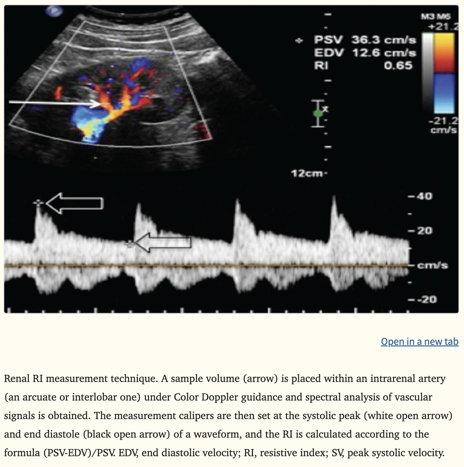
  • An elevated Renal Resistive-Index (RRI) > 0.7 is associated with impaired perfusion, development of AKI, and increased MR.
  • The kidney is a low vascular resistance organ sensitive to hypoperfusion in sepsis. 
  • 28-day all-cause MR and ventilator free days was decreased. 
  • RRI-guided MAP titration did not specifically decrease MR, incidence of AKI or need for CRRT.
  • Although a pilot trial, this study lends credence to more individualized sepsis hemodynamic management and the need for a larger study.
  • Use of RRI-guided MAP titration in sepsis may decrease overall 28-day MR.
  • How to Guide. https://ultrasoundpaedia.com/renal-arteries-normal/
Show References

Wang C, et al. Renal resistive index-guided mean arterial pressure titration in sepsis: a prospective single-center, single-blind, parallel group randomized controlled trial. Nature Communications. 2026; 17:8759.


Title: Bova Score Refresher

Category: Pulmonary

Posted: 9/6/2026 by Robert Flint, MD

Reminder that the Bova Score (2 points each for heart rate over 110, SBP less than 90, RV strain on CT or echo, and elevated troponin) along with an elevated lactate and BNP can help predict outcomes in hemodynamically stable PE patients. 

"Low risk (score <2): 
Proceed with standard anticoagulation therapy. Standard outpatient or inpatient monitoring, as appropriate.

Intermediate risk (score 3–4):  Consider closer monitoring (e.g., stepdown/ICU). Discuss the possibility of advanced therapies (e.g., thrombolysis) in select cases, especially if clinical status worsens. 

High risk (score >4): Maintain continuous ICU monitoring with a clear plan for rapid intervention if clinical status worsens (e.g., hypotension, worsening hypoxia, arrhythmias). Strongly consider multidisciplinary team involvement and potential advanced therapies (e.g., systemic thrombolysis or catheter-directed therapies)."

Show References

https://www.mdcalc.com/calc/4004/bova-score-pulmonary-embolism-complications


Title: Post termination family needs.

Category: EMS

Posted: 9/5/2026 by Robert Flint, MD (Updated: 9/17/2026)

A structured interview was conducted with families who had a loved one undergo a field terminated cardiac arrest in Denmark. What they found was pre-arrival instructions were very valued. And “After unsuccessful resuscitation, compassionate and unhurried communication was considered crucial.” I am sure this applies to prehospital and in hospital situations alike. Empathy and time are what these families need.

Show References

Skovbo, A., Jørck, C.L., Huniche, L. et al. When prehospital resuscitation attempts are unsuccessful: care for the relatives following termination of treatment in out-of-hospital cardiac arrest. Scand J Trauma Resusc Emerg Med(2026). https://doi.org/10.1186/s13049-026-01689-z


Title: Predicting Agitation in Pediatric Emergency Psychiatric Patients

Category: Pediatrics

Keywords: Agitation, Pediatrics, BRACHA-S, Aggression, Psychiatric emergencies, restraints (PubMed Search)

Posted: 9/4/2026 by Mary Girgis, MD (Updated: 9/4/2026)

BRACHA-S is a 5 item scoring tool that demonstrated moderate discrimination and identified clinically meaningful risk gradients for agitation requiring intervention (ARI) that may support early safety planning.

Show Additional Information

Objectives: To validate the 5 item Brief Rating of Aggression in Children and Adolescents- Short (BRACHA-S) completed by ER nurses for patients presenting with mental and behavioral health encounters for predicting agitation requiring intervention.

Background: MBH concerns account for about 13% of all pediatric ER visits and approximately 10% require the use of physical or pharmacologic restraints.

Methods: They conducted a prospective observational prognostic validation study in 2 pediatric EDs. After patient arrival with MBH concern, ED nurses completed the BRACHA-S. Discrimination was assessed with AUROC and performance was estimated for a 3 tier model. 

Results: Among 472 encounters, 55 (11.7%) had ARI. BRACHA-S scores were associated with ARI (AUROC, 0.72; 95% CI, 0.64–0.79). ARI incidence increased with score: 0, 3.3% (2/60); 1, 6.3% (6/96); 2, 6.4% (7/110); 3, 12.4% (17/137); 4, 31.8% (14/44); and 5, 36.0% (9/25). Scores 0 to 1 had a negative predictive value of 96.7% but a specificity of 13.9%. Scores 4 to 5 had a specificity of at least 89% and a positive predictive value of greater than 33%. Compared with scores 0 to 2, relative risk of ARI was 2.2 (95% CI, 1.1–4.3) for score 3 and 5.9 (95% CI, 3.3–10.7) for scores 4 to 5.

Conclusions: In pediatric ED MBH encounters, the nurse-completed triage BRACHA-S demonstrated moderate discrimination and identified clinically meaningful risk gradients for ARI that may support early safety planning.

Show References

Lynn Babcock, Lily Klein, Yin Zhang, Nancy Daraiseh, Ryan Siders, Beatrice Thomas, Ryan Murphy, Holly R. Hanson, Bijan Ketabchi, Shelby Tanguay, Drew Barzman, Wendy J. Pomerantz; A Brief Tool to Predict Agitation in Pediatric Emergency Psychiatric Patients. Pediatrics August 2026; 158 (2): e2026076729. 10.1542/peds.2026-076729


Title: Get the Lead Out

Category: Toxicology

Keywords: Lead poisoning, Chelation, Neurotoxin (PubMed Search)

Posted: 9/3/2026 by Kathy Prybys, DO

Bottom Line:

Lead has no biologic role in the human body. Lead toxicity and accumulation in humans remains a major health concern. Children < 6 years old are especially vulnerable to lead poisoning.  Lead is a potent neurotoxin and even low levels can cause irreversible harmful neurologic effects in children. Blood lead level of 3.5 µg/dL requires action in children.

Show Additional Information

  • Lead has no biologic role in the human body unlike essential metals (ie. iron, calcium, magnesium, zinc).
  • Children < 6 years old are especially vulnerable to lead poisoning due to environmental and physiological variables
    • Increased hand to mouth activity, 4-5 times better GI absorption, developing nervous systems, bone growth
  • Lead is toxic to every organ system and can present as a multisystem disease with nonspecific clinical manifestations 
  • The nervous system is particularly sensitive to lead and even low levels can cause irreversible harmful neurologic effects in children
    • Decreased IQ, ability to pay attention, and negative behavioral effects
  • Severe acute neurotoxicity is seen at high levels (75-100 ug/dL) causing encephalopathy, seizure, coma
  • No safe Blood Lead Level (BLL) in children has been identified 
  • BLL of 3.5 µg/dL requires action in children
    • Serial monitoring
    • Environmental investigation to identify potential sources of lead
    • Abdominal x-ray and decontamination for BLL > 20-44 ug/dL
  • BLLs do not accurately predict total body burden or duration of exposure but are surrogate markers indicating exposure and absorption 
  • Lead distributes to different body compartments. T1/2 of Lead:
    • Blood = 28 days (99% bound to RBCs)
    • Soft tissue = 40 days
    • Bone =  25 years or greater
  • 90% of total body lead burden is stored in bone which can later  be released 
  • Chelation therapy is indicated for treatment of severe symptoms or markedly elevated BLL:
    • Adults: 100 ug/dL
    • Children: >45-75 ug/dL
  • Acute lead encephalopathy is a medical emergency requiring aggressive chelation therapy

Show References

Recommended Actions Based on Blood Lead Level. August 21, 2025. https://www.cdc.gov/lead-prevention/hcp/clinical-guidance/index.html

The effect of lead exposure on IQ test scores in children under 12 years: a systematic review and meta-analysis of case-control studies. Heidari S, Mostafaei S, Razazian N, et al. Syst Rev. 2022 May 30;11(1):106. doi: 10.1186/s13643-022-01963-y. PMID: 35637522; PMCID: PMC9150353.

Pediatric Lead Chelation Managed During Critical Medication Shortages: Case Report and Literature Review. Singh MK, Kane JM, Said SJ. The Journal of Pediatric Pharmacology and Therapeutics. 2024 Sep;29(5):544–549. doi:10.5863/1551-6776-29.5.544.


Title: What size blade should be used for intubation?

Category: Critical Care

Keywords: airway, laryngoscopy, blade size, critical care (PubMed Search)

Posted: 9/1/2026 by Kristyn McLeod, MD

While many intubations in critically ill patients go smoothly, they are a high risk procedure that EM and ICU physicians seek to continuously improve upon. Blade size is one aspect of the procedure that has received attention over the past several years. We don’t have prospective trials addressing this question, but there are several retrospective analyses that have investigated it.

  • For Direct Laryngoscopy:
    • The 2022 MacSize-ICU study and a 2023 study by Landefeld and colleagues with the Pragmatic Critical Care Research Group suggested that when using Macintosh geometry blades for direct laryngoscopy there was improved first pass success with size 3 blade. Secondary outcomes for these studies point to a worse glottic view with a size 4 blade.
  • For Video Laryngoscopy:
    • This finding did not hold up in video laryngoscopy.  A 2023 study by Park et al. and now a 2026 retrospective study again by Landefeld et al. that looked at blade size for video laryngoscopy with Macintosh geometry blades found no difference in first pass success with size 3 vs size 4 blades.

Bottom Line: Continue to use your best clinical judgment when selecting a blade size, especially when performing video laryngoscopy. When performing direct laryngoscopy, if you are on the fence, consider using size 3 blade.

*These studies do not address intubation with hyperangulated designs

Show References

  • Godet, Thomas, et al. "Impact of Macintosh blade size on endotracheal intubation success in intensive care units: a retrospective multicenter observational MacSize-ICU study." Intensive Care Medicine 48.9 (2022): 1176-1184.
  • Landefeld, Kevin R., et al. "Effect of laryngoscope blade size on first pass success of tracheal intubation in critically ill adults." Critical Care Explorations 5.3 (2023): e0855.
  • Park, Jeongyong, et al. "Effect of blade size on the first-pass success rate of endotracheal intubation using the C-MAC video laryngoscope." Journal of Clinical Medicine 12.22 (2023): 7055.
  • Landefeld, Kevin R., et al. "Blade Size and Outcomes with Standard-Geometry Video Laryngoscopy in Emergency Tracheal Intubations: Secondary Analysis of Two Randomized Controlled Trials." CHEST Critical Care (2026): 100257.


Title: Hyperbaric Oxygen: A beneficial adjunct in burn care

Category: Trauma

Keywords: hyperbaric, HBOT, HBO2, burn, thermal burn (PubMed Search)

Posted: 8/31/2026 by TJ Gregory, MD (Updated: 9/17/2026)

Bottom Line: Burn care is directed by the managing surgeons at regional burn centers. Where available, hyperbaric oxygen treatment can be a beneficial adjunct.

Studies and clinical experience of hyperbaric oxygen treatment in burns has shown modulation of burn shock, dampening of the inflammatory response, reduction of edema, reversal of the zone of stasis, reduction of ischemia and ischemic necrosis, prevention of progression of partial- to full-thickness injury, modulation of inflammation, effecting modulation of hyper-metabolic syndrome, lessening of the capillary leak, preservation of dermal elements, a reduced need for grafting, shortened hospital stay, reduced physiological post-discharge problems, and a reduction in cost of care.

Show References

Cianci P, Sato RM, Faulkner J. Adjunctive Hyperbaric Oxygen in the Treatment of Thermal Burns. Undersea Hyperb Med. 2026 Second Quarter;53(2):363-390. PMID: 42365959.


Title: To admit or not:syncope

Category: Cardiology

Keywords: Syncope (PubMed Search)

Posted: 8/30/2026 by Robert Flint, MD (Updated: 9/17/2026)

This secondary review of a prospective observational group of 1200 syncope/presyncope patients over age 40 found 5.6% experienced a significant adverse outcome within 30 days. These patients had no diagnosis after their ED evaluation. Their conclusion was admission to the hospital “increased the diagnostic yield for SAOs and accelerated time to diagnosis.”

Show References

https://doi.org/10.1111/acem.70393


Title: Gout: mono or polyarticular?

Category: Orthopedics

Posted: 8/29/2026 by Brian Corwell, MD (Updated: 9/17/2026)

Polyarticular joint pain and swelling. Is it gout?

Polyarticular initial attacks occur in roughly 3-14% of patients.

Bilateral gout is uncommon at presentation.

However, this presentation is more frequent with longstanding , poorly controlled disease.

Show Additional Information

Common initial presentation is textbook: monoarticular, classically the first MTP (podagra) or may involve a single lower limb joint. 

Over time recurrent flares become:

  1. Longer lasting
  2. Polyarticular
  3. Affect upper-limb joints (wrist, elbow for example)

As gout becomes longstanding (~15 years), urate crystal deposition at a given joint was most strongly associated with symmetric involvement of the SAME joint on the contralateral side 

OR 26.1 in hands/wrists, 46.9 in feet/ankles, 9.9 in knees

Erosive lesions in the feet/ankles were also highly symmetric (OR 91.4)

Take home:

  1.  Consider acute gout as a lower limb monoarticular disease. 
  2. Conceptualize chronic gout as a bilateral/symmetric polyarthropathy
  3. An initial presentation of an acute symmetric polyarthritis should prompt consideration of alternative diagnoses (CPPD or rheumatoid arthritis for example).

Show References

Yokose C, Dalbeth N, Wei J, Nicolaou S, Simeone FJ, Baumgartner S, Fung M, Zhang Y, Choi HK. Radiologic evidence of symmetric and polyarticular monosodium urate crystal deposition in gout - A cluster pattern analysis of dual-energy CT. Semin Arthritis Rheum. 2020 Feb;50(1):54-58.


Title: The title is deceiving

Category: Trauma

Posted: 8/27/2026 by Robert Flint, MD (Updated: 9/17/2026)

This study conclusion states: “Prehospital epinephrine administration in patients with traumatic cardiac arrest was associated with increased survival to hospital discharge and prehospital ROSC.” When you investigate the methods, only 809 of the 22,105 patients in traumatic cardiac arrest actually received epinephrine. In the study country, EMS gets orders from base station physicians for epinephrine. Why did these 809 get the epinephrine and the other 97% did not? It is hard to know what to do with this data. It certainly doesn’t say epinephrine saves patients in traumatic cardiac arrest. More research is needed.

Show References

Hyun Seok Chai, Gwan Jin Park, Young Min Kim, Sang Chul Kim, Hoon Kim, Suk Woo Lee,

Prehospital epinephrine as a bridge to survival in traumatic cardiac arrest: A nationwide propensity score-matched analysis,

The American Journal of Emergency Medicine,

Volume 109,

2026,

Pages 52-57,

ISSN 0735-6757,

https://doi.org/10.1016/j.ajem.2026.06.039


Title: Diagnostic Excellence – More Than I Know It When I See It

Category: Administration

Keywords: diagnosis, error, patient safety, quality (PubMed Search)

Posted: 8/26/2026 by Steve Schenkel, MD, MPP (Updated: 9/17/2026)

What is diagnostic excellence in the Emergency Department?

Based on a modified Delphi panel of 11 experts, these authors developed this definition:

“[T]he use of optimal, evidence-based practice to attain an accurate and timely explanation about a patient’s condition based on the information available at the time and communicate that explanation to the patient/family. Diagnostic excellence is patient-centered and equitable. Diagnostic excellence includes avoidance of divergent practices. It leads to better choices in management, prevents missed opportunities, and reduces the risk of preventable patient harm.”

Click below to find the definition of missed diagnostic opportunity. Go to the article https://www.annemergmed.com/article/S0196-0644(26)00247-7/fulltext to read more about the work.

Show Additional Information

The authors developed this definition for a missed diagnostic opportunity:

“a measurable divergence from optimal, evidence-based practice that may lead to: an inaccurate or delayed explanation of a patient’s condition, despite information available at the time, or a failure in shared communication related to the diagnosis with the patient/family. A missed diagnostic opportunity may lead to suboptimal management, preventable patient harm, or inequitable outcomes.”

Show References

Berdahl C, Schiff G, Venkatesh A, et al. Defining Diagnostic Excellence and Missed Diagnostic Opportunity for the Emergency Department Setting. Annals of Emergency Medicine, 2026; 88, 376-393.


Title: Linezolid: Crossing the Line for Severe CAP

Category: Critical Care

Posted: 8/25/2026 by Jon Hurst, MD

Severe Community Acquired Pneumonia (SCAP) is broadly defined as CAP that requires ICU admission. Although MRSA pneumonia is overall quite rare (1), it is associated with a high mortality rate. Therefore, empiric MRSA coverage is commonly used for patients with SCAP. Common empiric agents include vancomycin and linezolid. Linezolid has both pharmacologic and clinical data that suggest it may be a preferred option for many patients.

When approaching a patient with SCAP, a key consideration is whether empiric MRSA coverage is needed. Risk factors for MRSA pneumonia include prior MRSA infection or colonization, recurrent skin infections, post-influenza pneumonia, recent hospitalization or antibiotic use (1). 

If empiric MRSA coverage is determined to be needed, Linezolid offers several advantages for the treatment of SCAP. Highlights below:

  • Linezolid has 100% oral bioavailability which can be especially useful for those patients with difficult IV access
  • Linezolid has better lung epithelial lining penetration compared to vancomycin (6)
  • In direct comparison between linezolid and vancomycin for confirmed MRSA pneumonia, linezolid was shown to have improved microbiologic cure rates without an improvement in mortality (2, 7)
  • Linezolid was shown to have less nephrotoxicity than vancomycin (2, 7)

Side effects to consider with linezolid include:

  • Serotonin syndrome, although extremely rare (5). May consider discussion with your pharmacist if taking additional serotonergic agents.
  • Myelosuppression (typically thrombocytopenia), although usually with longer treatment courses. This study shows no significant difference in rates of thrombocytopenia compared to vancomycin (4).

If providing linezolid for treatment of SCAP:

  • Dose: Linezolid 600mg IV or PO q12 hours
  • Ideally should obtain blood cultures, sputum culture, MRSA nares prior to (or closely following) antibiotic administration

Show References

  1. Aliberti, Stefano, et al. "Global initiative for meticillin-resistant Staphylococcus aureus pneumonia (GLIMP): an international, observational cohort study." The Lancet Infectious Diseases 16.12 (2016): 1364-1376.
  2. Jiang, H., R-N. Tang, and J. Wang. "Linezolid versus vancomycin or teicoplanin for nosocomial pneumonia: meta-analysis of randomised controlled trials." European journal of clinical microbiology & infectious diseases 32.9 (2013): 1121-1128. 
  3. Nair, Girish B., and Michael S. Niederman. "Updates on community acquired pneumonia management in the ICU." Pharmacology & therapeutics 217 (2021): 107663.
  4. Nasraway, Stanley A., et al. "Linezolid does not increase the risk of thrombocytopenia in patients with nosocomial pneumonia: comparative analysis of linezolid and vancomycin use." Clinical infectious diseases 37.12 (2003): 1609-1616.
  5. McCreary, Erin K., et al. "Antibiotic myths for the infectious diseases clinician." Clinical Infectious Diseases 77.8 (2023): 1120-1125.
  6. Stein, Gary E., and Elizabeth M. Wells. "The importance of tissue penetration in achieving successful antimicrobial treatment of nosocomial pneumonia and complicated skin and soft-tissue infections caused by methicillin-resistant Staphylococcus aureus: vancomycin and linezolid." Current medical research and opinion 26.3 (2010): 571-588.
  7. Wunderink, Richard G., et al. "Linezolid in methicillin-resistant Staphylococcus aureus nosocomial pneumonia: a randomized, controlled study." Clinical Infectious Diseases 54.5 (2012): 621-629.


Title: VL bears DL

Category: Trauma

Posted: 8/24/2026 by Robert Flint, MD (Updated: 9/17/2026)

In a randomized pragmatic study of critically ill trauma patients,  video laryngoscopy was successful in 88% of first pass attempts versus 68% in direct laryngoscopy.

Show References

Journal of Trauma and Acute Care Surgery 101(2):p 359-365, August 2026. | DOI: 10.1097/TA.0000000000005021


Title: CT on arrival

Category: Trauma

Posted: 8/23/2026 by Robert Flint, MD (Updated: 9/17/2026)

This Japanese hospital has a CT scanner hybrid resuscitation room. They did a retrospective study comparing their blunt trauma patients to a Japanese trauma database. Their patients had a shorter time to operative or interventional procedure, slightly less PRBC use over 24 hours and similar 24 hour mortality. 
Interesting concept. Not ready for adoption.

Show References

Matsumoto S, Senoo S, Aoki M, Funabiki T, Shimizu M. CT-first resuscitation for severe blunt trauma: A propensity score-matched cohort study. J Trauma Acute Care Surg. 2026 Jul 1. doi: 10.1097/TA.0000000000005105. Epub ahead of print. PMID: 42385208.


Previous |  1 |  2 |  3 |  4 |  5 |  6 |  ... |  239 |  Next
University of Maryland School of Medicine logo
  • Departments
  • Programs
  • Research Centers
  • Institutes
  • Graduate Education
  • CIBR Cores
  • Offices of the Dean
    • University of Maryland, Baltimore
    • University of Maryland Medical Center
    • Medical Alumni Association of the University of Maryland
    • Health Sciences and Human Services Library
University of Maryland School of Medicine
655 W. Baltimore Street
Baltimore, MD 21201
Contact Us

© University of Maryland School of Medicine

    • Webmaster
    • Web Accessibility
    • Site Index
    • UMB Hotline
    • Employment
    • Map