
University of Maryland School of Medicine logo
Emergency Medicine
All Departments
UMSOM Assistant
UMEM Educational Pearls
  • Home
  • Departments
  • Emergency Medicine
  • CME
  • UMEM Educational Pearls
  • CME
  • Courses and Conferences
  • Free Open Access Medical Education
  • UMEM Educational Pearls

Search

121-140 of 321 results by Haney Mallemat

Previous  |  1 |  2 |  3 |  4 |  5 |  6 |  7 |  8 |  9 |  10 |  11 |  12 |  ... |  17 |  Next

Title: What's the Diagnosis?

Category: Visual Diagnosis

Posted: 1/20/2014 by Haney Mallemat, MD

Question

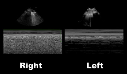
50 year-old male intubated for respiratory distress. Ultrasound is used post-intubation to confirm tube placement and the following images are obtained. What's the diagnosis?
Show Answer
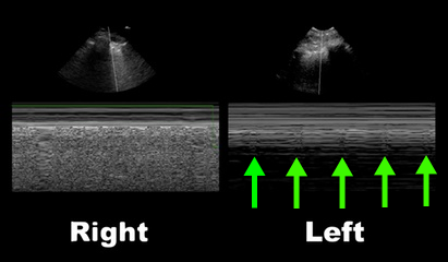
Right main-stem intubation as demonstrated by presence of lung-pulse on the left side

Lung-Pulse

  • Normal lung sliding creates an artifact at the pleural line, as the visceral and parietal pleura slide against one another; this is seen in the M-mode image below for the right lung
  • When a pneumothorax occurs the two pleural become separated and there is loss of this artifact; this is called the stratosphere sign (not shown here)
  • However, when the two pleura are in contact but there is no movement of the pleura (e.g. main-stem intubation on the non-ventilated side) lung-pulse is observed (seen below with arrows)
  • Although there is no ventilation of the affected lung, the lung pulse occurs because there is still cardiac activity that "pushes" the lung into the chest wall from underneath. This creates small periodic movements of the visceral and parietal pleura, creating small amounts of artifact
  • Lung pulse can differentiate pneumothorax from other causes of lung hypoventilation:
    • Mucus plug in main-stem bronchus
    • Foreign body in main-stem bronchus
    • Main-stem intubation

Show References

Follow me on Twitter (@criticalcarenow) or Google+ (+criticalcarenow)


Title: What's the Diagnosis? Case by Dr. Phillip Stafford

Category: Visual Diagnosis

Posted: 1/13/2014 by Haney Mallemat, MD

Question

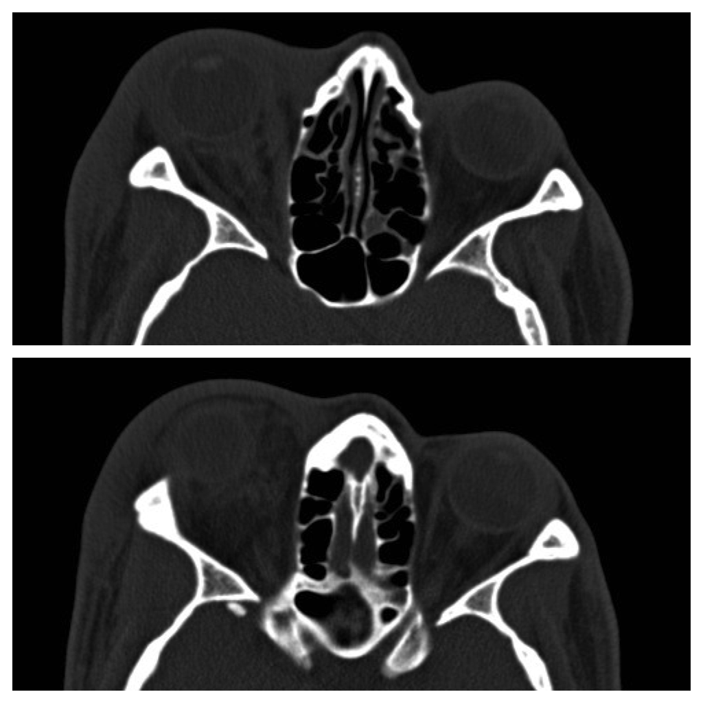
42 year-old male s/p assault complains of right sided facial pain, swelling, and decreased vision. Physical exam reveals subconjunctival hemorrhage, proptosis, afferent pupillary defect, and a firm globe. What's the diagnosis and what's the emergent treatment?
Show Answer
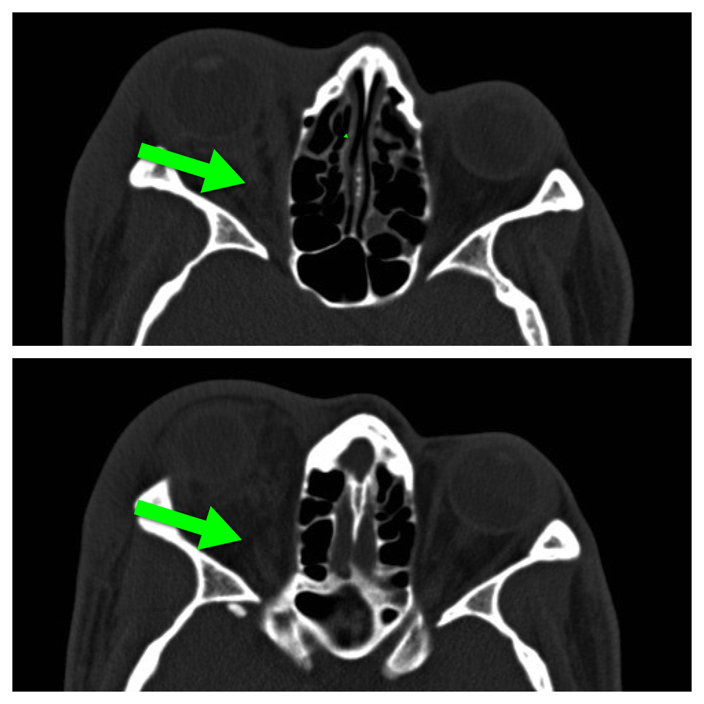
  • Retrobulbar hematoma (a compartment syndrome of the eye) with proptosis.
  • Treatment is emergent lateral canthotomy; lowering intraocular pressure with medical therapy (e.g., steroids, mannitol, etc.) is a temporizing option but should never delay canthotomy. 
  • Here's a video narrated by our own Dr. Mak Moayedi demonstrating the procedure.

Show References

Follow me on Twitter (@criticalcarenow) or Google+ (+criticalcarenow)


Title: What's the Diagnosis? Case by Dr. Tu Carol Nguyen

Category: Visual Diagnosis

Posted: 1/6/2014 by Haney Mallemat, MD

Question

37 year-old male presents after sustaining a burn from a pot of boiling water. He states that his skin started to blister a few hours after and it’s quite painful. What type of burn does he likely have? 
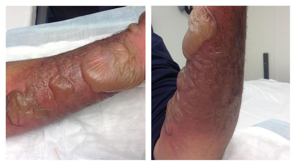
Show Answer
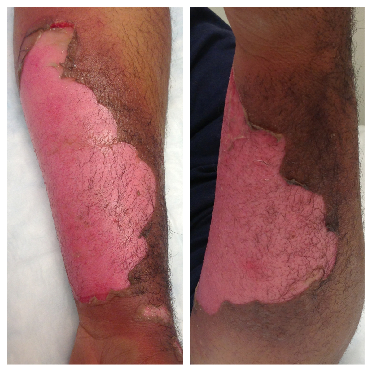
A non-circumferential, superficial partial-thickness burn; it was treated with Silvadene (silver sulfadiazine)

Burn Classification:

  • The traditional 1st, 2nd, 3rd degree classification-system was replaced by:
    • Superficial: skin is dry, red, and blanches with pressure; painful sensation (e.g. sunburn)
    • Superficial partial-thickness: skin has blisters, red, moist, weeping, and blanches with pressure; painful sensation
    • Deep partial-thickness: skin has blisters, wet/waxy dry, variable color (patchy to yellow-white to red), and does not blanch with pressure; pressure-like sensation
    • Full-thickness: skin is waxy-white to leathery-gray to charred and black; dry, elastic, and does not blanch with pressure; deep-pressure sensation
    • Fourth-degree: Burn extends into fascia, muscle, bone; deep-pressure sensation
  • Always be concerned with extremity burns because they can become edematous, causing superficial/deep partial-thickness to convert to full-thickness burns 
  • Treat with:
    • Extremity elevation 
    • Topical antibiotic creams/ointments
    • Blister debridement is controversial
    • Observation for compartment syndrome if circumferential
    • Consult with burn center as necessary
  • See image below demonstrating blisters after debridement

Show References

Follow me on Twitter (@criticalcarenow) or Google+ (+criticalcarenow)

Tintinalli, Judith E. (2010). Emergency Medicine: A Comprehensive Study Guide (Emergency Medicine). New York: McGraw-Hill Companies. pp. 1374–1386.


Title: What Does Dr. Fox Say? What's the Diagnosis by Dr. Zachary Dezman

Category: Visual Diagnosis

Posted: 12/30/2013 by Haney Mallemat, MD

Question

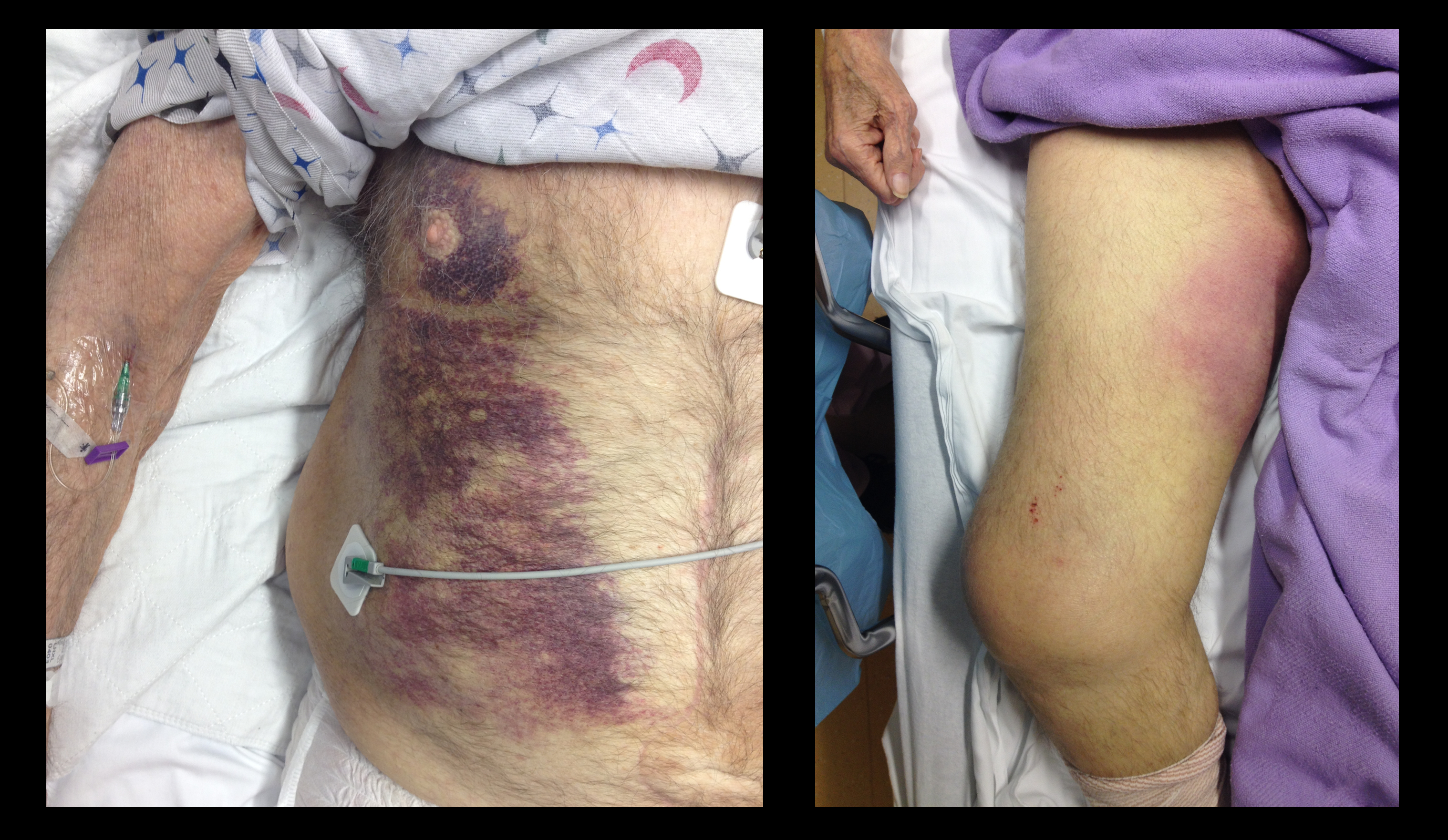
68 year-old male presents with weakness after surgical repair of his abdominal aorta. What’s the diagnosis and name at least one eponym for the signs displayed (there are five total)?
Show Answer
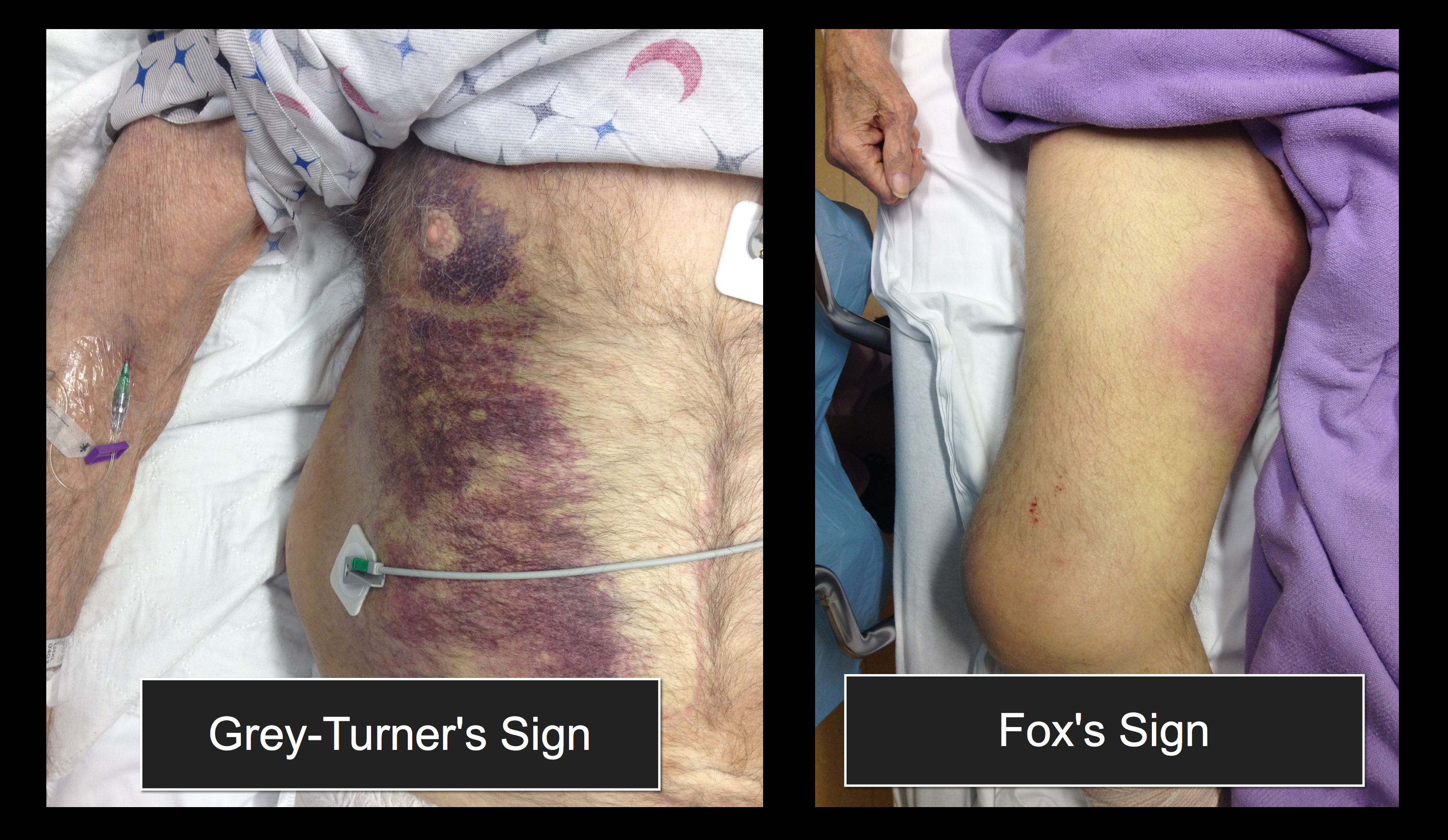
Grey-Turner and Fox's sign; these signs indicate retroperitoneal hemorrhage.

There are five signs suggesting retroperitoneal bleeding. They generally appear 24 hours after bleeding starts, occurring when blood extravasates along ligamentous connections between the retroperitoneal space and skin surface.

1. Grey-Turner Sign: Named for the surgeon who identified it in 1920; a bluish hematoma across the lateral abdominal wall when blood from the pararenal space leaks along the quadratus lumborum.

2. Fox’s Sign: Described by Dr. Fox in 1966 in two patients, (ruptured AAA and pancreatitis); ecchymosis over anteromedial thigh secondary to blood seeping along the fascia of psoas and iliacus.

3. Cullen’s Sign: Dr. Cullen (gynecologist), described this in 1918 as a sign of a ruptured ectopic pregnancy; bruise around the umbilicus from retroperitoneal blood tracking along falciform ligament.

4. Bryant’s Sign: Dr. Bryant initially described this sign; ecchymosis of the scrotum from blood tracking down the spermatic cord.

5. Stabler’s sign: Ecchymosis over the inguinal ligament

Show References

Follow me on Twitter (@criticalcarenow) or Google+ (+criticalcarenow)

  • Turner GG. Local discoloration of the abdominal wall as a sign of acute pancreatitis. Br J Surg, 1920;7:394–395
  • Fox JA. A diagnostic sign of extraperitoneal hemorrhage. Br J Surg 1966;53:193–195
  • Cullen TS. A new Sign in Ruptured Extrauterine Pregnancy, Amer J Obstet, 1918,78:457
  • James M. Dargin, Robert A. Lowenstein, Ruptured Abdominal Aortic Aneurysm Presenting as Painless Testicular Ecchymosis: The Scrotal Sign of Bryant Revisited, The Journal of Emergency Medicine, Volume 40, Issue 3, March 2011, Pages e45-e48
  • Stijn, Heyman, and Vervloessem, Bryant's and Stabler's Signs after a Difficult Delivery,N Engl J Med 2011; 365:1824


Title: One for All or Two for Some? Double-coverage for potential pseudomonal infections?

Category: Critical Care

Posted: 12/24/2013 by Haney Mallemat, MD

The morbidity and mortality from pseudomonas aeruginosa infections is high and empiric double-antibiotic coverage (DAC) is sometimes given; quality evidence for this practice is lacking.

Although there is little supporting data, the following reasons have been given for DAC:

  • DAC provides better empiric coverage through differing mechanisms of antibiotic action
  • DAC prevents the emergence of antibiotic resistance during therapy

The potential harm of antibiotic overuse cannot be ignored, however, and include adverse reaction, microbial resistance, risk of super-infection with other organisms (e.g., Clostridium difficile), and cost.

There may be a signal in the literature demonstrating a survival benefit when using DAC for patients with shock, hospital-associated pneumonia, or neutropenia. The IDSA guidelines, however, do not support DAC for neutropenia alone; only with neutropenia plus pneumonia or gram-negative bacteremia.

Bottom line: Little data supports the routine use of DAC in presumed pseudomonal infection. It may be considered in patients with shock, hospital-associated pneumonia, or neutropenia (+/- pneumonia), but consult your hospital’s antibiogram or ID consultant for local practices.

Show References

Freifeld, A. et al Clinical practice guideline for the use of antimicrobial agents in neutropenic patients with cancer: Clin Infect Dis 2011 Feb 15;52(4):e56-93.

Johnson, SJ et al. Is double coverage of gram-negative organisms necessary? Am J Health Syst Pharm 2011 Jan 15;68(2):119-24

Follow me on Twitter (@criticalcarenow) or Google+ (+criticalcarenow)


Title: What's the Diagnosis? Case by Dr. Thuy Pham and Dr. Stephen Shaheen

Category: Visual Diagnosis

Posted: 12/23/2013 by Haney Mallemat, MD

Question

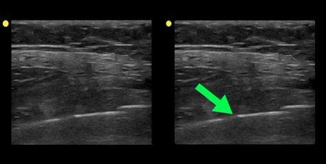
Patient with a history of osteogenesis imperfecta presents with right lateral chest pain following a sneeze. The ultrasound of his chest is shown (hint: arrow points to a rib). What's the diagnosis? 
Show Answer
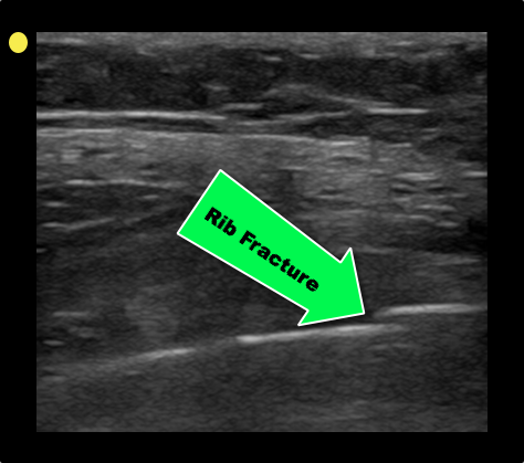
Rib fracture

  • Rib fractures may be missed on traditional diagnostics such as chest X-ray.
  • Ultrasound can be used to not only detect rib fractures but more importantly to detect co-existing injury such as pneumothorax or pulmonary contusion.
  • The ultrasound exam is easy and fast to perform:
    • The linear probe is used to evaluate
    • Ask the patient to place the probe on the point of maximal tenderness.
    • Evaluate the bright cortex of the bone in either cross-section or longitudinal plane (as is shown in case)
  • Look for other pathologies (e.g., pneumothorax) if rib fracture is discovered
  • Watch this video for a demonstration of the technique http://vimeo.com/1086731


Show References

Turk F. et al. Evaluation by ultrasound of traumatic rib fractures missed by radiography. Emerg Radiol. 2010 Nov;17(6):473-7. doi: 10.1007/s10140-010-0892-9

Follow me on Twitter (@criticalcarenow) or Google+ (+criticalcarenow)


Title: What's the Diagnosis? Case by Dr. Tristan Meador

Category: Visual Diagnosis

Posted: 12/16/2013 by Haney Mallemat, MD

Question

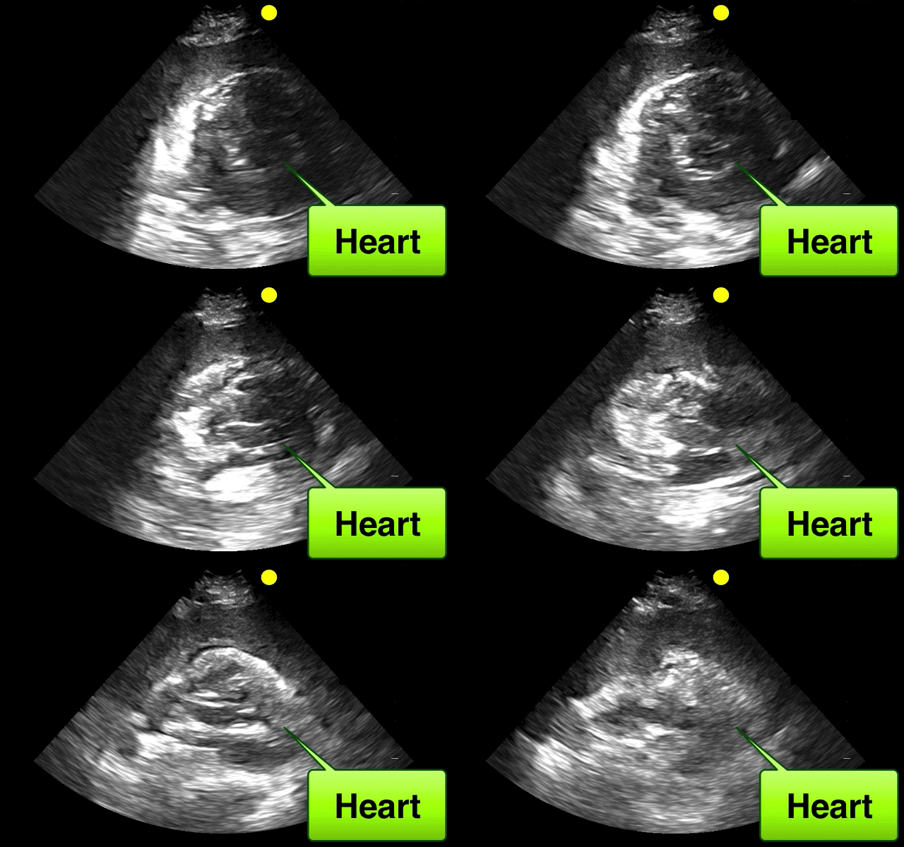
46 year-old female found unresponsive at a party. EMS transports the patient in cardiac arrest. A parasternal-long axis view of the heart is obtained during the pulse check. What's the diagnosis?
 

Show Answer
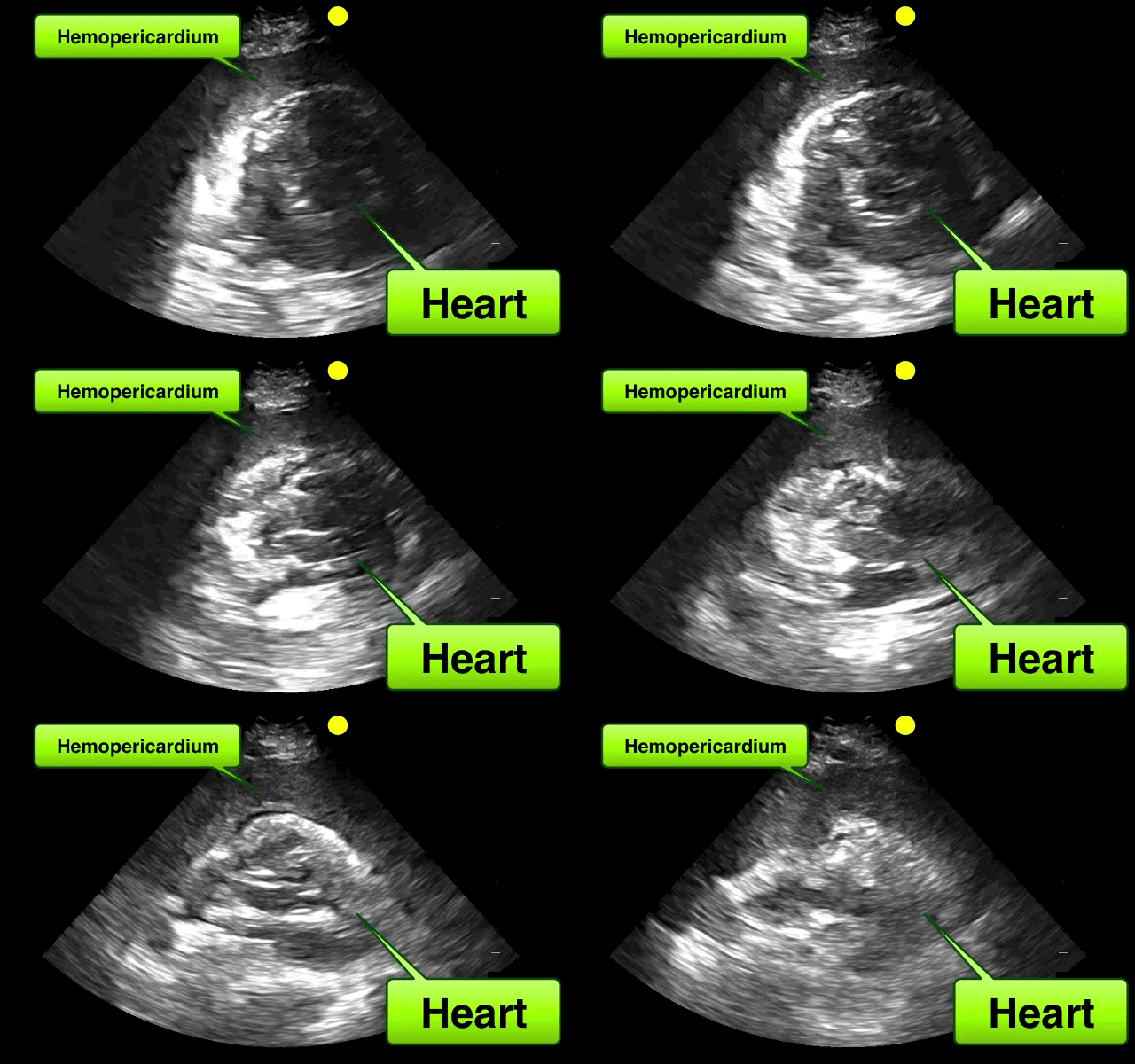
Hemopericardium

The heterogeneous appearance of the pericardial fluid indicates that is likely a complex pericardial effusion; the fluid could be blood, pus, or a malignant effusion.

Differential diagnosis of hemopericardium includes:

  • Ascending aortic dissection
  • Cardiac free-wall rupture
  • Blunt / penetrating trauma
  • Coagulopathies
  • Metastasis to the pericardium

Based on this initial ECHO, a pericardiocentesis was performed and blood was aspirated.

Show References

Follow me on Twitter (@criticalcarenow) or Google+ (+criticalcarenow)


Title: What's the Diagnosis?

Category: Visual Diagnosis

Posted: 12/9/2013 by Haney Mallemat, MD

Question

37 year-old male presents with cough and a fever. What's the diagnosis and name three risk factors assiciated with disease?
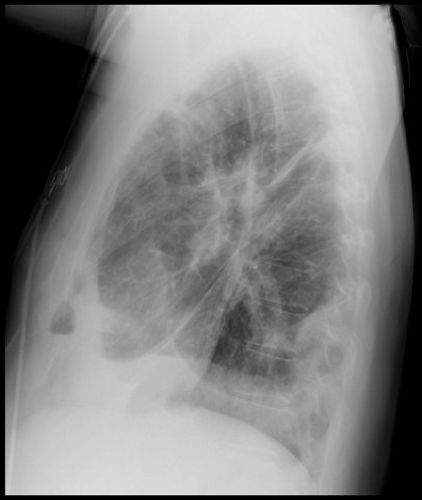
Show Answer
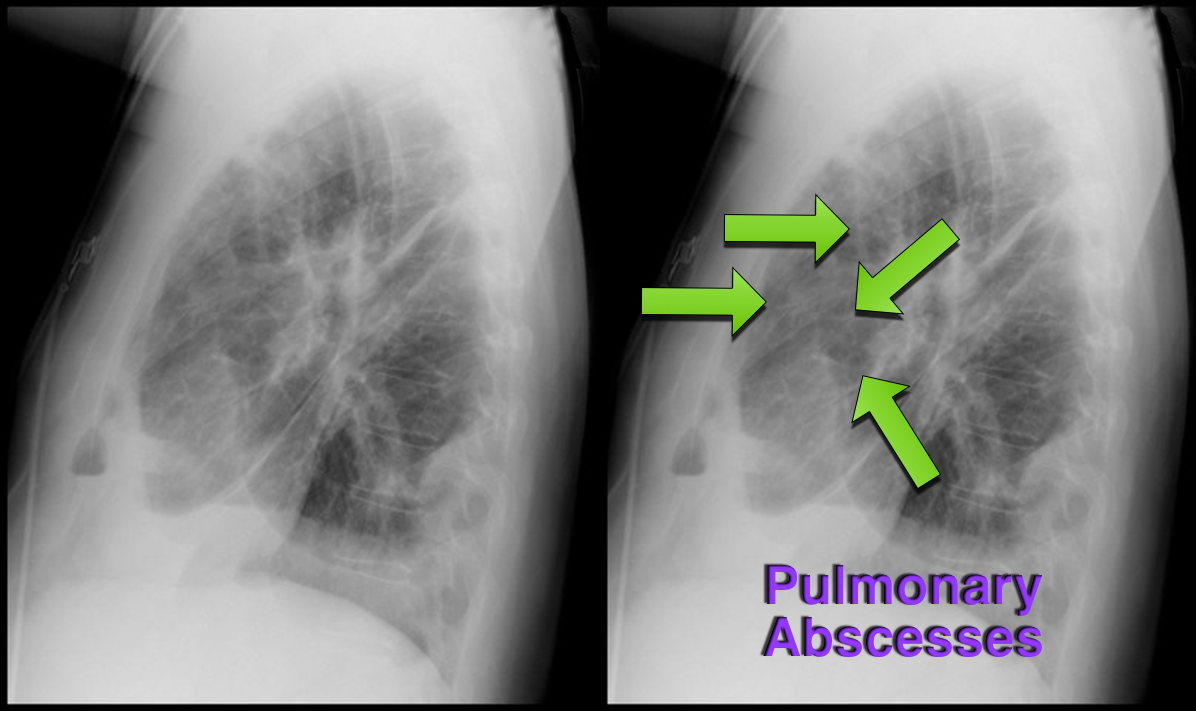
Answer: Lung Abscesses

Lung Abscess

  • Necrosis of lung parenchyma with pus and debris-filled cavities
  • Caused by
    • direct injury (e.g., aspiration pneumonia)
    • secondary causes (e.g., tricuspid  endocarditis, bacteremia, etc.)
  • Risk factors for disease:
    • Loss of airway reflexes (e.g., CVA, seizures, alcohol / narcotic abuse, etc)
    • Poor dentition
    • Immunosuppression (Diabetes, chronic steroids, etc.)
    • IVDA
  • Gram positives (especially Staph), negatives, and anaerobic bacteria have been implicated.
  • CXR may suggest, but CT scan better identifies abscess, necrotic tissue, empyema, or other pathology
  • Start broad-spectrum antibiotics and address potential underlying cause 


Show References

Follow me on Twitter (@criticalcarenow) or Google+ (+criticalcarenow)


Title: Echocardiography 101: Which ultrasound view of the heart is this?

Category: Visual Diagnosis

Posted: 12/2/2013 by Haney Mallemat, MD

Question

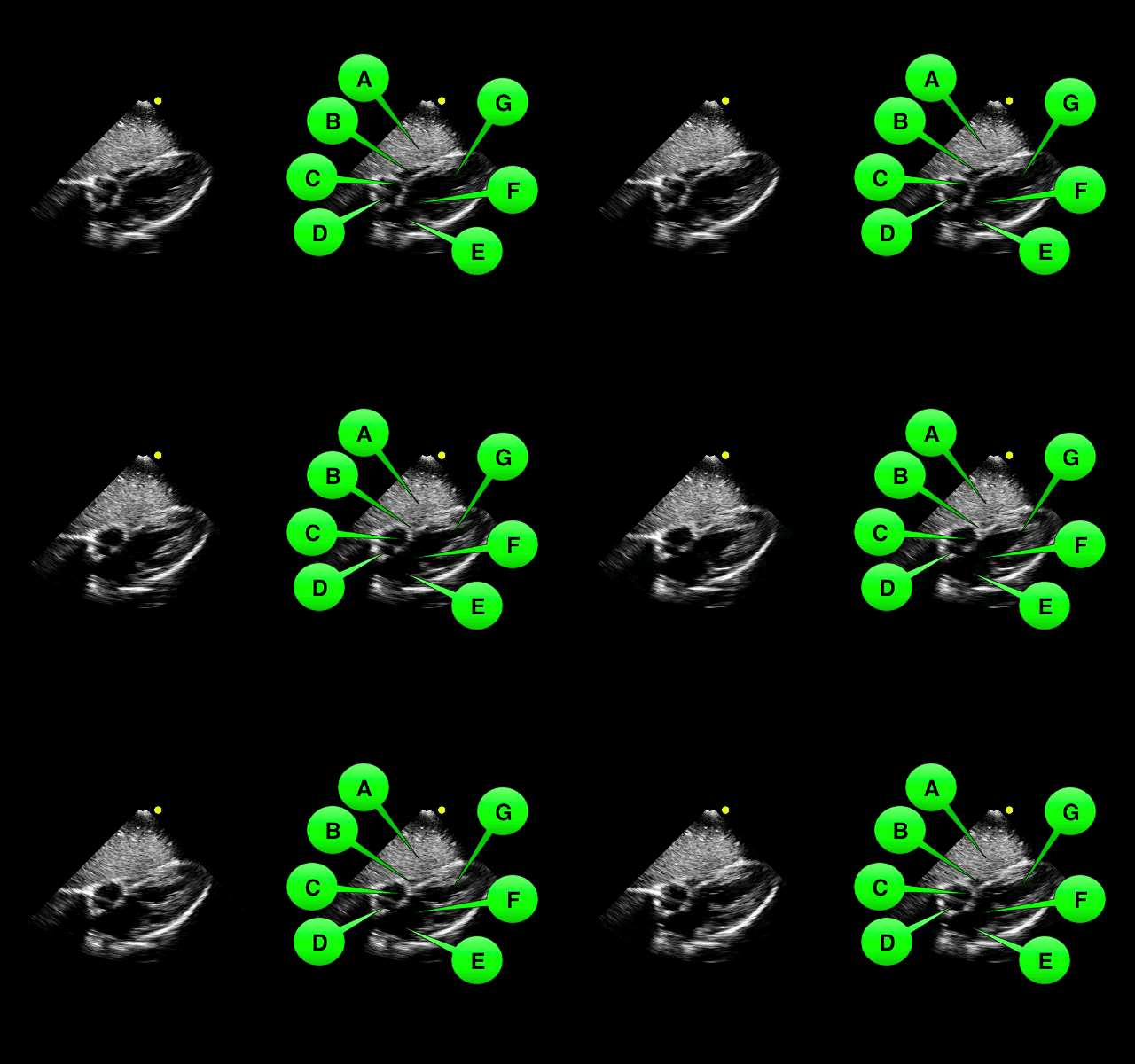
Which view of the heart is this and can you name the structures from A-G?
Show Answer

1. Subcostal or Subxiphoid view; this view is obtained by placing the probe under the ribs with the patient supine. The liver is used as an acoutic window to image the heart.

2. Name the items labeled A-G:

  • A = Liver
  • B = Right ventricle
  • C= Tricuspid valve
  • D= Right atrium
  • E = Left atrium
  • F = Mitral valve
  • G = Left ventricle

Show References

Follow me on Twitter (@criticalcarenow) or Google+ (+criticalcarenow)


Title: Dexmedetomidine...Better Than (Just) Benzos?

Category: Critical Care

Posted: 11/26/2013 by Haney Mallemat, MD

The management of alcohol withdrawal syndrome (AWS) includes supportive care focusing on the ABC’s and administration of benzodiazepines (BDZ). 

While BDZ are effective in the treatment of AWS, some patients may require very high doses of BDZ to control symptoms (tachycardia, hypertension, diaphoresis, etc.); unfortunately, high-doses of BDZ may lead to suppression of the respiratory drive and endotracheal intubation.

Dexmedetomidine (DEX) is a sedative agent that is an intravenous alpha2-agonist (it's like clonidine); it reduces sympathetic outflow from the central nervous system and it may help treat withdrawal syndromes. The major benefit of DEX is that it does not suppress the respiratory drive, thus intubation is not required.

Smaller trials and case series have shown that patients with AWS who were treated with BDZ in addition to DEX had better symptom control, lower overall BDZ doses, and less respiratory depression/intubation.

Bottom-line: While more trials are needed, consider adding DEX for patients with AWS who require high-doses of BDZ.

Show References

  • Tolonen, J. et al. Dexmedetomidine in addition to benzodiazepine-based sedation in patients with alcohol withdrawal delirium. Eur J Emerg Med. 2013 Dec; 20(6): 425-7
  • Rayner, S. et al. Dexmedetomidine as adjunct treatment for severe alcohol withdrawal in the ICU. Annals of intensive care, 2012

Follow me on Twitter (@criticalcarenow) or Google+ (+criticalcarenow)


Title: Echocardiography 101: What's the diagnosis?

Category: Visual Diagnosis

Posted: 11/25/2013 by Haney Mallemat, MD

Question

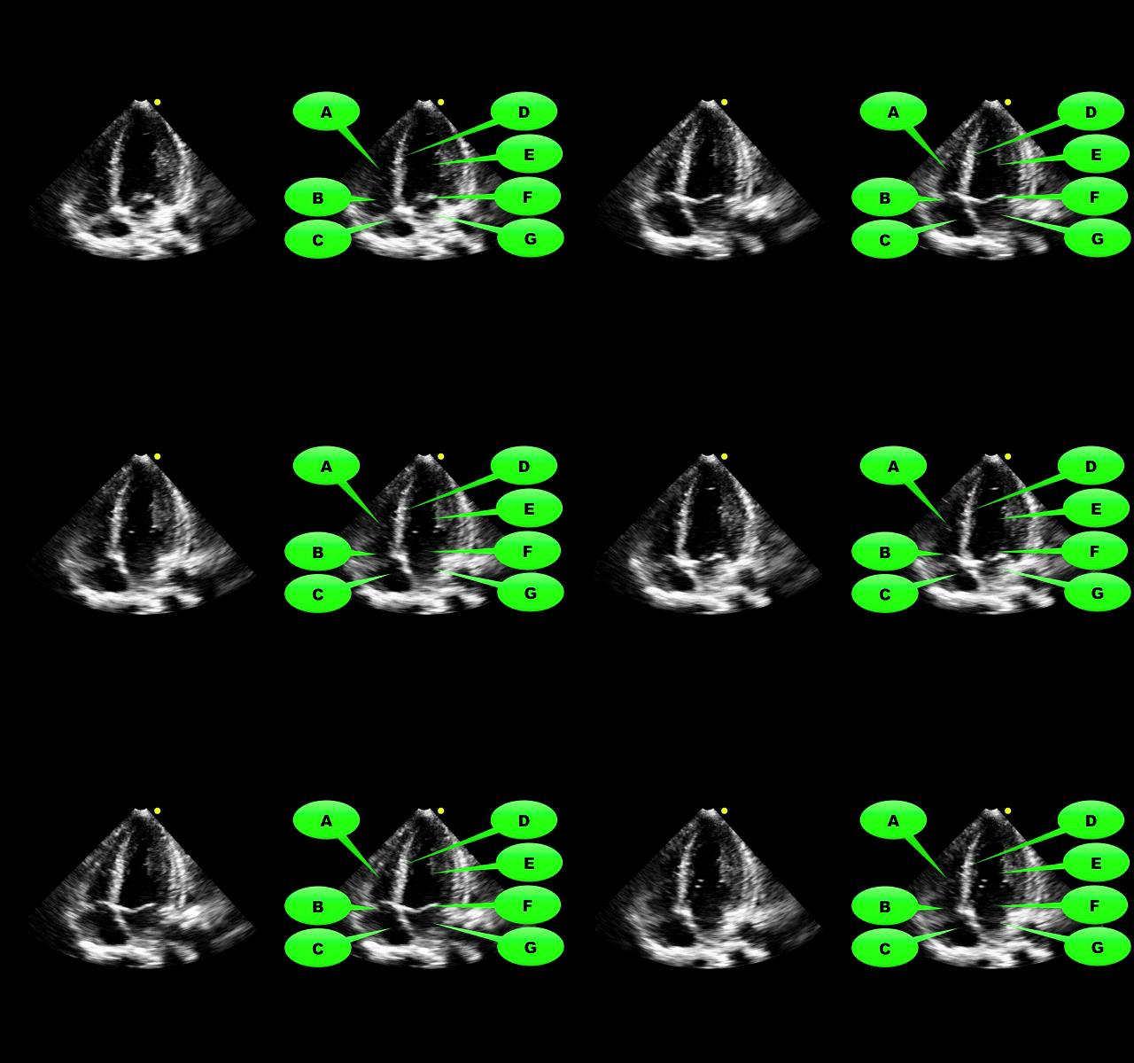
What view of the heart is this and can you name everything from A-G?
Show Answer

1. Apical-four chamber view; this view is obtained by placing the probe in the 4-5th intercostal space at the anterior axillary-line. The patient can be placed in left lateral decubitus to improve imaging.

2. Name the items labeled A-G:

  • A = Right ventricle
  • B = Tricuspid valve
  • C= Right atrium
  • D= Interventricular septum
  • E = Left ventricle
  • F = Mitral valve
  • G = Left atrium

Show References

Follow me on Twitter (@criticalcarenow) or Google+ (+criticalcarenow)


Title: What's the Diagnosis? Case by Dr. Ari Kestler

Category: Visual Diagnosis

Posted: 11/18/2013 by Haney Mallemat, MD

Question

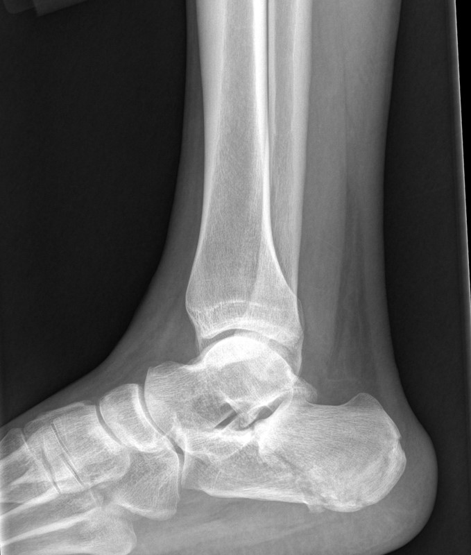
48 year-old presents after falling 15 feet following a “misunderstanding” with police. What's the diagnosis? ...and for a bonus question, why is this called a “Lover’s Fracture”? 
 

Show Answer
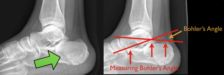
Calcaneus fracture

Answer to Bonus Question: Historically called a “Lover’s Fracture” for “lovers” jumping out of bedroom windows (to evade suspicious spouses) who then land directly on their feet.

Calcaneus fractures

  • Most commonly fractured tarsal bone
  • 2 types:
    • Extra-articular fracture (direct blow), twisting, and repetitive forces 
    • Intra-articular fracture from axial loading secondary to fall >6 feet, motor vehicle crash, etc.; (classic “lover’s fracture”)
  • 10% of axial loaded intra-articular fractures associated with:
    • Bilateral calcaneus fractures and/or,
    • Thoracic or lumbar compression fractures and/or,
    • Proximal femur or tibial plateau fractures
  • Ankle Xray is diagnostic and to measure Bohler’s angle (see figure below)
    • Normally 20-40 degrees
    • <20 degrees increases suspicion for intra-articular fracture
  • Ankle CT in select cases as Xray may underestimate some injuries
  • Extra-articular fractures treated with closed reduction and casting and intra-articular fractures treated by open reduction and internal fixation (closed reduction in select cases)

Show References

Follow me on Twitter (@criticalcarenow) or Google+ (+criticalcarenow)


Title: What's the Diagnosis? Image by Dr. Tu Carol Nguyen

Category: Visual Diagnosis

Posted: 11/11/2013 by Haney Mallemat, MD

Question

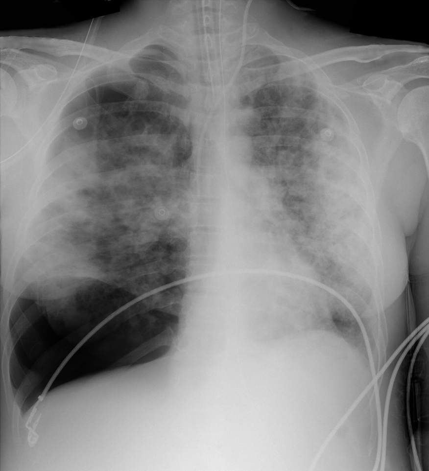
28 year-old cachectic female presents in respiratory distress and is immediately intubated on arrival to Emergency Department. What's the diagnosis and what are some potential etiologies?
Show Answer
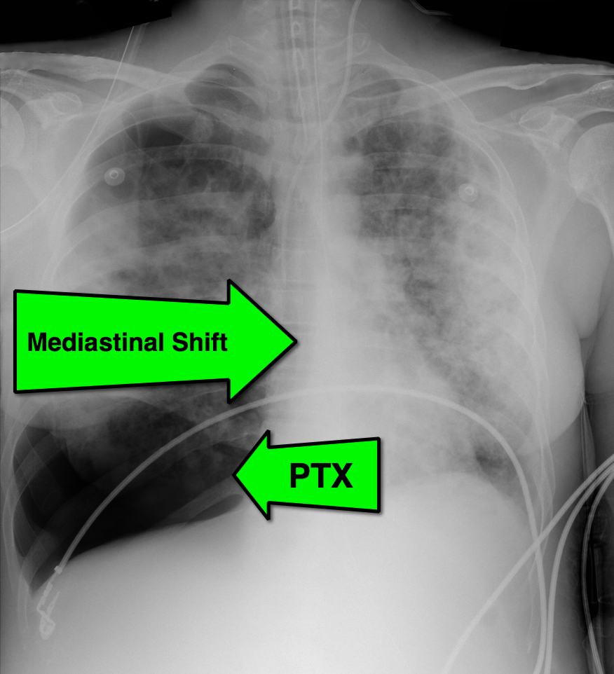
Pneumothorax with mediastinal shift

Differential Diagnosis

  • Atypical bacterial PNA
  • Acute Respiratory Distress Syndrome
  • Interstitial lung disease
  • Pneumocystis Jirovecii PNA (PJP)

The patient in this case had undiagnosed HIV/AIDS and presented with PTX secondary to PJP. The lifetime risk of PTX with HIV is 6% and 85% of those cases are secondary to PJP.

Show References

Follow me on Twitter (@criticalcarenow) or Google+ (+criticalcarenow)


Title: What's the Diagnosis? Basic ECHO Part 1

Category: Visual Diagnosis

Posted: 11/4/2013 by Haney Mallemat, MD

Question

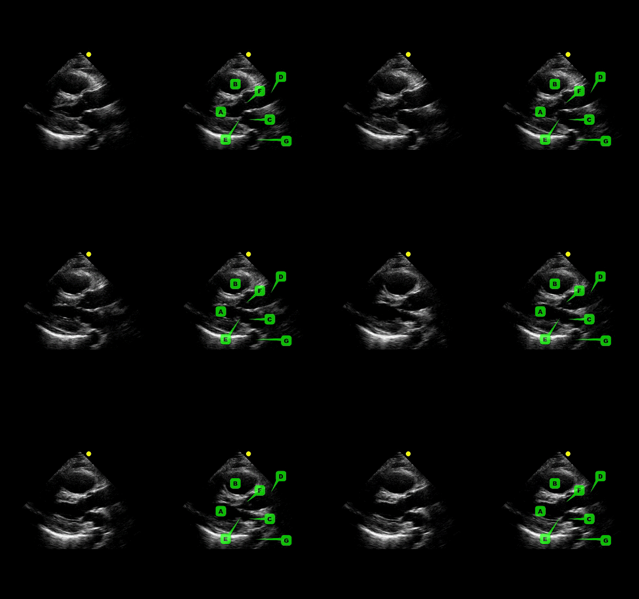
This week's visual pearl reviews the structures of the heart when being viewed in a parasternal long-axis view. What do the labels correspond to in the clip below (note: "E" and "F" are valves) and do you see any obvious abnormalities?  
 

Show Answer

The parasternal long-axis is obtained by scanning to the left (patient's left) of the sternum through the 2nd-5th intercostal space. Click here for a tutorial on the technique. 

  • A - Left Ventricle
  • B - Right Ventricular Outflow Tract (RVOT)
  • C - Left atrium
  • D - Ascending Aorta
  • E - Mitral Valve
  • F - Aortic Valve
  • G - Descending Aorta

Answer to Bonus Question: Dilation of the RVOT

  • Quick tip for evaluating the RVOT; use the left atrium as a size reference (assuming normal atrial size) for the RVOT
  • Normally, the RVOT and left atrium should be approximately equal in size


Show References

Follow me on Twitter (@criticalcarenow) or Google+ (+criticalcarenow)


Title: Simple tips for managing the critically-Ill pregnant patient

Category: Critical Care

Posted: 10/29/2013 by Haney Mallemat, MD

The pregnant patient normally has increased cardiac output and minute ventilation by the third trimester. Despite this increase, however, these patients have little cardiopulmonary reserve should they become critically-ill.

Remember the mnemonic T.O.L.D.D. for simple tips that should be done for the pregnant patient who presents critically-ill or with the potential for critical illness: 

  • Tilt: The supine-hypotension syndrome occurs after the 20th week of pregnancy as the gravid uterus compresses the IVC and aorta, reducing cardiac output by up to 30%. Placing a 30-degree right hip-wedge under the patient will relieve this obstruction.
  • Oxygen: the growing uterus pushes up on the base of the lungs reducing the functional residual capacity meaning there is less oxygen reserve and rapid oxygen desaturations. Supplemental oxygen may increase the patient's reserve.
  • Lines: The circulatory system reserve is reduced, so early and large bore venous access is important. Remember that lines should be placed above the diaphragm because the enlarging uterus compresses pelvic veins, reducing venous return to the heart.
  • Dates: Rapidly determine the gestational age of the fetus as 24 weeks is a critical date to remember (e.g., increased risk of supine-hypotension syndrome, fetal viability, etc.)
  • Delivery: Call labor and delivery early on, not only for the consultation, but also for the fetal monitoring that this service provides. 


Show References

Follow me on Twitter (@criticalcarenow) or Google+ (+criticalcarenow)


Title: What's the Diagnosis? Images by UMEM alumni Dr. Joy Kay

Category: Visual Diagnosis

Posted: 10/28/2013 by Haney Mallemat, MD (Updated: 10/28/2013)

Question

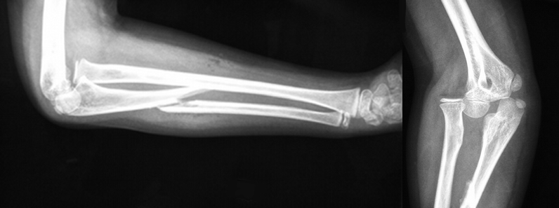
15 year-old right-hand dominant male received a direct blow to the right arm with a hockey stick. What’s the diagnosis?
Show Answer

Monteggia Fracture

  • Ulnar fracture with dislocation of proximal radio-ulnar joint; typically secondary to fall on outstretched hand (with arm in hyper-pronation) or with direct trauma to forearm as in a defensive injury (e.g., "Nightstick" injury).
  • Should not be confused with a Galeazzi fracture, a fracture of the radius with distal radio-ulnar dislocation
  • Remember the mnemonic GRUM; Galeazzi = Radius fracture / Ulnar fracture = Monteggia
  • Children may be treated with closed reduction and immobilization; adults usually require open reduction and internal fixation.
  • Radial head dislocations should be reduced within 6-8 hours because they can lead to articular damage and/or nerve injury

Show References

Follow me on Twitter (@criticalcarenow) or Google+ (+criticalcarenow)


Title: What's the Diagnosis?

Category: Visual Diagnosis

Posted: 10/21/2013 by Haney Mallemat, MD (Updated: 12/5/2023)

Question

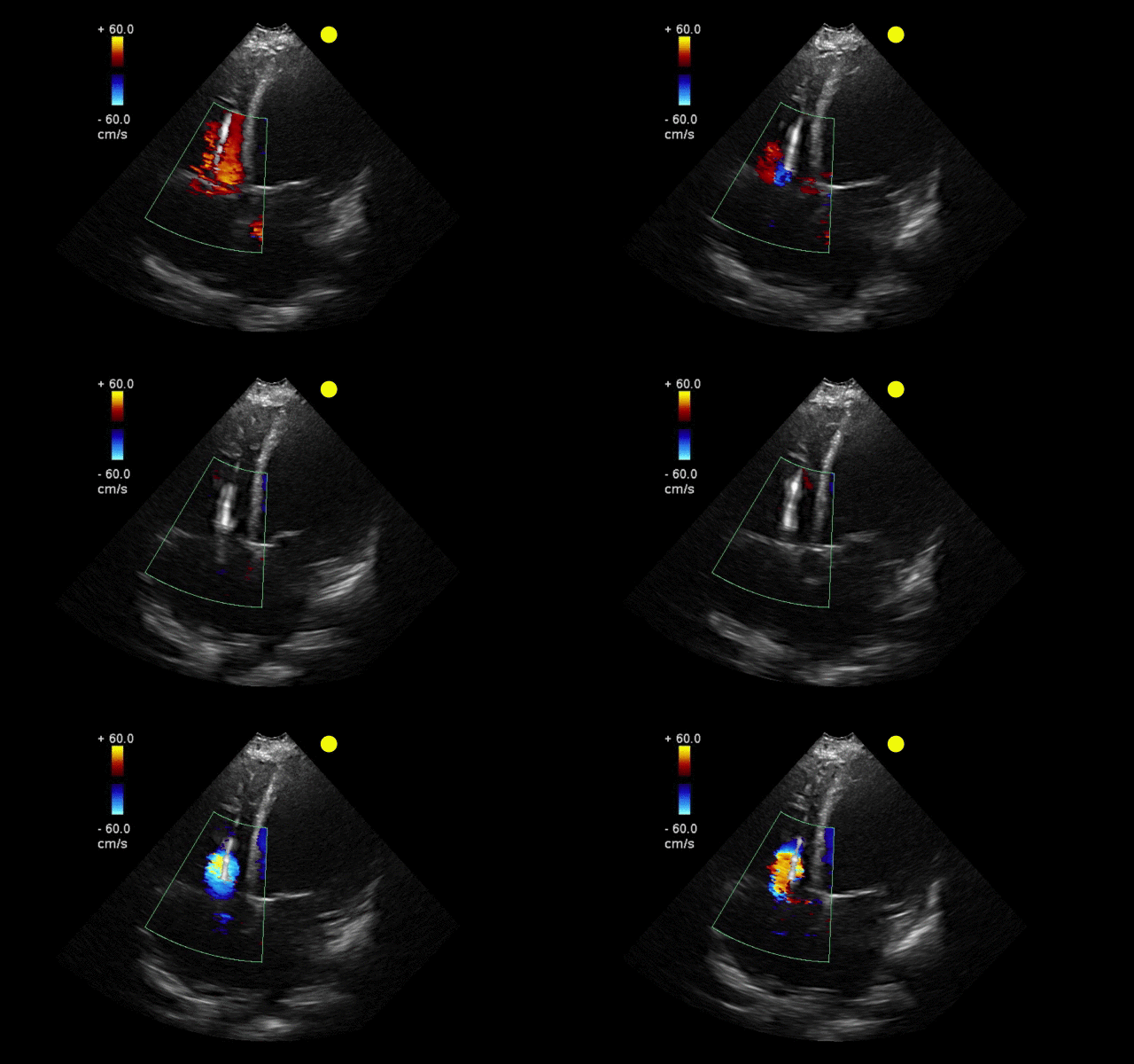
55 year-old male presents with chest pain. You take a look at his cardiac function with ultrasound and here's the patient's apical four-chamber view. What's in his right ventricle and why would it be there?
Show Answer
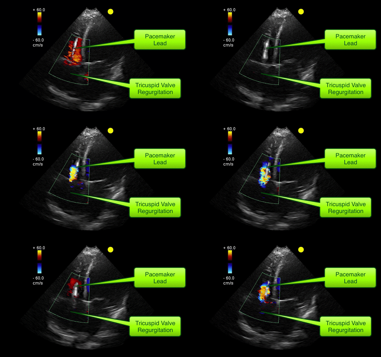
  • Automatic Implantable Cardioverter-Defibrillator (AICD); Pacemaker could also be the answer as it is difficult to distinguish on ECHO.
  • It was placed in this patient for primary prevention of VT/VF secondary to a chronically low left-ventricular ejection fraction. 

AICD

  • Placed for patients at risk for sudden cardiac death secondary to malignant arrhythmias (e.g., ventricular tachycardia / fibrillation)
  • Shown to improve mortality for patients with severe left ventricular dysfunction (EF<30%) and has been shown to improve mortality (as in this case)
  • Tricuspid regurgitation is caused by the intra-cardiac lead preventing complete closure of tricuspid valve leaflets.

Show References

Follow me on Twitter (@criticalcarenow) or Google+ (+criticalcarenow)


Title: High-Yield Pearls for the ICU Patient Bording in the ED

Category: Critical Care

Posted: 10/16/2013 by Haney Mallemat, MD

There have been so many great talks at ACEP 2013, but Dr. Michael Winters' talk "The ICU is NOT Ready for Your Patient" was chock full of great critical care pearls. Here are just a few:

  • Increased mortality for ICU patients boarding in the Emergency Department; the increase is 1.5% per each hour of delayed transfer.
  • Intubated patients should receive analgesia BEFORE sedation; fentanyl is recommended because hemodynamically stable, but you can use anything. Good analgesia will also reduce total sedative dosing
  • Use continuous capnography for the intubated patient; can detect equipment malfunction and allow titration of ventilation
  • Keep an eye out for abdominal compartment syndrome. Physical exam is not always conclusive, should obtain bladder pressures
  • Reduce the risk of ventilator-associated pneumonia by keeping endotracheal cuff pressures adequate and keeping the head of bed elevated 30-45 degrees

Show References

Follow me on Twitter (@criticalcarenow) or Google+ (+criticalcarenow)


Title: What's the Diagnosis? Case by Dr. Zachary Dezman

Category: Visual Diagnosis

Posted: 10/14/2013 by Haney Mallemat, MD

Question

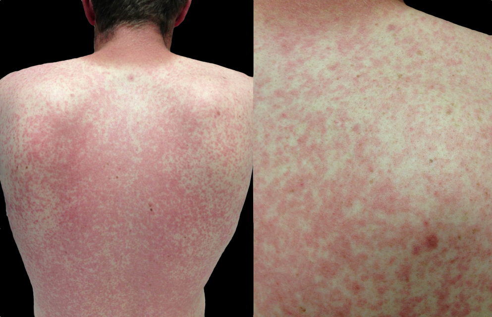
A 23 year-old male presents with the rash below. He originally presented to his primary care doctor for a sore throat and was given a prescription for a medication; this rash subsequently broke out. What's the diagnosis and which medication did he receive?
Show Answer

Rash secondary to Epstein-Barr pharyngitis treated with amoxicillin

  • Morbilliform rash occuring in up to 95% of patients given amoxicillin for EBV pharyngitis; the rash typically spreads to the trunk 
  • Other types of beta-lactams, cephalosporins, and macrolides may also cause this rash 
  • The exact mechanism is unknown but it is non-allergic; researchers have demonstrated that patients may receive amoxicilin after the rash resolves without adverse events.  
  • The rash spontaneously resolves in 1-2 weeks

Show References

Luzuriaga, K., Sullivan, J. Infectious Mononucleosis. N Engl J Med 2010; 362:1993-2000

Follow me on Twitter (@criticalcarenow) or Google+ (+criticalcarenow)


Title: What's the Diagnosis? Image by Dr. Christina Tupe

Category: Visual Diagnosis

Posted: 10/7/2013 by Haney Mallemat, MD

Question

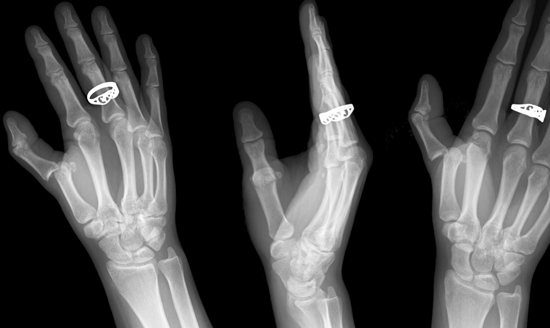
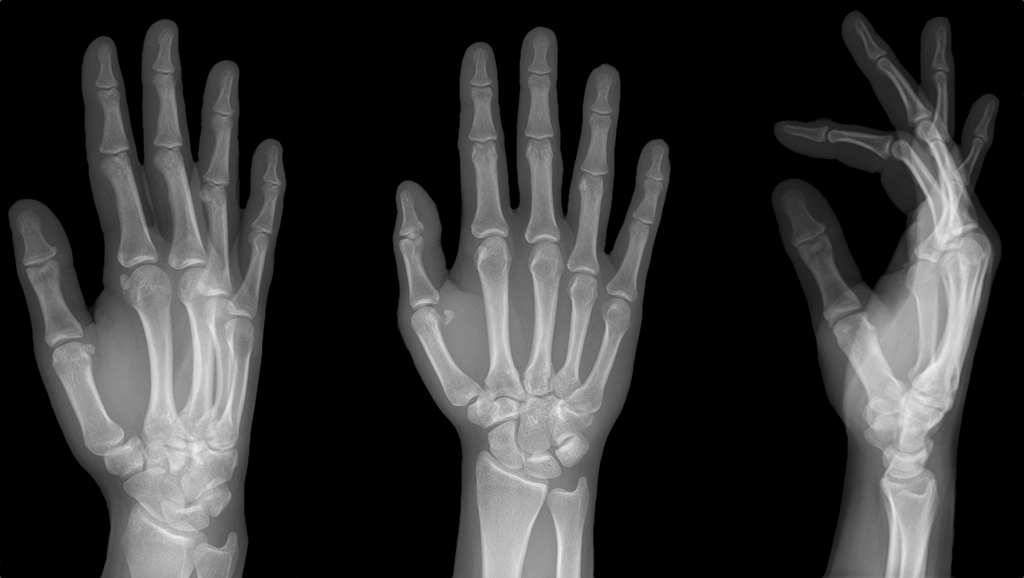
25 year-old female struck in the left hand by a football. Presents with pain, visible deformity, and the Xray below. What are the next step(s) in management?
Show Answer

Dorsal Metacarpophalangeal (MCP) Dislocation

  • MCP dislocations typically occur secondary to a hyperextension injury
  • Two categories of dislocations
    • Simple; joint spaces closely opposed and treated by closed reduction
    • Complex; volar plate is entrapped dorsal to metacarpal head with lumbricals and tendons interposed between joint surfaces; treated by operative reduction
  • To reduce a simple dislocation:
    • Provide a digital ring-block for analgesia
    • Maximally hyperextend the proximal phalanx with wrist in flexion
    • Push base of proximal phalanx distally while bending the joint into flexion
    • Test neurovascular integrity and joint-stability post-reduction by ranging the joint through a full range of motions
    • Place in thumb spica splint and refer to hand specialist

Show References

Follow me on Twitter (@criticalcarenow) or Google+ (+criticalcarenow)


Previous  |  1 |  2 |  3 |  4 |  5 |  6 |  7 |  8 |  9 |  10 |  11 |  12 |  ... |  17 |  Next
University of Maryland School of Medicine logo
  • Departments
  • Programs
  • Research Centers
  • Institutes
  • Graduate Education
  • CIBR Cores
  • Offices of the Dean
    • University of Maryland, Baltimore
    • University of Maryland Medical Center
    • Medical Alumni Association of the University of Maryland
    • Health Sciences and Human Services Library
University of Maryland School of Medicine
655 W. Baltimore Street
Baltimore, MD 21201
Contact Us

© University of Maryland School of Medicine

    • Webmaster
    • Web Accessibility
    • Site Index
    • UMB Hotline
    • Employment
    • Map