
University of Maryland School of Medicine logo
Emergency Medicine
All Departments
UMSOM Assistant
UMEM Educational Pearls
  • Home
  • Departments
  • Emergency Medicine
  • CME
  • UMEM Educational Pearls
  • CME
  • Courses and Conferences
  • Free Open Access Medical Education
  • UMEM Educational Pearls

Search

Title: What's the Diagnosis? Basic ECHO Part 1

Category: Visual Diagnosis

Posted: 11/4/2013 by Haney Mallemat, MD

Question

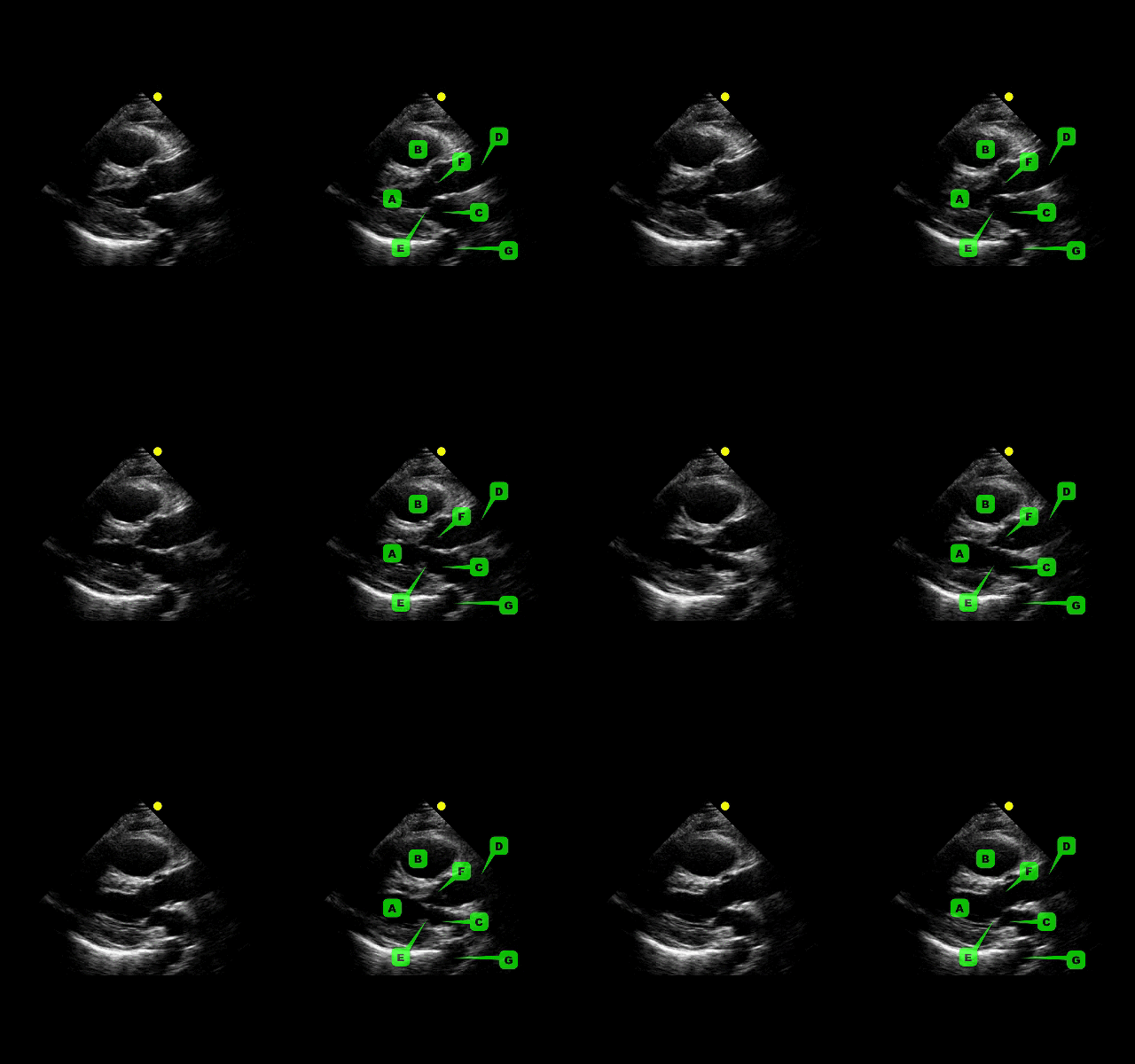
This week's visual pearl reviews the structures of the heart when being viewed in a parasternal long-axis view. What do the labels correspond to in the clip below (note: "E" and "F" are valves) and do you see any obvious abnormalities?  
 

Answer

The parasternal long-axis is obtained by scanning to the left (patient's left) of the sternum through the 2nd-5th intercostal space. Click here for a tutorial on the technique. 

  • A - Left Ventricle
  • B - Right Ventricular Outflow Tract (RVOT)
  • C - Left atrium
  • D - Ascending Aorta
  • E - Mitral Valve
  • F - Aortic Valve
  • G - Descending Aorta

Answer to Bonus Question: Dilation of the RVOT

  • Quick tip for evaluating the RVOT; use the left atrium as a size reference (assuming normal atrial size) for the RVOT
  • Normally, the RVOT and left atrium should be approximately equal in size


References

Follow me on Twitter (@criticalcarenow) or Google+ (+criticalcarenow)


University of Maryland School of Medicine logo
  • Departments
  • Programs
  • Research Centers
  • Institutes
  • Graduate Education
  • CIBR Cores
  • Offices of the Dean
    • University of Maryland, Baltimore
    • University of Maryland Medical Center
    • Medical Alumni Association of the University of Maryland
    • Health Sciences and Human Services Library
University of Maryland School of Medicine
655 W. Baltimore Street
Baltimore, MD 21201
Contact Us

© University of Maryland School of Medicine

    • Webmaster
    • Web Accessibility
    • Site Index
    • UMB Hotline
    • Employment
    • Map