
Since the first description of acute respiratory distress syndrome (ARDS), various consensus conferences (including American-European Consensus Conference (AECC) and the Berlin Conference) have produced definitions focused on adult lung injury but have limitations when applied to children.
This prompted the organization of the Pediatric Acute Lung Injury Consensus Conference (PALICC), comprised of 27 experts, representing 21 academic institutions and eight countries. The goals of the conference were 1) to define pediatric ARDS (PARDS); 2) to offer recommendations regarding therapeutic support; and 3) to identify priorities for future research in PARDS.
Although there were several recommendations from the group, some notable ones, in contrast to the Berlin definition focused on adults, include: 1) use the Oxygenation Index (or, if an arterial blood gas is not available, the Oxygenation Severity Index) rather than the P/F ratio; 2) elimination of the requirement for “bilateral” pulmonary infiltrates (may be unilateral or bilateral) 3) elimination of specific age criteria for PARDS.
Tune in next month for pearls on management for children with PARDS...
Show References
Hyperkalemia is a potentially life threatening problem which can lead to cardiac dysrhythmias and death. Drug interactions inducing hyperkalemia are extremely common such as the combination of ACE inhibitors and spironolactone or ACE inhibitors and trimehoprim sulfamethoxazole. Hyperkalemia can also occur with a single agent and is a relatively common complication of therapy with trimethoprim sulfamethoxazole. The following drugs can cause hyperkalemia:
- Ace inhibitors
- Beta blockers
- Cyclosporine
- Digitalis
- Non-steroidal Anti-inflammatory Drugs
- Pentamidine
- Potassium supplement
- Succinylcholine
- Tacrolimus
- Trimethoprim sulfamethoxazole
Show References
The blue-ringed octopus (genus Hapalochlaena) is normally found in the Great Barrier Reef and other coastal waters and tide pools around Australia and other Western Pacific islands. Though not an aggressive animal, when it does bite, such as stepped upon, it can inject tetrodotoxin along with a number of other toxic compounds.
Tetrodotoxin can cause paralysis, leading to respiratory failure and death, though the blockage of voltage-gated fast sodium channel conduction, blocking peripheral nerve conduction. Treatment is supportive, as the venom usually wears off within 4 to 10 hours.
Show References
Post-Arrest Tidal Volume Setting
- Most patients with ROSC from out-of-hospital cardiac arrest undergo endotracheal intubation and mechanical ventilation.
- Optimal management of mechanical ventilation for the post-arrest patient is currently not well defined.
- A recent retrospective cohort study sought to determine if a lower tidal volume (Vt) was associated with improved neurocognitive outcome at hospital discharge.
- Of 256 patients included in the study, investigators found:
- 38% were ventilated with Vt > 8 ml/kg predicted body weight
- Lower Vt was significantly associated with favorable neurocognitive outcome, decreased duration of mechanical ventilation, and decreased ICU length of stay
- Take Home Pearl: Pay attention to Vt in the post-arrest patient.
Show References
Elective surgeries with general anesthesia are often cancelled when the child has an upper respiratory tract infection. What are the adverse events when procedural sedation is used when the child has an upper respiratory tract infection?
Recent and current URIs were associated with an increased frequency of airway adverse events (AAE). The frequency of AAEs increased from recent URIs, to current URIs with thin secretions to current URIs with thick secretions. Adverse events not related to the airway were less likely to have a statistically significant difference between the URI and non-URI groups
AAEs for children with no URI was 6.3%. Children with URI with thick/green secretions had AAEs in 22.2% of cases. Children with URIs did NOT have a significant increase in the risk of apnea or need for emergent airway intervention. The rates of AAEs, however, still remains low regardless of URI status.
Show Additional Information
Show References
During the past several years, several new classes of diabetic medications were introduced for clinical use, including SGLT2 inhibitors (canagliflozin, dapagliflozin and empagliflozin).
SGLT2 inhibitors prevent reabsorption of glucose in the proximal convoluted tubules in the kidney and does not alter insulin release.
A recent retrospective study (n=88) of 13 poison center data from January 2013 to December 2016 showed
- 91% of the patients were asymptomatic.
- 7% developed minor symptoms (tachycardia, nausea/vomiting, abdominal pain, & confusion)
- 2% developed moderate symptoms (metabolic acidosis, hypertension [166/101], & hypokalemia)
- Hypoglycemia was not reported.
49 patients were evaluated in a health care facility (HCF) with 18 admissions. Referral to HCF was more common in pediatric patients. This was likely due to unfamiliarity with a new mediation and lack of toxicity data.
Other case reports have shown higher incidence of DKA with the therapeutic use of SGLT2 vs. other classes of DM medications.
Bottom line:
Limit data is available regarding the toxicologic profile of SGLT2 inhibitors.
Based upon this small retrospective study, hypoglycemia may not occur and majority of the patient experience minimal symptoms.
Show References
IV vs. Non-IV Benzodiazepines for Cessation of Seizures
- A meta-analysis by Alshehri et al. included 11 studies with a total of 1633 patients, comparing IV vs. non-IV benzodiazepines from any route (buccal, intranasal, intramuscular) for seizure cessation in status epilepticus.
- They found that non-IV benzodiazepine is more effective than IV benzodiazepine in patients presenting without IV access.
- The largest and highest quality study included in the meta-analysis was the RAMPART study, which was also the only study to include adults.
- When considering pediatric studies only, there is no difference between IV vs. non-IV benzodiazepine in seizure cessation for status epilepticus.
Show Additional Information
Show References
Negative-pressure pulmonary edema (NPPE) is a well-documented entity that occurs after a patient makes strong inspiratory effort against a blocked airway. The negative pressure causes hydrostatic edema that can be life-threatening if not recognized, but if treated quickly and appropriately, usually resolves after 24-48 hours. These patients may have any type of airway obstruction, whether due to edema secondary to infection or allergy, laryngospasm, or traumatic disruption of the airway, such as in attempted hangings.
Management:
1. Alleviate or bypass the airway obstruction.
· Usually via intubation; may require a surgical airway
· If obstruction in an intubated patient is due to biting on tube or dyssynchrony, add bite-block (if not already in place), sedation, and even paralysis if needed.
2. Provide positive pressure ventilation and oxygen supplementation.
3. Use low tidal volume ventilation.
4. In severe hypoxemia without shock, add a diuretic agent and consider additional measures such as proning and even ECMO if the hypoxemia is refractory to standard therapy.
Show Additional Information
Show References
Viscosupplementation
Hyaluronic acid (HA) is a high-molecular weight polysaccharide
A major component of synovial fluid and of cartilage
Major role of HA is as a lubricant, shock absorption, antinociceptive effect
Used in veterinary medicine for decades
Multiple brands exist with differences based on the molecular weight and how they are produced
Use supported by the Cochrane database (2007, 2014) for knee OA
Post injection strength gains are due to pain relief
May have a role for those who cannot receive steroid injections
Inject in similar manner to intra articular steroids
Caution in those with known allergy to poultry /eggs
Risks: Local reaction (likely from preservative), injection site pain, infection, bleeding.
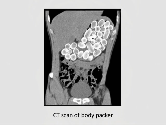
Radiographs studies can be valuable in poisoning diagnosis, management, and prognosis. Radiographic imaging should be utilized for the following toxins:
- Iron (gastrointestinal)
- Mercury (gastrointestinal, intravenous or subcutaneous)
- Lead (bullets intraarticular, gastrointestinal foreign bodies, lead lines)
- Zinc phosphide (gastrointestinal)
Container toxins - Body packers
- Drug packets and vials
Sustained Released preparations
- Potassium Chloride
Show Additional Information
Show References
There have been reports of “intoxication” or adverse effects among first responders and law enforcement due to exposure to a “powder” suspected to be fentanyl or its analog.
This has led to a significant concern among first responders and law enforcement when investigating or handling “powder” at the scene of overdose or drug enforcement related raids. (http://www.foxnews.com/health/2017/08/15/police-department-gets-hazmat-like-protective-gear-for-overdose-calls.html)
American College of Medical Toxicology and American Association of Clinical Toxicology recently published a position statement to help clarify the potential health risk associated with exposure to fentanyl and its analogs.
- Opioid toxicity is unlikely from incidental dermal exposure.
- Nitrile gloves provide sufficient protection against dermal exposure.
- N95 respirator provide sufficient protection against aerosolize fentanyl/opioids.
- Naloxone should be administered for patients with objective signs of opioid toxicity - hypoventilation and CNS depression – not for vague or subjective symptoms.
With the recent destruction by Hurricane Harvey and the impending impact of Hurricane Irma, it is important to recognize the historical death toll from natural disasters. While the list can vary, here is a top ten list from the library of the National Oceanic and Atmospheric Administration:
| Rank | Event | Location | Date | Death Toll (Estimate) |
| 1 | 1931 Yellow River flood | Yellow River, China | Summer 1931 | 850,000-4,000,000 |
| 2 | 1887 Yellow River flood | Yellow River, China | September-October 1887 | 900,000-2,000,000 |
| 3 | 1970 Bhola cyclone | Ganges Delta, East Pakistan | November 13, 1970 | 500,000- 1,000,000 |
| 4 | 1201 Earthquake | Eastern Mediterranean | 1201 | 1,000,000 |
| 5 | 1938 Yellow River flood | Yellow River, China | June 9th, 1938 | 500,000 - 900,000 |
| 6 | Shaanxi Earthquake | Shaanxi Province, China | January 23, 1556 | 830,000 |
| 7 | 2004 Indian Ocean earthquake/tsunami | Indian Ocean | December 26, 2004 | 225,000-275,000 |
| 8 | 1881 Haiphong Cyclone | Haiphong, Vietnam | 1881 | 300,000 |
| 9 | 1642 Kaifeng Flood | Kaifeng, Henan Province, China | 1642 | 300,000 |
| 10 | Tangshan Earthquake | Tangshan, China | July 28, 1976 | 242,000* |
* Official Government figure. Estimated death toll as high as 655,000.
Show References
Alpha-blockers (tamsulosin, alfuzosin, doxazosin, and terazosin) are antagonists of alpha1A-adrenoreceptors, which results in the relaxation of ureteral smooth muscle. Current evidence suggests alpha-blockers may be useful when ureteral stones are 5-10 mm; however, there is no evidence to support the use of alpha-blockers with stones <5 mm. Patients with ureteral stones >10 mm were excluded from studies utilizing these medications.
The size of most ureteral stones will be unknown due to the lack of need for imaging able to measure stone size. Given that the median ureteral stone size is <5 mm, most patients will not benefit from the use of an alpha-blocker.
Also, keep in mind that the data for adverse events with alpha-blockers used for ureteral stones is limited and that these medications have a risk of hypotension.
Show Additional Information
Show References
Background:
There is an increased incidence of venous thromboembolic events (VTE) in pediatrics due to improved diagnosis and survival of children with VTE.
The mortality rate is estimated at 2%.
The most common etiologies vary by age - Central venous catheters in neonates and infants, and inherited thrombophilia in children and adolescents.
Learning Points:
-
With neonates and infants, carefully assess medical history from neonatal period. Umbilical lines? PICC? Broviac? History of these is likely to be the cause.
-
In children and adolescents, unprovoked VTE is most likely due to inherited thrombophilia, and can be DVT, PE, Portal venous thrombus, etc.
-
Antithrombin deficiency: The first discovered inherited thrombophilia. The result is a lack of inhibition of coagulation factors – IIa, IXa, Xa, XIIa.
-
Protein C or/and S deficiency: The result is lack of inhibition of activated Factor V.
-
Factor V Leiden: Most common inherited thrombophilic defect. Resultant activated Factor V is resistant to normal Protein C and S activity.
-
Prothrombin Mutation: Second most common inherited thrombophilia. The result is increased levels of prothrombin, which increases the half-life of factor Va.
-
-
Initial treatment of clinically significant VTE can start with enoxaparin (1-1.5 mg/kg q12-24h, while checking Anti-Xa levels 4 hours after administration for therapeutic dosing.)
Pearl: Testing for thrombophilia is not always appropriate when diagnosing pediatric patients with their first VTE, but in children and adolescents with first diagnosed, unprovoked VTE, it is worthwhile to send off the initial hypercoaguability work up as this can affect the duration of treatment and need for testing or evaluation. Enoxaparin is a recommended medication to start therapeutic treatment of VTE, even in pediatric patients.
Show References
- Most poisonous substance known to man
- LD50 oral dose 1 mcg/kg
- Heat labile single polypeptide chain undergoes proteolytic clevage irreverisibly binds and blocks cholinergic transmission causing a deadly neuroparalytic syndrome
- Rx: Botulin antitoxin (equine derived against Clostriduim botulinum A,B,E)
- Highly toxic, restricted availability is rapidly absorbed and metabolized to methylmercury crosses CNS
- LD50 of 50 mcg/kg means a dose as little as 0.1ml can result in severe poisoning
- Death of Darmouth inorganic chemist Karen Wetterhahn who spilled a few drops on back of her latex gloved hand, quickly permeated, and absorbed causing severe neurotoxocity and death 10 months later
- Rx: Chelation
VX ("venomous agent X")
- Organophosphate nerve agent has been used as chemical weapon
- Colorless, odorless, low volatility, and high lipophilicity
- LD50 of 0.04mg/kg (10 mg). Death can occur within 15 minutes after absorption
- Blocks acetylcholinesterase enzyme causing excess accumulation of acetylcholine at the neurojunction and cholinergic poisoning
- Rx: Decontamination, Atropine, 2-PAM
- 100 fresh and salt water varieties (pufferlike fish/blue ringed octopus, frogs)
- Heat stable, water soluble found in fish skin, liver, ovaries,intestine, and muscle
- 25 mg (0.000881 oz) expected to be lethal to a 75 kg person
- Neurotoxicity by inhibition of Na-K pump and blockade neuromuscular transmission
- Rx: Supportive measures
LD50 expresses the dose at which 50% of exposed population will die as a result of exposure.
Show References
Background: Sedation and analgesia are key components for mechanically ventilated patients. While significant data exists regarding how to manage sedation and analgesia in the ICU setting, very little data exists on management in the ED.
Data: A prospective, single-center, observational study of mechanically-ventilated adult patients used linear regression to identify ED sedation practices and outcomes, with a focus on sedation characteristics using the Richmond Agitation-Sedation Scale (RASS).
Findings:
- 15% of intubated patients had no sedation or analgesia ordered
- 64% of intubated patients were documented as deeply-sedated (RASS -3 to -5)
- Deep sedation was not only associated with more ventilator days, but also increased mortality, with an adjusted OR of 0.77 (95% CI 0.54-0.94) favoring patients with lighter sedation.
Bottom line: Avoid early deep sedation in your intubated patients as this may be directly associated with increased mortality. Instead, a goal RASS of 0 to -2 should be appropriate for most non-paralyzed, mechanically-ventilated ED patients, extrapoloating from ICU guidelines.
Show References
There is tremendous interest in identifying factors that may influence outcome from sports related concussion.
The strongest predictor of slower recovery is the severity of symptoms in the 1-2 days post injury
-Fewer Sx's in this time period predict a quicker recovery
Pre injury history of mental health problems, depression or migraine headaches predict a longer recovery course
Teenagers might be more vulnerable to having persistent symptoms with greater risk for girls than boys
Having a prior concussion is a risk for having a future concussion
The large majority of injured athletes recover from a clinical perspective within the first month of injury many within the first 10 days
Show References
Anaphylaxis is a life threatening emergency with mortality of up to 2% [1]. Early recognition is imperative and administration of timely Epinephrine is the single most important intervention [2]. While providers may be hesitant to administer epinephrine in older patients due to fear of precipitating adverse cardiovascular events, they may also hesitate in younger patients due to fear of overdose.
Iimmediate administration with any dose available is recommended because:
- the risks of untreated anaphylaxis are greater than the risk of over-treating with epinephrine.
- 20% of Anaphylaxis patients require a second dose of Epinephrine [3].
- The recommended IM dose of 0.01mg /kg was determined arbitrarily.
- The vast majority of epinephrine overdoses are via IV injection at doses 100 - 1000 fold the recommended IV dose [4]
Bottom line:
There are no absolute contraindications (including age) for epinephrine in patients with anaphylaxis. Give the initial dose IM into the anterolateral thigh.
Show References
Full cohort analysis idarucizumab for dabigatran associated bleeding was recently published in NEJM.
This study evaluated the laboratory correction of elevated ecarin clotting time or diluted thrombin time induced by dabigatran and time to either cessation of bleeding (Group A: patients with GI bleeding, traumatic bleeding, or ICH) or time to surgery (Group B: patients requiring surgical intervention within 8 hours).
Findings
Group A (n=301): Median time to the cessation of bleeding was 2.5 hours in 134 patients.
HOWEVER:
- Bleeding cessation could not be determined in 67 patients
- Cessation of bleeding could not be assess in 98 patients with ICH
- Bleeding stopped spontaneously in 2 patients.
Group B (n=202): Median time to intended surgery after infusion of idarucizumab was 1.6 hours.
- Normal hemostasis in 184 patients (93.4%), mildly abnormal in 10, and moderately abnormal in 3.
- Many received PRBC and other blood products during surgery
Laboratory markers:
100% reversal of abnormal ecarin clotting time or diluted thrombin time within 4 hours after the administration
Mortality
- 5 Day: Group A: 6.3% vs. Group B: 12.6%
- 30 Day: Group A: 13.5% vs. Group B: 12.6%
- 90 Day: Group A: 18.8% vs. Group B: 18.9%
Conclusion
Authors concluded thate idaurcizumab is an "effective" reversal agent for dabigatran.
Overall, the findings are more promising compared to the interim analysis that was published in 2015.