
Acetaminophen (APAP) overdose is the leading cause of liver failure in the U.S. and Europe. Large APAP ingestion can result in hepatotoxicity despite the early initiation of n-acetylcysteine (NAC).
A recently published study from Austrialia investigated the effect of activate charcoal and increasing the NAC dose for large APAP overdose patients (3rd bag: 100 to 200 mg/kg over 16 hours) during first 21 hours of NAC therapy
acetaminophen ratio (first APAP level taken between 4 to 16 hour post ingestion / APAP level on the Rumack nomogram line at that time point) was determined to compare APAP levels at different time points among study sample
e.g.
first APAP level at 4 hour post ingestion = 400
APAP level on the Rumack APAP nomogram at 4 hour post ingestion = 150
APAP ratio = 400/150 = 2.67
Findings:
- Activated charcoal (AC): if given within 4 hours, AC significantly decreased the APAP ratio (OR: 1.4 vs. 2.2)
- Increased dose of NAC during the first 21 hour significantly decreased the risk of hepatotoxicity (OR: 0.27; 95% CI: 0.08 - 0.94).
Conclusion:
- Administration of AC in patients with history of large APAP overdose (>=40 gm) within 4 hour of ingestion can still be beneficial.
- Increasing NAC dosing (3rd bag in first 21 hour thearpy) may decrease the risk of hepatotoxicity.
Note: Any increase in NAC dosing from the standard 21 hour therapy should be performed after consulting your regional poison center.
Show References
- Reversible cerebral vasoconstriction syndrome (RCVS) is the second most common cause of thunderclap headache after aneurysmal subarachnoid hemorrhage (SAH) and the most common cause of recurrent thunderclap headaches.
- Up to 40% of patients with RCVS have a history of migraine.
- It is associated with selective serotonin reuptake inhibitors (SSRIs), triptans, cocaine, marijuana, tacrolimus, oral contraceptives, as well as the peripartum period.
- Symptoms are often triggered by emotional stress, sexual activity, showering, straining, and physical exertion.
- Although the vasoconstriction is reversible, it can cause intracranial hemorrhage, seizures, stroke, and coma.
- Diagnosis is by history, cerebral angiography and exclusion of aneurysmal SAH.
Bottom Line: Consider RCVS in the differential of thunderclap headache and in patients who present with worse than usual migraine headache.
Show References
Septic Cardiomyopathy
- Cardiac dysfunction is common in patients with sepsis.
- Though mulitiple definitions exist, sepsis cardiomyopathy (SCM) is generally defined as an "acute syndrome of cardiac dysfunction that is unrelated to ischemia in patients with sepsis".
- Depending on the study, the incidence of SCM ranges anwywhere from 7% to 70%.
- Risk factors for SCM include:
- Male
- Younger age
- High lactate at admission
- History of heart failure
- The best approach to treating patients with SCM is to maximize your treatment of sepsis.
- Dobutamine is no longer routinely recommended for SCM based solely on measurements of ScvO2.
Show References
Patients with severe asthma exacerbations that are unresponsive to inhaled beta-agonists may require the use of epinephrine to control their symptoms. When patients get to this point what route of administration should be used for the administration of epinephrine?
The most recent asthma guidelines (published in 2007) recommend the use of SubQ epinephrine 0.3-0.5 mg every 20 minutes for 3 doses. Drug references typically list SubQ or IM epinephrine 0.01 mg/kg (~0.3-0.5 mg) every 20 minutes as appropriate routes of administration. There is currently no data demonstrating that one route of administration is better than the other in patients with asthma; however, in other disease states, such as anaphylaxis, IM epinephrine is preferred due to the more rapid and reliable absorption over SubQ administration.
Auto-injectors that administer IM epinephrine 0.3 mg are available. These auto-injectors may decrease the risk of medications error; however, they can be expensive. SubQ administration requires the use of a syringe and a vial/ampule of 1 mg/mL epinephrine.
Bottom Line: Either SubQ or IM epinephrine administration is appropriate for patients with severe asthma exacerbations. The preferred method at a given institution will be dictated by historical practice, risk of medication dosing errors, and drug cost.
Show Additional Information
Show References
A recent study was undertaken to validate the 4A's Test for the assessment of delirium in the elderly, with particular focus on inpatient geriatric patients; it revealed that the tool had high sensitivity in detecting delirium, particularly in those with dementia or language barriers, in whom this diagnosis can often be difficult to make. Further studies would be useful in a similar demographic of emergency department geriatric patients to confirm that this straightforward test is generalizable to the emergency department geriatric patient population.
Show Additional Information
Show References
In addition to suspicion of NAT with traumatic brain injury and burns, remember these other high risk injuries and features:
- Duodenal injuries in children <4 y/o
- Frena injuries in non-ambulating children
- Proximal and midshaft humeral fractures > supracondylar fractures
- Any bruising on the trunk, ears, neck, or with larger size or pattern
- Delay in seeking care, inconsistent history, mechanism inconsistent with developmental age, and blame of a sibling or other child inflicting harm are all historical features also high risk.
Show Additional Information
Show References
Although the data is limited, current published rates of in-hospital, non-operating room peri-intubation cardiac arrest (PICA) range from 2 to 6%.1,2,3
Several risk factors associated with PICA have been identified and include:
- Preintubation hemodynamic instability (shock index ≥ 1 or systolic blood pressure < 90mmHg)1,2,3
- Elevated Body Mass Index (and increased risk with every 10kg body weight)1
- Use of succinylcholine as paralytic3
- Intubation occurring within one hour of nursing shift change3
Other common findings:
- Most PICA occurs within 10 minutes of rapid sequence induction (RSI)1,2
- PEA is the initial recorded rhythm 80-100% of the time.1,2,3
- Even if ROSC obtained, PICA is associated with higher rates of in-hospital mortality compared to patients requiring emergent intubation who do not experience cardiac arrest.1,2,3
Bottom Line: Endotracheal intubation is one of the riskiest procedures we regularly perform as emergency physicians.
- Resuscitate hypotensive patients prior to or concomitantly with RSI and/or have a vasopressor at the ready in patients with higher risk of cardiovascular collapse.
- Consider use of vecuronium or rocuronium, rather than succinylcholine, in patients who require a paralytic for intubation but are at higher risk of hyperkalemia or have an unknown history.
Show References
Children less than 8 years, and especially infants, are more susceptible to upper cervical spine injury. Moreover, validated decision rules for suspected cervical spine injury imaging have not been proven to be as sensitive or specific for children less than 8 years of age.
The pediatric cervical spine has greater elasticity of the ligamentous structures, while the cartilaginous structures are less calcified. An infant's neck musculature is underdeveloped, with a disproportionally large head. These factors increase the risk of cervical spine injury, and can make it difficult to properly place protective cervical collars in infants while assessing them for injury.
In very young children, consider placing padding under the shoulders to prevent abnormal flexion that can occur with placement of a cervical collar, and consider having a lower threshold to image if mechanism history or exam is concerning.
Children are not little adults! Clinicians must acknowledge the anatomic differences, varying age-related ability to cooperate with examination, pediatric specific injury mechanisms, and decreased reliability of validated decision rules for imaging in children, especially when younger than 8 years old.
Show References
Show References
The peak age for croup is 6 months to 3 years. The cornerstone of treatment is corticosteroids, traditionally dexamethasone. With oral administration, the peak onset is 1-2 hours. Steroids shorten the duration of symptoms, reduce the need for nebulized epinephrine and decrease the need for intubation.
Racemic epinephrine has been used for moderate to severe croup and can show an improvement in patient symptoms for up to 120 minutes. There is little evidence to suggest how long to observe the patient for recurrence of symptoms after racemic epinephrine was given. Previous studies have suggested both 2 and 4 hour observation.
299 patients were included in this study. 136 patients were observed for 3.1 to 4 hours. In the 3.1 to 4 hour group, 21 (7%) failed treatment, 19 of those patients required admission and 2 returned within 24 hours. No patients who were discharged home after 4 hours returned to the emergency department within 24 hours.
Bottom Line: Consider a 4 hour period of observation after giving racemic epinephrine in order to decrease bounce backs.
Show References
- The DAWN trial was a multicenter, randomized, open-label study comparing endovascular thrombectomy plus standard medical care with standard medical care alone for patients with:
- Acute stroke symptoms
- Last known well 6 to 24 hours earlier
- Evidence of intracranial ICA or proximal MCA occlusion
- Mismatch between clinical deficit and infarct volume on CTA or MRA
- The study found that patients receiving thrombectomy plus standard medical care had improved functional independence at 90 days as defined by modified Rankin Scale (mRS) of 0, 1, or 2 (49% vs 13%).
- The trial was stopped early based on prespecified interim analysis intended with the adaptive trial design.
- While the two treatment groups were similar, with median NIHSS score of 17, they had small infarct volumes and short time from symptom observation (4.8 vs 5.6 hours) compared to time of patient's last known well (12.2 vs 13.3 hours).
- 88% of the patients had unwitnessed stroke onset (including wake-up strokes), thus it is possible that these patients had actual ischemia times closer to 6 hours, thereby reproducing similar results as prior thrombectomy trials.
Bottom Line: The use of neuroimaging to identify an ischemic penumbra that may benefit from thrombectomy may be considered even for patients with time of last known well beyond 6 hours.
Show Additional Information
Show References
Sedating The Critically Ill Patient
- Sedating critically ill ED patients can be challenging.
- Excessive sedation is associated with a prolonged duration of mechanical ventilation, ICU LOS, and may increase mortality.
- Important pearls to consider when managing these patients include:
- Prioritize pain management first - may reduce the need for sedative medications
- When possible, target a calm and interactive patient shortly after intubation - consider adding a atypical antipyschotic with propofol or dexmedetomodine
- Use a validated tool (i.e., RASS) to dose opioids and sedative medications
- Avoid continuous infusions of benzodiazepines
Show References
CC: 12yo boy presents with pain to base of 5th metatarsal
Osteochondrosis overuse syndromes associated with development of secondary ossification or apophyseal centers
Iselin disease – Osteochondrosis of 5th MT base
Lateral 5th foot pain with weight bearing and activity in early adolescence
Child may limp or walk on inner part of foot
Adolescents: Girls >10, Boys >12
Commonly seen in soccer, basketball, gymnastics and dance
Exam: Tenderness to palpation at proximal 5th MT at peroneal brevis insertion
Area may show edema and redness
Pain with foot inversion and resisted eversion and dorsiflexion
XR: May be normal or show enlargement or fragmentation of epiphysis
Obliquely oriented small bony fleck at 5th MT base. Parallel to long axis of 5th MT. Best seen on oblique view. Unlike fractures which tend to be horizontally oriented.
Treatment: Immobilize for comfort if severe (walking boot) or simple activity modification if mild. Ice and calf muscle stretching.
http://https://images.radiopaedia.org/images/2343487/d3478d2024c845ba0f2fffffd7d51c_big_gallery.jpg

Loperamide (Imodium) is a common inexpensive over-the counter antidiarrheal agent. It acts peripherally at the mu opioid receptor to slow gastrointestinal motility and has no CNS effects at therapeutic doses due to it's low bioavailability and limited abillity to cross the blood brain barrier dependent on glycoprotein transport. In the past few years, reports of loperamide abuse causing serious cardio toxicity began to appear in the literature. Abused at daily doses of 25-200 mg to get high or and to treat symptoms of withdrawal. (therapeutic dose: 2-4 mg with a maximun of 8mg for OTC and 16mg for prescription). Loperamide has been called the "poor man's methadone".
At large doses, loperamide effects the cardiac sodium, potassium and calcium channels which prolongs the QRS complex and can lead to ventricular arrhythmias, hypotension, and death. Clinical features includes:
- QT prolongation
- QRS widening
- Ventricular arrythmias
- Hypotension
- Syncope
- CNS depression
Take Home Point:
Consider loperamide as a possible cause of unexplained cardiac events including QT interval prolongation, QRS widening, Torsades de Pointes, ventricular arrhythmias, syncope, and cardiac arrest. Intravenouse sodium bicarbonate should be utilized to overcome blockade and may temporize cardiotoxic events. Supportive measures necessary may include defibrillation, magnesium, lidocaine, isoproternol, pacing, and extracorporeal life support.
Show Additional Information
Show References
Community-associated Clostridium difficile infection (CA-CDI) represents 41% of all CDI cases annually. The association of specific outpatient exposures was assessed in a case control study by Guh, et al. They reviewed the CDC’s active surveillance reporting from 10 states through the Emerging Infections Program (Maryland participates).
Cases: ≥18, + C. difficile stool specimen collected as an outpatient or within 3 days of hospitalization, with no overnight stay in a health care facility in the prior 12 weeks, and no prior CDI diagnosis
Controls: matched 1:1 for age and sex within the same surveillance catchment area as the case patient on the date of the collection specimen. Exclusion criteria: prior diagnosis of CDI, diarrheal illness, overnight stay in health care facility in the prior 12 weeks
Data Collection: telephone interview, standardized questionnaire or comorbidities, medication use, outpatient health care visits, household and dietary exposures in the prior 12 weeks
Results: 452 participants (226 pairs), over 50% were ≥ 60 years of age, 70.4% female, and 29% were hospitalized within 7 days of diagnosis, no patients developed toxic megacolon or required colectomy.
Cases had more health care exposures, including the emergency department (11.2% vs 1.4% p <0.0001), urgent care (9.9% vs 1.8%, p=0.0003). In addition, cases also reported higher antibiotic exposures (62.2% vs 10.3%, p<0.0001) with statistically significant higher exposure to cephalosporins, clindamycin, fluoroquinolones, metronidazole, and beta-lactam and/or beta-lactamase inhibitor combination. The most common antibiotic indications were ear or sinus infections, URI, SSTI, dental procedure, and UTI. No differences were found in household or dietary exposures.
Take-home point: This study highlighted the risk for CA-CDI infection for patients presenting to an ED and reiterates that exposures to fluoroquinolones, cephalosporins, beta-lactam and/or beta-lactamase inhibitor combinations, and clindamycin significantly increases the risk of CA-CDI infection. Reducing unnecessary outpatient antibiotic prescribing may prevent further CA-CDI. 36% of case patients did not have any antibiotic or outpatient health care exposure; therefore, additional risk factors may exist.
Show References
Severe acute respiratory failure among patients with PCP pneumonia, especially among those newly diagnosed with AIDS, remains a disease of high morbidity and mortality. Among those requiring mechanical ventilator support, the mortality rate has been reported between 50-70%.
According to ELSO guidelines, pharmacologic immunosuppression (specifically neurtrophil <400/mL) is a relative contraindication. Furthermore, a status predicting poor outcome despite ECMO should also be considered a relative contraindication.
That said, there are several case reports now of successful use of ECMO in AIDS patients, particularly those suffering with PCP pneumonia.
In a case report and literature review published in BMJ in Aug 2017, 11 cases of ECMO (including 1 VA) in AIDS patients were described.
- 7 survived to hospital discharge (including 1 VA)
- 2 survived to decannulation, but ultimately died in hospital
- 2 died on ECMO
- Length of ECMO runs in survivors varied between 4 days (VA) to 31 days
Bottom Line: HIV/AIDS is not an absolute contraindication to VV ECMO therapy in ARDS and may be particularly useful in the treatment of severe PCP pneumonia. Initiation of ECMO in this patient population should be considered on an individual case by case basis.
Show References
Background:
We are all familiar with the Surviving Sepsis Campaign recommendation (& CMS core measure) for an initial 30ml/kg bolus of IV crystalloid within the first 3 hours for our patients with septic shock. There is minimal data, however, on how much IVF we should be giving our patients with BMIs ≥30.
A recent study in obese patients with septic shock retrospectively stratified the total fluids administered at 3 hours into 3 different weight categories, to categorize patients as having received 30mL per kg of ___ body weight, whether actual (ABW), adjusted (AjdBW), or ideal (IBW**).
AdjBW = (ABW – IBW) *40% + IBW
They found:
- Most patients received fluids based on actual body weight, BUT
- Patients at highest BMIs received ABW fluids less often
- 30ml/kg dosing according to adjusted body weight was associated with improved mortality compared to IVF per actual or ideal body weight.
Bottom Line:
- If the 30ml/kg IVF bolus seems clinically appropriate for your obese patient, consider administering according to Adjusted Body Weight first.
- As always, reevaluate your septic shock patients frequently to determine if additional fluids are necessary, and go to vasopressors early if they are not fluid responsive.
**IBW calculated using Devine’s formula for men and women:
- Males: IBW = 50 + 2.3*(# inches over 5 feet)
- Females: IBW = 45.5 + 2.3*(# inches over 5 feet)
Show References
Asymptomatic bacteriuria is common and increases with age, with an incidence of up to 50% in women over the age of 70. Asymptomatic bacteriuria does not carry an associated high morbidity or mortality if left untreated; it is usually transient and resolves spontaneously. In order to decrease polypharmacy and possible drug interactions in our elderly patients, they should only be diagnosed with and treated for a UTI if they have laboratory evidence of a UTI (bacteriuria and pyuria) and have two of the following:
· Fever
· Worsened urinary urgency or frequency
· Acute dysuria
· Suprapubic tenderness
· Costovertebral angle tenderness
Show References
Question
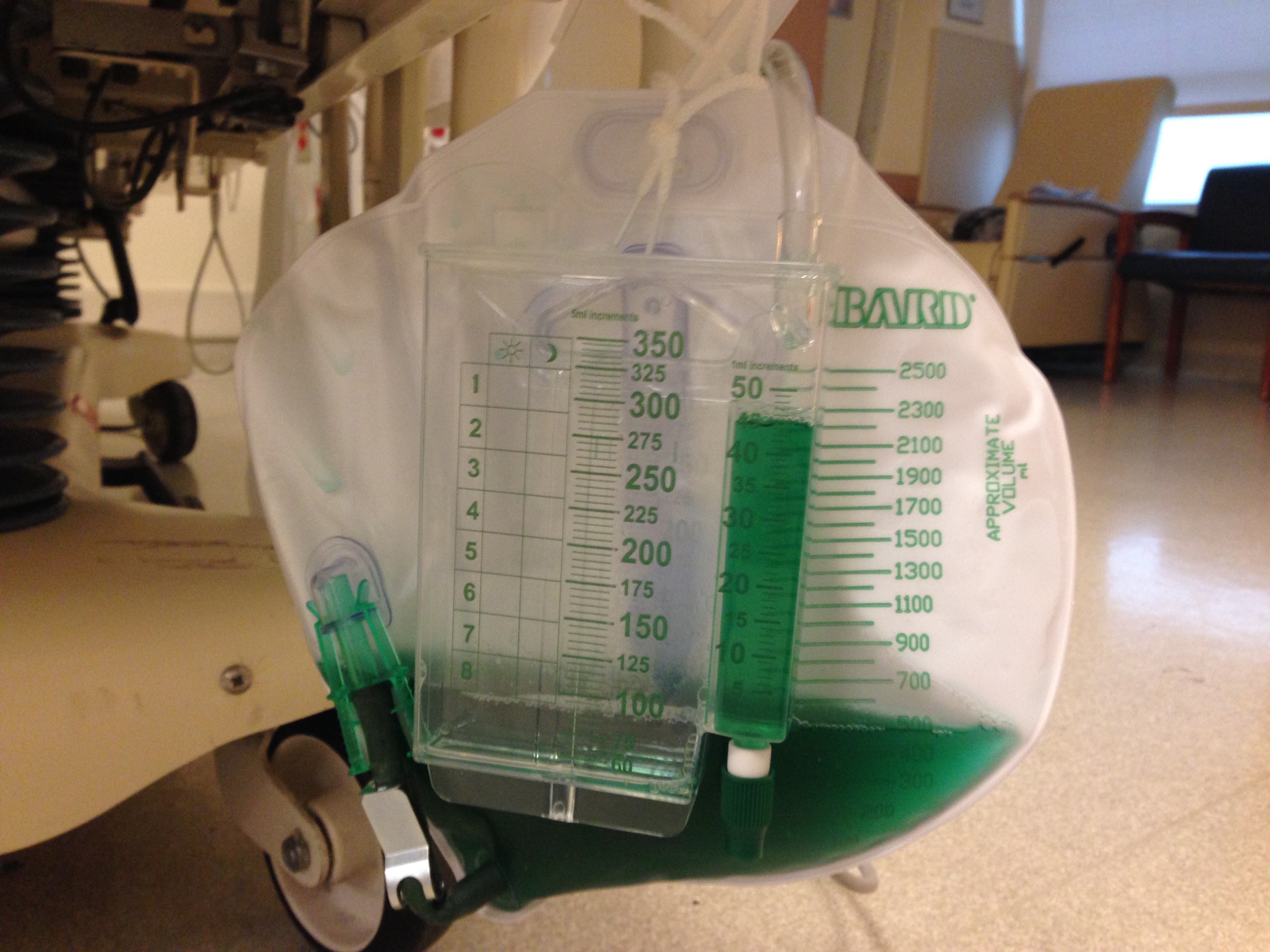
Different chemical, food or pharmaceutical agent exposure can change the color of the urine.
What could cause this patient's urine to turn green?