
- Trigeminal neuralgia is diagnosed by:
- Pain in 1 or more divisions of the trigeminal nerve
- Paroxysms of pain that are sudden, intense, usually few seconds in duration
- Pain triggered by innocuous stimuli in the trigeminal nerve territory (91-99% patients)
- 24-49% of patients experience continuous or long-lasting pain
- Exam may reveal forceful contraction of the facial muscles during a paroxysm (tic convulsif)
- Causes include:
- Intracranial vascular compression of the trigeminal nerve root (most common)
- Multiple sclerosis, cerebellopontine angle tumor
- Idiopathic (10% of cases)
- Carbamazepine and oxcarbazepine are first-line treatments
- They may be poorly tolerated due to side effects including dizziness, diplopia, ataxia, CNS depression, and hyponatremia
- They also have limited efficacy on continuous pain
- Acute exacerbations may warrant admission for hydration, acute pain control, and titration of antiepileptic drugs
- Botulinum toxin A was recently added as a treatment option
Bottom Line: New onset trigeminal neuralgia needs workup for its etiology. Carbamazepine and oxcarbazepine can be effective for symptom management though continuous or long-lasting pain exacerbations are difficult to treat.
Show References
Ketamine In the Critically Ill Patient
- Ketamine has become a popular agent in the ED for both RSI and procedural sedation.
- Given the sedative, analgesic, dissociative, antidepressant, and anti-inflammatory properties, ketamine has also been used in a number of other critical illness conditions including:
- Acute pain management
- Status asthmaticus
- Alcohol withdrawal syndrome
- Status epilepticus
- Acute agitated delirium
- The authors of a recent review in Critical Care Medicine found that the evidence supporting the use of ketamine in the critically ill is most robust for adjunctive analgesia in the intubated patient. Surprisingly, the data is very limited to support the use of ketamine in these other conditions.
- Pearl: ketamine does have a myocardial depressant effect, which can be unmasked in states of catecholamine depletion and result in hypotension and bradycadia.
Show References
Diagnostic performance of Ultrasonography for detection of pediatric elbow fracture
Elbow fractures account for approximately 15% of pediatric fractures
Fat pads are traditionally taught as a marker of fracture
In a cadaveric study:
Elbow effusions of 1-3 mL could be identified with ultrasound
Elbow effusions of 5-10 mL could be identified with plain film
Pediatric plain films are sometimes challenging to obtain and interpret compared to adults
-More likely to be uncooperative in obtaining required views
-Non-ossified epiphyses
Ultrasound may be used to detect
-Cortical disruption and irregularity
-Growth plate widening
-Hematoma interposed between fracture fragments
-Elevated posterior fat pad
Absence of elbow fracture was indicated by
-Lack of cortical disruption
-Absence of posterior fat pad sign
Meta-analysis of 10 articles totaling 519 patients using ultrasonography to detect elbow fractures
Sensitivity 96%
Specificity 89%
False negative rate 3.7%
For comparison, plain radiographs
Interpreted by peds EM physicians (87.5% sensitive and 100% specific)
Interpreted by radiology (96% sensitive, 100% specific)
Consider using ultrasound as a noninvasive, radiation-free modality for accurate diagnosis of pediatric elbow fractures.
Show References
Definition: Congenital anomaly where the hymen is completely obstructing the vaginal opening
Demographic: Incidence 0.05-0.1% of females
History: Most are asymptomatic and diagnosed on physical exam or incidentally when there is lack of menarche. Symptoms in adolescents can include: Abdominal pain (50%), urinary retention (20%), abnormal menstruation (14%), dysuria (10%), frequency, renal failure, UTI and back pain.
Physical exam: bulging, blueish hymenal membrane
Complications: Late detection can lead to infections, fertility problems, endometriosis, hydronephrosis, and rarely renal failure
ED treatment: If abdominal pain is significant or there is urinary obstruction, a urinary foley can be placed. GYN should be consulted.
Definitive treatment: Hymenectomy, hymenotomy, carbon dioxide laser treatments or foley insertion through the hymen (done by a specialist).
Show References
Baclofen is a presynaptic GABA-B receptor agonist in the spinal cord that is primarily used for muscle spasms/spasticity. In large overdose, baclofen can produce CNS depression, respiratory depression, bradycardia/hypotension, hypothermia, seizure and coma.
Baclofen is primarily eliminated by the kidney. In patients with end-stage kidney disease/acute kidney failure, hemodialysis (HD) has been used to enhance baclofen clearance. However, it is unclear if there is a benefit of using HD in patients with normal kidney function.
In a recently published case report, HD was implemented in an attempt to shorten the anticipated prolonged ICU course.
Case: 14 year old (51 kg) woman ingested 60 tablets of baclofen (20 mg tablets)
Her symptoms were:
- Coma/CNS depression
- Tonic-clonic seizure
- Transient hypotension (95/47 mmHg – resolved with IV fluids)
- Flaccid extremities
- Initially intubated for airway protection --> no spontaneous breathing on mech. ventilation.
Baclofen level: 882 ng/mL (therapeutic range: 80 – 400 ng/mL)
Baclofen clearance from hemodialysis vs. urine
- 24 hour urine output: 2810 mL --> total baclofen urinary elimination: 42 mg
- 3 hours of HD #1: 3.05 mg removed. Total of 3 HD session performed.
Patient’s mental status improved on hospital day 6 and was extubated. She was discharged to psychiatry on hospital day 14.
Conclusion:
- Although this is a single case report, it appears that hemodialysis does not remove baclofen effectively.
Show References
Another week, another COVID-19 study...
On August 12th, the Metcovid study was e-published ahead of print in Clinical Infectious Diseases. This was another study looking at steroids in COVID-19 pneumonia, this time performed in Brazil. Metcovid was a parallel, double-blind, randomized, placebo-controlled phase IIb clinical trial which enrolled 416 patients at a single academic center for the evaluation of methylprednisolone (MP; 0.5 mg/kg BID x 5 days) vs placebo. As with all COVID studies, Metcovid has some significant limitations, and some equivocal findings. However, Metcovid was largely in line with RECOVERY and other trials looking at steroids in COVID-19, which lends it some face validity. Metcovid found no significant difference in the primary outcome (mortality at day 28), but did find a difference in mortality in patients over 60 years old (a post-hoc analysis). Metcovid was probably underpowered (sample size was based on a 50% reduction in mortality), and did have a very small trend towards reduced mortality in the MP group (37.1% vs 38.2%, p=0.629).
Bottom Line:
- Steroids (methylprednisolone 0.5 mg/kg BID x 5 days in this case) may have some mild benefit in severe cases of COVID-19 pneumonia, especially in patients who are elderly or have more aggressive inflammatory responses (as measured by CRP here).
- Steroids in COVID-19 may be associated with some theoretical downsides like reduced viral clearance, but are relatively safe. Main side effect is the well known hyperglycemia induced by corticosteroids.
- When using steroids in COVID pneumonia, both to stick with the evidence and for theoretical pharmacologic reasons, it may make sense to use dexamethasone or methylprednisolone, as these medications have a higher glucocorticoid:mineralocorticoid activity ratio. It is hypothesized that using high mineralocorticoid steroids (like cortisone or hydrocortisone) may lead to increased water retention, which could be deterimental in ARDS. This is purely theoretical.
- There was a signal towards harm in younger and less sick patients in this study, and it probably remains prudent to reserve steroids for older, sicker COVID-19 pneumonia patients, similar to the RECOVERY trial.
Show References
What physical exam findings are associated with nitrous oxide abuse?
Nitrous oxide (NO) inhalation abuse, also called “whip-its” or “whippets”, inactivates vitamin B12 and create a vitamin B12 deficiency state. Chronic abuse of nitrous oxide can result in neurologic deficits/findings affecting the posterior/dorsal column of the spinal cord.
Physical exam findings:
- Truncal ataxia
- Decreased vibratory sensation and proprioception in lower extremities
- Impaired coordination and rapid alternative movements
- Lhermitte’s sign: paresthesia of the upper and lower extremities associated with flexion of the head/neck.
- Rossolimo’s sign: exaggerated flexion of the toes when the tips of the toes are percussed
- Spontaneous cerebrospinal fluid (CSF) rhinorrhea is rare and usually related to a combination of thinning of the bone and dura and fluctuating intracranial pressure.
- CSF rhinorrhea can be associated with idiopathic intracranial hypertension, skull base tumors, neurosurgical and otolaryngology procedures, and trauma.
- Trauma with fracture of the anterior skull base is the most common cause of CSF rhinorrhea.
- CT and MRI can identify bony defects, whereas cisternography can diagnose occult leaks.
- Fluid containing CSF is classically described to make a “halo” or “double-ring” pattern on gauze or linen.
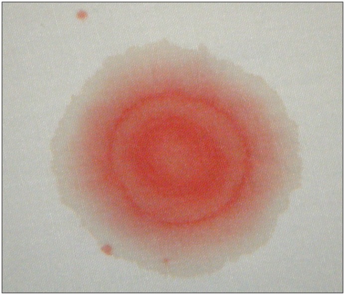
- However, this sign is not specific to CSF, as mixtures of blood with saline, tears, or rhinorrhea can also produce halos.
- Beta-2 transferrin is a protein found almost exclusively in CSF* thus can be used to diagnose CSF rhinorrhea.
Bottom Line: Beta-2 transferrin is more accurate than the halo sign to identify CSF containing fluid.
Show References
While chest X ray (CXR) is routinely obtained in the setting of traumatic injury, ultrasound (US) is a fast and reliable way to evaluate for life-threatening traumatic injuries requiring emergent intervention, and is supported by the Eastern Association for the Surgery of Trauma (EAST) guidelines. A recent Cochrane Review compared the test characteristics of chest US vs CXR for detection of traumatic pneumothorax when using Chest CT or thoracostomy as the gold standard.
- Primary end point: sensitivity and specificity for pneumothorax
- US performed by nonradiologists.
- 9 studies, 1271 patients, 410 of which had a pneumothorax
- Summary sensitivity: US 0.91 (95% CI 0.85-0.94), ranging from 0.82-0.98 in the included studies, vs. CXR 0.47 (95% CI 0.31- 0.63) ranging from 0.09 to 0.75
- Summary specificity: US 0.99 (95% CI 0.97-1.00, ranging from 0.96-1.00 vs. CXR 1.00 (95% CI 0.97- 1.00), ranging from 0.98 to 1.00
There possible weaknesses of this study, including blinding in the original studies, and several studies may or may not have been at risk for bias as their risk of bias was ‘unclear’. However, the results were consistent across the studies analyzed and remained similar after sensitivity analysis.
Several anatomical as well as patient care issues may confound US findings for pneumothorax such as the presence of bleb, prior thoracic surgery or pathology, as well as main stem intubation.
Show References
Pronator Teres Syndrome
A compressive neuropathy of the median nerve in the region of the elbow
The median nerve passes through the cubital fossa and passes between the superficial and deep heads of the pronator teres muscle.
Rare compared to other compressive neuropathies such as carpal tunnel syndrome.
More common in women and in fifth decade of life
May be seen with weight lifters, arm wrestlers, rowers, tennis, archery, professional cyclists, dentists, fiddlers, pianists, harpists
Also associated with well-developed forearm muscles
History:
Forearm pain – unlike carpal tunnel
Paresthesias in median distribution
No night symptoms – unlike carpal tunnel
Physical exam:
Sensory loss in medial nerve distribution.
Involves the thenar eminence!
Unlike carpal tunnel syndrome which doesn’t involve sensory loss in thenar eminence.
Pain may be made worse with resisted forearm pronation
Compression/Tinel’s sign over pronator mass reproduces symptoms
Treatment:
Splinting which limits pronation and NSAIDs
Steroid injection
Surgical nerve decompression is non operative treatment fails after greater than 6 months (rare)
Prior to this study, a Cochrane review and meta-analysis of TXA for upper GI bleeds with 7 trials (1654 patients), showed a large reduction in mortality with TXA (RR 0.61, 95% CI 0.42-0.98, p=0.01)
Design:
-Randomized, international, multicentre, placebo-controlled trial at 164 hospitals in 15 countries Juy 2013-2019
->16/18 years old with upper or lower GI bleeding
-1 g TXA IV over 10 minutes followed by maintenance dose 3 g TXA over 24 hours
Results:
-Main outcome death due to bleeding within 5 days
-4% (222/5994) died in TXA group vs 4% (226/5981) placebo risk ratio RR 0.99, 95% CI 0.82-1.18
-Arterial thromboembolic events MI/CVA similar in both groups (0.7% vs 0.8%)
-Venous thromboembolic events PE/DVT higher in TXA group (0.8% vs 0.4%)
Pitfalls:
-Initially calculated all cause mortality until realization that over half deaths were due to non-bleeding causes, changed to death related to bleeding, allowing study appropriate power to detect difference
-Majority of patients had UGIB/variceal bleeding due to liver disease, over 75% deaths in those with liver disease
-Only 16% patients randomized in <3 hours, most >8 hours (CRASH-2 trial found benefit TXA in trauma patients only <3 hrs to administration)
Takeaway:
-TXA should not be used in the management of GI bleeds
-Increased venous thromboembolic events associated with TXA administration for GI bleeds
Show Additional Information
Show References
The liver performs an essential role in the metabolism and clearance of many drugs. Liver damage due to cirrhosis can decrease first-pass metabolism of oral medications and increase free-drug concentrations of protein-bound medications due to decreased albumin production. In the absence of cirrhosis, patients with chronic hepatitis or hepatic cancer may only have a small decrease in drug clearance. Hepatic dose adjustments are not as prevalent or readily available as renal dose adjustments, which can create difficulty in finding the balance between pain relief and adverse effects.
The most common medications used for pain control in the emergency department are acetaminophen, NSAIDs, and opioids.
Acetaminophen
It is sometimes misconceived that acetaminophen should never be used in patients with cirrhosis due to the common knowledge that acetaminophen overdoses can cause hepatotoxicity. Alcoholics may have an increased risk of hepatotoxicity due to induction of CYP2E1 and decreased glutathione stores. However, acetaminophen is safe in patients with cirrhosis when used at appropriate doses. Limit the total daily dose of acetaminophen to 2 g daily in patients with cirrhosis and avoid acetaminophen in patients that are actively drinking. Also, educate patients that over-the-counter (OTC) and prescription medications may contain acetaminophen.
NSAIDs
In patients with cirrhosis, NSAIDs have increased bioavailability due to decreased CYP metabolism and decreased protein binding. In addition, prostaglandin inhibition can precipitate renal failure and sodium retention, worsening ascites and increasing the risk of hepatorenal syndrome, and increase the risk of gastrointestinal bleeding. Thrombocytopenia from NSAID use can further increase the risk of bleeding. Thus, avoid NSAID use in patients with cirrhosis. Topical NSAIDs can be considered.
Opioids
Opioid metabolism is altered in patients with cirrhosis and can contribute to complications with cirrhosis, such as precipitating encephalopathy. Generally, the bioavailability is increased and half-life is extended; thus, lower doses of immediate-release (IR) formulations at extended dosing intervals should be utilized. Common opioids for acute pain control in the emergency department are fentanyl, hydrocodone/oxycodone, hydromorphone, and morphine.
- Fentanyl: Largely unaffected by cirrhosis. High potency so utilize only in appropriate clinical situations.
- Hydrocodone/Oxycodone: Metabolized by CYP to active metabolites (hydromorphone/oxymorphone). Due to decreased CYP metabolism, analgesia may be less potent and clearance decreased. Also, be aware that some formulations are combined with acetaminophen.
- Hydromorphone: Metabolized by glucuronidation to inactive metabolite. Metabolism and clearance less affected by cirrhosis.
- Morphine: Increased bioavailability and concentration due to decreased first-pass metabolism. Decreased clearance and longer half-life. Avoid use in renal impairment and hepatorenal syndrome due to risk of neurotoxic metabolite accumulation.
- Tramadol, codeine, meperidine, methadone, and buprenorphine not recommended for acute pain control in the emergency department.
Take Home Points
| Drug/Class | Preferred Agent | Considerations |
| Acetaminophen | Max daily dose 2 g/day | Avoid if actively drinking. Be cautious if patient also taking OTC or combination products. |
| NSAIDs | None; Avoid | Topical NSAIDs may be considered. |
| Opioids | Hydromorphone, Fentanyl | Start with IR products at lower doses and extended intervals. |
Show References
Patient
-
Single Center, double-blinded, randomized trial.
-
Patients with cancer and septic shock
Intervention
-
4% albumin + lactate ringer bolus in 10 minutes
-
180 patients
Comparison:
-
Lactate ringer bolus in 10 minutes
-
180 patients
Outcome:
-
Primary: Any mortality within 7 days of randomization.
-
Secondary: Mortality within 28 days, renal replacement therapy (RRT)
Results:
-
7-day mortality: 46 (25%) for LR + albumin vs. 40 (22%) for LR only
-
28-day mortality: 96 (53%) for LR + albumin vs. 83 (46%) for LR only
-
RRT: 16 (9%) for LR + albumin vs. 12 (7) for LR only
Conclusion:
Adding albumin for early resuscitation to crystalloids did not improve mortality in cancer patients with septic shock.
Show References
There is no well validated clinical decision rule similar to NEXUS or the Canadian Cervical Spine rule in children for clearing the cervical spine. Clinical clearance versus imaging first is a complicated decision. Certain risk factors may predispose children to injury and should be taken into account when deciding about clinical clearance versus imaging (XR).
High Risk Criteria for Cervical Spine Injury in Pediatrics
| Mechanism
| High risk MVC Intrusion > 12 inches at the occupant site Intrusion > 18 inches at any site Partial or complete ejection Death in the same passenger compartment Vehicle telemetry consistent with high speed Fall > 10 feet Nonaccidental trauma Diving injury |
| History
| Down’s Syndrome 22.q11.2 deletion Klippel-Fiel syndrome |
| Physical Exam
| Altered mental status Intoxication Hypotension Focal neurological exam Neck pain Torticollis |
Show References
Tianeptine is an antidepressant with mu-opioid receptor agonism. It is available in several European countries for therapeutic use, but not available in the US.
There has been an increase in tianeptine exposure in the US since August 2019. Recently a retrospective observation study was done to characterize the clinical features associated with tianeptine exposure.
Result
- 48 cases of tianeptine exposure were identified from January 1, 2015 to March 15, 2020 from a single poison center
- 37 cases (77%) occurred from May 2019 to March 2020.
| Intoxication (n=11) | Withdrawal (n=27) |
| Symptoms · Lethargy: 7 (63%) · Agitation: 3 (27%) · Tachycardia: 3 (27%) · GI distress: 2 (18%) · Myoclonic/hallucination: 2 (18) | Symptoms · Anxiety: 12 (44%) · GI distress: 3 (33%) · Hypertension: 8 (30%) · Agitation: 8 (30%) · Tachycardia: 7 (26%) |
| Treatment · Naloxone: 3 (27%) · Benzodiazepines: 2 (18%) · Antipsychotics: 2 (18%) · Antimuscarinic: 1 (9%)
| Treatment · Benzodiazepine: 10 (37%) · Opioids: 6 (22%) · Alpha-2-agonist: 5 (19%) · Antipsychotics: 5 (19%) · Antimuscarinic: 5 (19%) |
| Disposition · ICU: 6 (55%) · Non-ICU: 2 (18%) · Discharged home: 2 (18%)
| Disposition · ICU: 4 (15%) · Non-ICU: 7 (26%) · Psych: 1 (4%) · Discharged home: 10 (37%) |
Conclusion
- Tianeptine exposure is increasing in the US .
- Intoxication frequently results in lethargy and/or agitation.
Show References
Design
-Two-center prospective observational study with 157 patients admitted to the ICU for pneumonia and being treated with HFNC
-ROX (Respiratory rate-OXygenation) index = ratio of SpO2/FIO2 to RR
Results:
-ROX index ≥4.88 at 12 hours after HFNC onset with a sensitivity of 70.1%, a specificity of 72.4%, PPV of 89.4%, NPV of 42%, LR+ of 2.54, and LR- of 0.41 in predicting treatment failure
Validation study: Roca, 2019
-results similar, but ROX index ≥4.88 at 12 hour with LR+ of only 1.82
-also found that a ROX index of <3.85 at 12 hours had a sensitivity of 23.5%, specificity of 98.4%, PPV of 88.9, NPV 69.9, LR+ of 14.47, and LR- 0.78
Pitfalls:
-decision to intubate was not made based on ROX index
-criteria for intubation was also part of the ROX index
-NIV was not part of their treatment algorithm
-created and validated prior to current COVID-19 pandemic
Takeaways:
- The ROX index can be a tool to help predict whether a patient with pneumonia on HFNC may need mechanical ventilation or higher level of care
- May be most helpful with patients with pneumonia on HFNC boarding in the ED
- At 12 hours of HFNC, ROX index of >4.88 suggests patient likely to succeed with HFNC vs. <3.85 which suggests likely need for mechanical ventilation
Show References
The RECOVERY (Randomized Evaluation of COVid-19 thERapY) investigators recently published a non-peer reviewed article on their findings utilizing dexamethasone to treat patients with COVID-19.
Rx: Dexamethasone 6mg daily* x 10 days (PO or IV) *or steroid equivalent
- 2104 in the dexamethasone group vs 4321 in the “usual care” group
- Did not exclude children or pregnant/breastfeeding mothers
- Follow-up at 28 days, hospital discharge, or death
Primary outcome: All-cause mortality at 28-days
Secondary outcomes:
- Major arrhythmia
- Time to discharge from hospital
- Duration of mechanical ventilation
- Need for renal replacement therapy
- In patients not ventilated at enrollment, need for intubation/ECMO & death
Results:
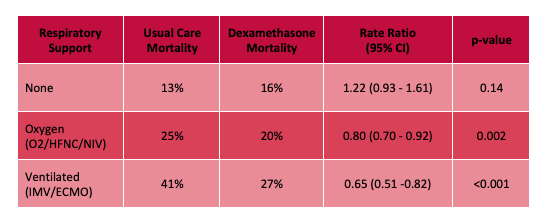
- Decrease in overall mortality at 28-days with 3% absolute risk reduction.
- NNT of 25 in patients requiring O2, HFNC, or NIV
- NNT of 8 in patients requiring invasive mechanical ventilation
- More mortality benefit seen the higher the respiratory support required, with no benefit and apparent trend towards increased mortality in the group not requiring any respiratory support at all.
- When stratified by symptoms < or > 7 days, mortality benefit only seen in the >7 days group (which was more of the ventilated patients).
- Less progression to intubation, shorter hospital duration, greater likelihood of hospital discharge.
Limitations:
- Not yet peer-reviewed, haven't seen all the data, additional analyses could be helpful in determining if treatment effect is real
- Unblinded study
- 7% of control group received dexamethasone
Bottom Line: Strongly consider admininstering dexamethasone to your patients with known COVID-19 who require respiratory support, and look for the peer-reviewed publication from the RECOVERY Trial investigators.
Show References
Treatment for carpal tunnel syndrome (CTS)
The management of CTS depends of the severity of the disease
If symptoms or on the mild to moderate range, a trial of conservative treatment is encouraged.
Possible therapeutic approaches can include splinting in wrist neutral position. Some even extend to keep the CMP joints extended. Extreme flexion and extension can increase pressure within the carpal tunnel. Usually for nighttime use only. May be used during day based on work and activity demands.
Has been shown to improve electrophysiologic findings after 12 weeks of use in moderate CTS.
Formal hand physical therapy (by an experienced therapist) may also be of some benefit including carpal bone mobilization, ultrasound and nerve glide exercises.
There is small evidence for the benefit of prednisone (20mg/d) as it has been shown to be more effective than placebo with improvements lasting an average of 8 weeks.
There is no benefit to NSAIDs or diuretics.
There is poor evidence for therapeutic ultrasound and acupuncture.
While more invasive than the above modalities, steroid injections may decrease inflammation and pressure in the carpal tunnel. Patients randomized to steroid injection may do better than those randomized to nighttime splinting.
Early referral in those with positive electrodiagnostic findings is encouraged as they do best with earlier surgical release and have better recovery.
If however the patient has severe, progressive or persistent symptoms or there is known evidence of nerve injury on diagnostic testing, referral for surgical decompression is warranted.
Question
What is the name of the toxin found in this seed/bean and its mechanism of toxicity?
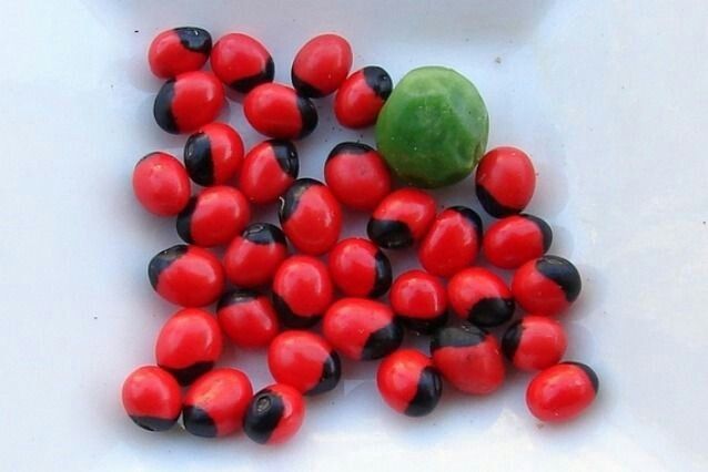
Show Answer
Show References
Attachments
Idiopathic intracranial hypertension (IIH) is a vision-threatening illness with significant morbidity and needs to be considered as a possible headache diagnosis in the ED. Most often, this occurs in women of childbearing age with a BMI >30, but atypical varieties exist.
Symptoms: Headache (90%), visual disturbance, pulsatile tinnitus, horizotal diplopia.
Signs: Papilledema, 6th cranial nerve (abducens) palsy.
Evaluation: Neuroimaging including CTV or MRV to identify alternate cause including cerebral venous outflow obstruction, lumbar puncture with opening pressure >30 cmH2O (25-30 cmH2O is gray zone), blood work per clinical presentation, CSF analysis.
Treatment: No clear consensus, but typically acetazolamide. Severe or refractory symptoms may require surgical intervention such as optic nerve sheath fenestration, VP shunt, venous sinus stenting.