
17-18% of cases of syncope are attributable to arrhythmias
The greatest predictors of arrhythmias as the cause of syncope are:
a. Abnormal ECG (odds ratio 8.1)
b. History of CHF (odds ratio 5.3)
c. Age older than 65 (odds ratio 5.4)
[Sarasin, et al. Academic Emergency Medicine 2003]
Show References
Supraspinatus: “Empty can” test. Have the patient abduct the shoulders to 90 degrees in forward flexion with the thumbs pointing downward. The patient attempts to lift the arms against the examiner’s resistance.
http://bjsportmed.com/content/42/8/628/F2.large.jpg

Infraspinatus and teres minor: These muscles are responsible for external rotation of the shoulder. Have the patient flex both elbows to 90 degrees while the examiner provides resistance against external rotation.
http://www.physio-pedia.com/images/4/4b/Infraspinatus_test.jpg
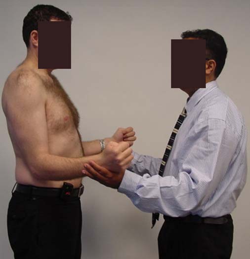
Subscapularis: “Lift-off” test. The patient rests the dorsum of the hand on the lower back (palm out) and then attempts to move the arm and hand off the back. Patients with tears may be unable to complete test due to pain.
http://www.aafp.org/afp/2008/0215/afp20080215p453-f4.jpg

Show References
As RSV season approaches, remember these key points in managing bronchiolitis:
- Diagnosis is clinical - labs and XRays will not help you, unless you want to rule out a specific alternate diagnosis. It's all about the H&P.
- Supportive care, including bulb suction of secretions, placing the child in a position of comfort, and possibly providing humidified air, is the mainstay of treatment.
- Ribavirin, corticosteroids, and antibiotics are not indicated. Don't use them.
- Bronchodilators have no benefit in bronchiolitis alone, and non-response to bronchodilators supports the diagnosis of bronchiolitis. If a trial does work, know what you are treating - some children with bronchiolitis may have an underlying component of reactive airway disease, and should be treated accordingly.
- Before disposition be sure that the child can tolerate PO. A fussy, tachypneic child may require admission for IV hydration if they are unable to tolerate feeds - recall that infants are obligate nose breathers.
- Finally, beware the RSV bronchiolitis bounceback - the peak incidence of respiratory failure in RSV bronchiolitis is after 3-4 days of illness, when most children should be improving.
Show References
In the setting of acute cyanide poisoning, it is virtually impossible to obtain a timely cyanide level to help assess toxicity. However, there are two diagnostic tests that can help confirm your diagnosis.
- Anion gap metabolic acidosis with elevated lactate
- Narrowing of the venous-arterial PO2 gradient
Remember cyanide halts cellular respiration meaning the cells cannot utilize oxygen. Therefore, the venous PO2 should be about the same as the arterial PO2. The cells then switch to anaerobic metabolism, thereby producing lactate.
How to Perform a Median Nerve Block
- The most common emergency department indication for performing median nerve blocks is to anesthetize its hand distribution (i.e. volar surface of hand) for pain control and/or to perform procedures such as laceration repair and dislocation reductions.
- The median nerve is located at the proximal flexor crease of the wrist, between the palmaris longus (PL) and flexor carpi radialis (FCR) tendons. The FCR lies radial to the PL tendon.
- Use a 25 or 27 gauge needle, inserted to a depth of 1 cm, to inject 3-5 mL of plain lidocaine proximal to the distal wrist flexor crease, just ulnar to the PL tendon.
- If the PL tendon is absent, as is the case in 25% of people, direct the needle in line with the ring finger.
- If distal paresthesias result, withdraw and reposition the needle as this suggests that the median nerve was directly struck, which should be avoided.
Show References
Pulmonary Contusion and Ventilator Management
- Pulmonary contusion is the most common injury in blunt thoracic trauma.
- Patients with pulmonary contusion often present with hypoxia, hypercarbia and increased work of breathing.
- Importantly, patients with pulmonary contusion have a low cardiopulmonary reserve. Maintain a low threshold for initiating mechanical ventilation is these patients.
- When starting mechanical ventilation, think about the following:
- Patients are at high risk for developing ARDS
- Most centers use a low tidal volume ventilatory strategy
- Higher levels of PEEP may be necessary to recruit collapsed alveoli
- High frequency oscillatory ventilation (HFOV) and airway pressure release ventilation (APRV) are modes of ventilation that are gaining in popularity for ventilating patients with pulmonary contusions.
Show References
Acute use of cocaine increases risk of acute MI due to tachydysrhythmias, vasospasm, and increased platelet aggregation. There is a 24-fold increased risk of MI in the first hour after use of cocaine. 6% of patients presenting with cocaine-chest pain rule in for acute MI.
[Weber, Acad Emerg Med 2000]
Show References
Hydrofluoric acid is a weak acid used primarily in industrial applications for glass etching and metal cleaning/plating. It is contained in home rust removers. Although technically a weak acid, it is very dangerous and burns can be subtle in appearance while having severe consequences.

Wilkes G. Hydrofluoric Acid Burns. Jan 28, 2010.
http://emedicine.medscape.com/article/773304-overview
- 2 mechanisms that cause tissue damage*
- corrosive burn from the free hydrogen ions
- chemical burn from tissue penetration of the fluoride ions
- Clinical features*
- Cutaneous burns - absent findings to white-blue appearance
- Pulmonary edema
- Hypocalcemia, hyperkalemia, hypomagnesemia
- Treatment*
- Decontaminate by irrigation with copious amounts of water.
- With any evidence of hypocalcemia, immediately administer 10% calcium gluconate IV.
- Cutaneous burns:
- Apply 2.5% calcium gluconate gel to the affected area. If the proprietary gel is not available, constitute by dissolving 10% calcium gluconate solution in 3 times the volume of a water-soluble lubricant (eg, KY gel). For burns to the fingers, retain gel in a latex glove.
- If pain persists for more than 30 minutes after application of calcium gluconate gel, further treatment is required. Subcutaneous infiltration of calcium gluconate is recommended at a dose of 0.5 mL of a 5% solution per square centimeter of surface burn extending 0.5 cm beyond the margin of involved tissue (10% calcium gluconate solution can be irritating to the tissue).
- Do not use the chloride salt because it is an irritant and may cause tissue damage.
*Extracted from emedicine article.
Show References
Radiologic evaluation of the elbow (Part 2)
Helpful clues in the evaluation of elbow trauma:
- The Anterior humeral line and the Radiocapitellar line
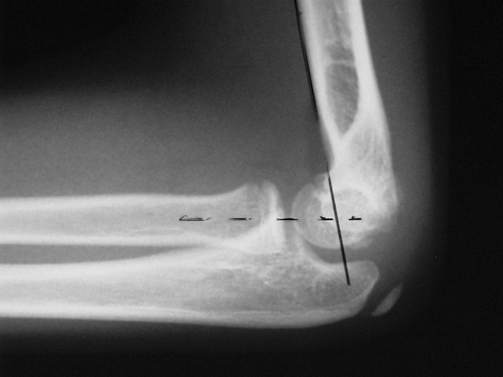
- The anterior humeral line: On a true lateral film, this line is drawn along the anterior aspect of the humeral shaft on the lateral radiograph This line passes through the middle one third of the capitellum in bones that are not injured. It is very useful for detecting subtle fractures.
- Fractures (i.e. supracondylar) usually result in displacement of the capitellum posteriorly.
- Thus, the anterior humeral line passes through the anterior one third or entirely anterior to the capitellum.
- The Radiocapitellar line: Since the radius articulates with the capitellum, a line is drawn through the middle of the radius shaft and extended proximally through the joint should bisect the capitellum on all views (AP & lateral).
- http://img.medscape.com/pi/emed/ckb/radiology/336139-415822-5412.jpg
- http://nypemergency.org/images/v2c18n.jpg
-
Improper alignment indicates a radial head dislocation (which may be very subtle)




Show References
A recent study examined the effects of accidental digital epinephrine injection from auto-injectors. 127 cases with complete follow-up had the following effects:
- no effects were reported in 10%
- minor effects in 77%
- moderate effects in 13%
- major effects in 1 case
Pharmacologic vasodilators were used in 23%. Four patients had possible digital ischemia. All patients had complete resolution of symptoms, most within 2 hours. No patient was admitted, received hand surgery consultation, or had surgical care.
Although this speaks for the safety of digital anesthesia using epinephrine, it underscores the importance of providing education to patients who are prescribed epinephrine auto-injectors.
Show References
- Ulnar nerve blocks are relatively easy to perform and excellent for anesthetizing the ulnar nerve distribution, particularly of the hand.
- Ulnar nerve blocks can be performed at the level of the wrist (dorsal or volar side) or at the elbow. Volar side blocks at the wrist tend to be easier to perform and associated with less risk.
- Using a 27 gauge needle, infiltrate 2 to 3 mL's of lidocaine between the flexor carpi ulnaris tendon and the distal-most aspect of the ulnar bone. The needle should be inserted 1 to 2 cm's at about a 40 degree angle, at the proximal-most wrist crease.
- Do not puncture the actual ulnar nerve or the ulnar artery. Should needle insertion cause distal hand paresthesias or blood withdrawal, do not inject and immediately remove the needle, as this suggests that the ulnar nerve or artery was struck, respectively. The objective is to allow the lidocaine to infiltrate into the nerve, not to inject it directly into the nerve.
Show References
Hyponatremia plagues many neurosurgical patients due to the syndrome of inappropriate secretion of ADH (SIADH) or the cerebral salt wasting syndrome (CSW). Both diseases may appear similar (hyponatremia, increased urine osmolarity, increased urine sodium, normal adrenal, renal and thyroid function), but there is one BIG difference. Patients with SIADH are euvolemic or hypervolemic (excess ADH causes fluid retention) whereas patients with CSW are fluid depleted (impaired renal handling of sodium and water). To differentiate, look for signs of hypovolemia: orthostatics, dry mucus membranes, hemoconcentration, pre-renal azotemia, and/or hemodynamics (IVC collapse anyone?).
Bottom line: Distinguish SIADH from CSW because the treatments are exact opposites:
SIADH: Fluid restrict
CSW: Give water and salt (i.e., 0.9% saline)
Show References
Hypertensive Encephalopathy (HE) is a clinical diagnosis and can look like many other disease entities.
HE refers to a relatively rapidly evolving syndrome of severe hypertension in association with severe headache, nausea, and vomiting, visual disturbances, convulsions, altered mental status and, in advanced cases, stupor and coma.
The key is the presence of severe hypertension. Remember, though, that 160/105 mm Hg may be high for an individual patient. Most patients with the syndrome will have diastolic pressures well in excess of 120-130 mm Hg. The only way you will know if the diagnosis is correct is to treat the BP (carefully control), work up other etiologies, and see of symptoms improve with BP control.
Beware the patient with severe HTN and seizure. Seizure may be the first, and only, symptom of hypertensive encephalopathy.
Some confusion exists regarding proper distinction and treatment between the different tachydysrhythmias associated with WPW. Here's the scoop:
1. orthodromic SVT: narrow regular tachycardia, looks just like a routine SVT, treat just like any other SVT (AV nodal blockers work fine)
2. antidromic SVT: wide regular tachycardia, looks just like VTach, treat like VTach (amiodarone, procainamide, shock; lidocaine won't work, though won't harm either)
3. atrial fibrillation: very different!! irregularly irregular, morphologies of the QRS complexes vary between narrow and wide, some areas may have rates as high as 250-300/min, MUST avoid all AV nodal blockers (which includes adenosine, CCBs, BBs, digoxin, amiodarone); treat with procainamide or sedation+cardioversion
Show References
Adhesive Capsulitis -- Frozen Shoulder
- Characterized by pain and loss of motion or stiffness in the shoulder.Normally not seen below the age of 40, affects ~2% of the population and diabetics are at increased risk.
- Due to thickening and contracture of the capsule surrounding the shoulder joint.
- Can occur after trauma to the shoulder if the shoulder is not moved early enough, but is also know to occur idiopathically.
- X-rays are only helpful to rule out other causes of the shoulder pain and are typically normal in Adhesive capsulitis.
- Typically will get better on its own over 2-3 years.
- Physical Therapy and home exercises aimed at restoring ROM can shorten the duration of pain and stiffness.
- Surgery can be done if there is no improvement with medical management and physical therapy.
- Prevention strategies include early ROM exercises in those with shoulder injuries especially in the elderly diabetic.
Show References
- most common cause of low platelets in children
- immune-mediated destruction of circulating platelets
- acute ITP peak incidence between 2-5 years of age; chronic ITP peaks in adolescence
- recent history (1-6 weeks) of viral infection or immunization is common
- no hepatosplenomegaly
- low platelets with megathrombocytes on smear, with normal hemoglobin (which differentiates from TTP, HUS, and DIC)
- nearly 90% of children will have normal platelet counts in 6 months
- treatment reserved for platelet counts <20,000 or significant bleeding: IVIG (best response rate of 95%), corticosteroids (79% resposne rate), anti-rH (D) immunoglobulin (82% reesponse reate)
Show References
In a previous pearl we were discussing the need to perform EGD for any suicidal patient with a history of ingestion of a caustic to grade injury and assess chance of perforation and/or stricture formation. Suicidal patients are intentionally ingesting the caustic and can thus justify the risk/benefit ratio more easily than the pediatric unintentional ingestion. The concerned parent will bring the child in with a possible ingestion of a caustic. The container could be simply in the same room, spilled on the child and never be ingested. Even if ingested, the amount is less if the child tastes the caustic and will reflexively cause spitting. The literature is scant in regards to this type of patient but seems to point to this general algorithm:
Child displays 2 or more of the following symptoms there is enough evidence from case series that there will be a clinically signficant lesion found on EGD.
Vomiting, Drooling, Stridor, Presence of Oropharyngeal Burns
That being said, many clinicians would elect for EGD and assessment of airway with stridor alone. Do not be fooled into thinking if you see no oral lesions that there is no way the child ingested the caustic. Each case series showed a lack of correlation of physical exam findings to EGD findings.
Show References
- When examining the hand, care should be taken to thoroughly assess both the sensory and motor function on both the dorsal and palmar surfaces.
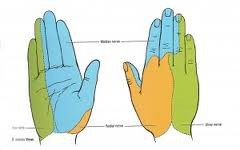
- The dermatomes of the hand provide sensation and are comprised of the ulnar, median, and radial nerves (see diagram below).
- (1) Light touch, (2) sharp touch (i.e. pinprick), (3) temperature, (4) propioception (joint position sense), (5) vibration, and (6) 2-point discrimination in the following nerve distributions should be assessed:
-- ulnar nerve >>> supplies palmar surface and dorsal tips of little finger and medial half of ring finger, including
adjacent parts of hand.
-- median nerve >>> supplies palmar and dorsal aspects of thumb, index finger, middle finger, and lateral half
of ring finger, including adjacent parts of hand.
-- radial nerve >>> supplies most of dorsal surface of hand.
Hemostatic Therapy for ICH - Updated Guidelines
- The AHA/ASA just published updated guidelines for the diagnosis and treatment of acute spontaneous intracerebral hemorrhage (ICH).
- Regarding hemostatic therapy, new/revised recommendations from the 2007 AHA/ASA guidelines include:
- Patients with severe thrombocytopenia or factor deficiency should receive platelets or factor replacement
- Patients with ICH due to oral anticoagulants (warfarin) should receive intravenous vitamin-K and vitamin-K dependent factor replacement
- Prothrombin complex concentrates (PCCs) are being increasingly used and are considered a reasonable alternative to FFP. To date, studies have not shown improved outcome with PCCs.
- Recombinant factor VIIa (rFVIIa) is not recommended as a sole agent for warfarin-related ICH
- rFVIIa is not recommended in unselected patients
- Usefulness of platelet transfusions for patients using antiplatelet medications is unclear and currently investigational.
Show References
Beta Blockade in Treating Acute Aortic Dissection
Medical therapy for acute aortic dissection is aimed at decreasing shear stress within the aorta. Although there are many agents to choose from when treating hypertension in patients with acute aortic disease, all regimens should include a beta blocker (like esmolol) unless contraindicated. Initiation of a beta blocker before another antihypertensive agent is added is crucial as this will prevent reflex tachycardia associated with vasodilators and other afterload reducers. Reflex tachycardia may worsen the dissection.