
Tranexamic acid (TXA) is an antifibrinolytic that prevents clot breakdown by inhibiting plasminogen activation and plasmin activity
The CRASH-2 trial enrolled 20,211 adult trauma patients with significant hemorrhage (SBP <90 or HR 110) or at significant risk of hemorrhage
Patients were randomized to 1 gram TXA over 10 minutes followed by an infusion of 1 gm over 8 hours vs placebo
There was a significant reduction in the relative risk off all cause mortality of 9% (14.5% vs 16%, RR 0.91, CI 0.85-0.97, p = 0.0035)
The patients that benefited most were those most severely injured, and in those treated in less than 3 hours of injury.
Show References
Background Information: While the concept of Emergency Medicine is fairly well understood in the United States, it is less clear in countries where the concept is not as well established. This has caused quite a bit of confusion and hindered progress and collaboration.
Pertinent Study Design and Conclusions: In a recent consensus conference held at SAEM several definitions were standardized.
- Acute Care: all promotional, preventive, curative, rehabilitative, and palliative actions, whether oriented toward individuals or populations, whose primary purpose is to improve health and whose effectiveness depends largely on time-sensitive and frequently rapid intervention.
- Emergency Medicine: a named field of specialty practice for which formal training prepares a candidate whose competence is officially standardized and regulated (thus EM is a subset of acute care)
- Emergency services: the sum of all efforts to deliver effective health action in response to extreme risk under intense time pressure
- Emergency care: the subset of emergency services focused on delivery of curative interventions targeting severe clinical cases
Bottom Line:
It is imperative that the same terminology be used when discussing the delivery of care on a time-sensitive basis.
University of Maryland Section of Global Emergency Health
Author: Andi Tenner, MD, MPH, FACEP
Show References
VAD thrombosis: A Must Know VAD Complication
The HeartMate left ventricular assist device (LVAD) is one of the most frequently placed LVADs today. Originally, it was thought to have a lower incidence of thrombosis due to its mechanical design. However, a recent multi-center study published in the NEJM reported a dramatic increase in the rate of thrombosis since 2011 in the HeartMate II device. The report found:
-
An increase in pump thrombosis at 3 months after implantation from 2.2% to 8.4%
-
The median time from implantation to thrombosis was 18.6 months prior to March 2011, to 2.7 months after.
Pump thrombosis is a major cause of morbidity and mortality (up to almost 50%!!) and is a can't miss diagnosis. It's important to keep thrombosis on the differential for any VAD patient presenting with:
-
Power spikes or low pump flow alarms on the patient's control box
-
Pump (VAD) failure
-
Recurrent/new heart failure
-
Altered mental status
-
Hypotension (MAP < 65)
-
Signs of peripheral emboli (including acute CVA)
Useful lab findings suggestive of thrombosis include:
-
Evidence of hemolysis
-
LDH > 1,500 mg/dL or 2.5-3 times the upper limit of normal
-
Hemoglobinuria
-
Elevated plasma free hemoglobin
Bottom Line: In the patient with suspected VAD thrombosis, it is important to contact the patient's VAD team immediately (CT surgeon, VAD coordinator/nurse, VAD engineer). Treatment should begin with a continuous infusion of unfractionated heparin, while other treatment options can be discussed with the VAD team.
Show References
Question
68 year-old male presents with weakness after surgical repair of his abdominal aorta. What’s the diagnosis and name at least one eponym for the signs displayed (there are five total)?
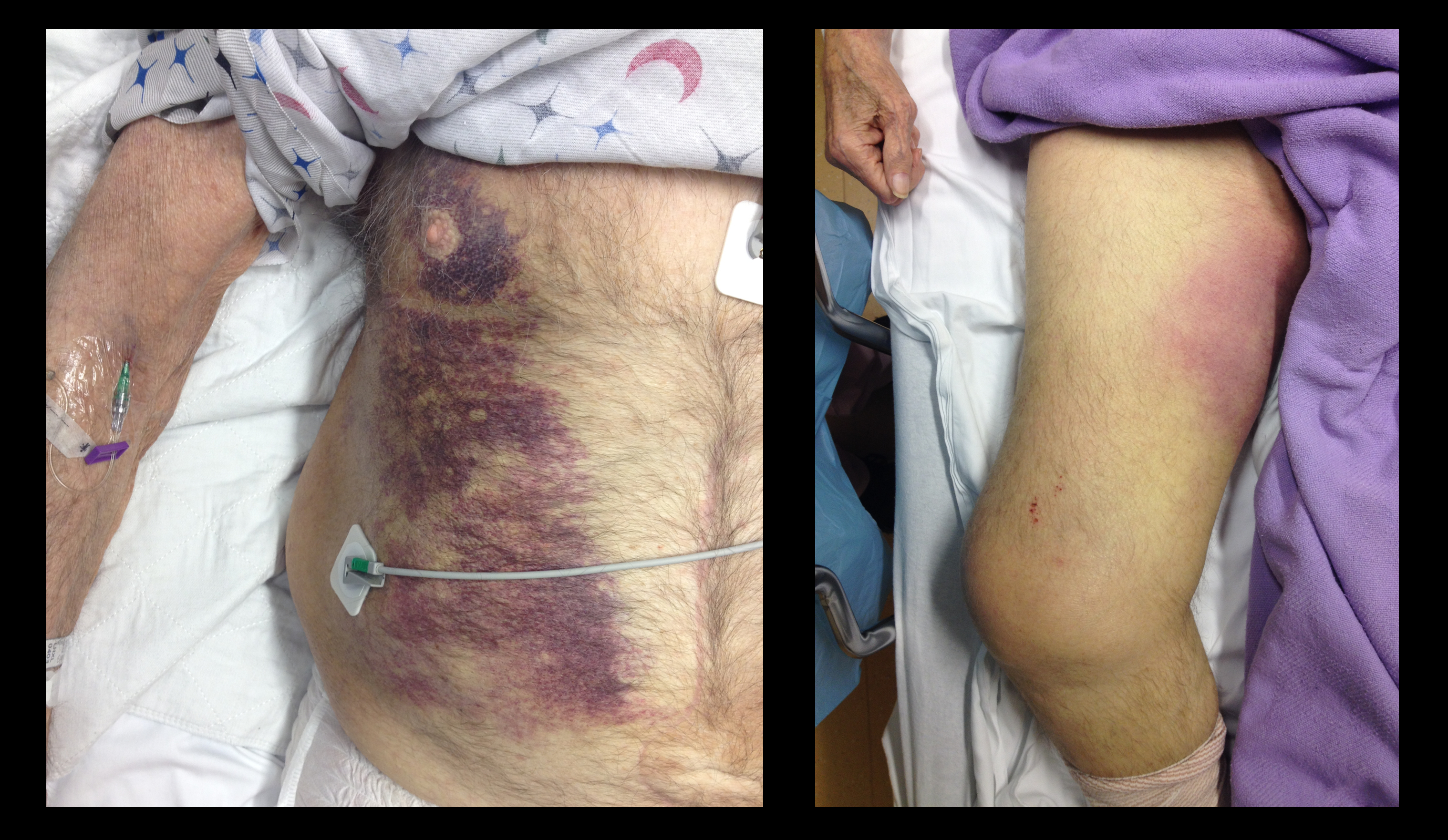
Show Answer
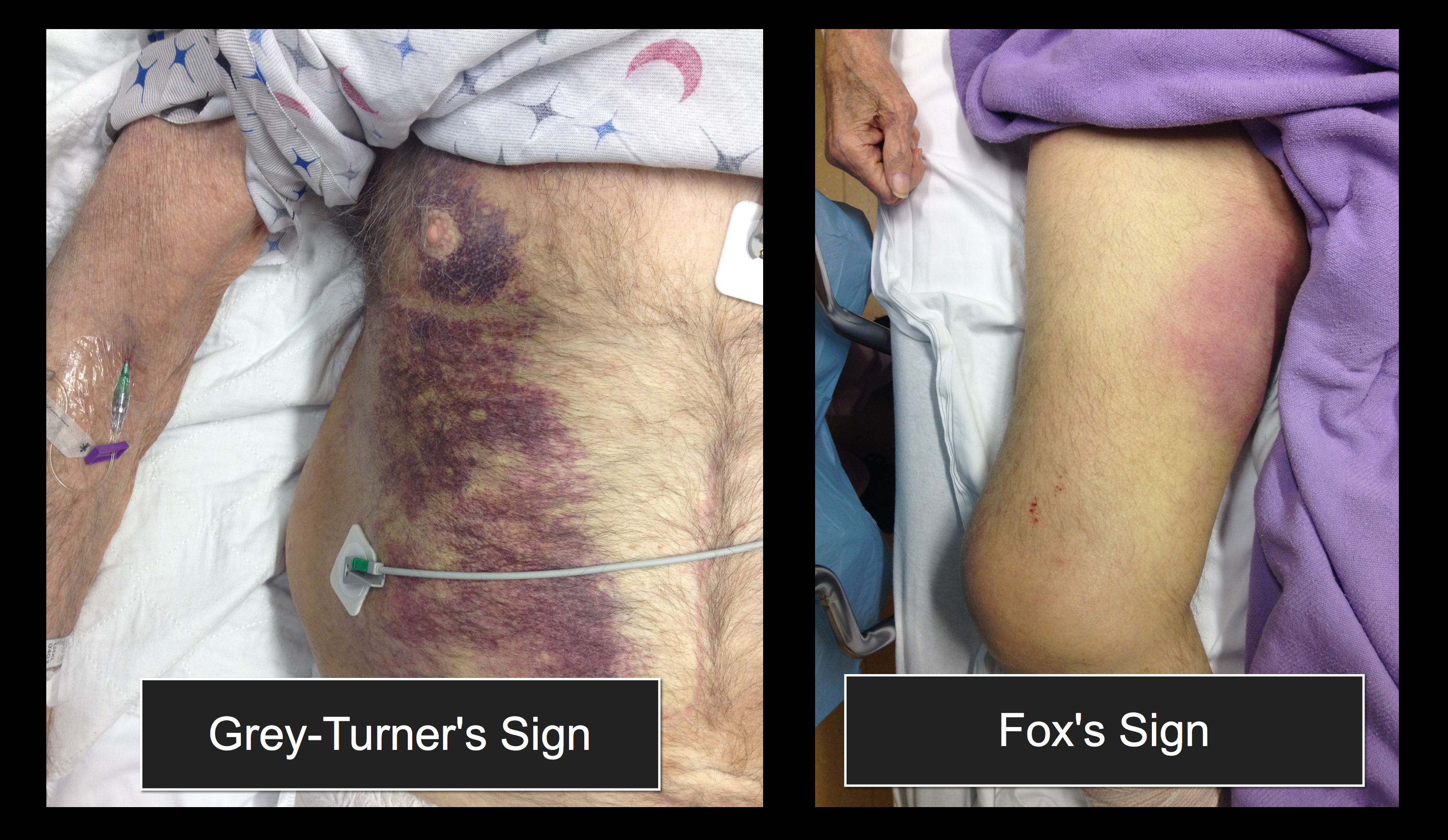
Show References
There is debate and confusion regarding where and how to measure ST elevation (STE). Do you measure the STE at the J-point? Or at 40 msec after the J-point? And how much STE is considered significant? The current guidelines have clarified this issue.
- STE should be measured at the J-point.
- STEMI is defined by STE ≥ 1 mm in at least 2 contiguous leads, with the exception of leads V2-V3.
- STEMI is defined by STE ≥ 2 mm in leads V2-V3 in men.
- STEMI is defined by STE ≥ 1.5 mm in leads V2-V3 in women.
For more cardiology pearls from the 2013 literature , check out Amal Mattu's Articles You've Gotta Know!
Want more emergency cardiology pearls? Follow me @alifarzadmd
Show References
No single feature of the history of physical examination reliably rules out ostemyelitis
Aids in making the diagnosis include:
An ulcer area larger than 2 cm2 (LR 7.2),
A positive probe to bone test (LR 6.4),
An ESR greater than 70 mm/h (LR 11)
Show References
Though an uncommon exposure, it can occur from chronic mercury exposure. One mode of exposure that I have seen is with elemental mercury thermometers that were broken to collect the beads of mercury - for entertainment. This occurred in a child's room and were forgotten. One child presented with personality changes and pink hands and feet. The patient suffered from severe mercury poisoning and acrodynia due to prolonged exposure to the mercury vapor.
Acrodynia or Pink Disease includes:
Irritability, shyness, photophobia, pink discoloration of the hands and feet and polyneuritis.
Case Presentation:
A 10 year old boy presents with a dog bite sustained 3 days ago, during a family trip to India. He has no prior history of vaccination and, at the time, he was taken to a local clinic where the wound was irrigated and he received a rabies vaccine.
Clinical Question:
Now that his has come to your ED 3 days later, is there anything further to be done?
Answer:
This patient should also receive rabies immunoglobulin (RIG) and complete his post-exposure prophylaxis. Post-exposure prophylaxis is a combination of rabies vaccine and rabies immunoglobulin (RIG).
RIG:
- Infiltrate the wound and surrounding tissue RIG 20 IU/kg (if human RIG) or 40 IU/kg (if equine RIG).
- Can be administered up to 7 days after the first vaccine.
Vaccine:
- Several vaccine regimens are approved by the WHO. Based on the CDC guidelines, vaccination should be administered at day 0, 3, 7 and 14.
- Had the patient received rabies immunization prior to travel, he would only need 2 vaccines should be given on days 0 and 3.
- Thus our patient needs RIG today and 3 more vaccinations (one today and then one at days 7 and 14)
Bottom Line:
- Travelers at highest risk are individuals visiting families in endemic areas.
- Often times, rabies IG is not available but can be administered up to 7 days after initial vaccination.
University of Maryland Section of Global Emergency Health
Author: Jenny Reifel Saltzberg
Show References
The morbidity and mortality from pseudomonas aeruginosa infections is high and empiric double-antibiotic coverage (DAC) is sometimes given; quality evidence for this practice is lacking.
Although there is little supporting data, the following reasons have been given for DAC:
- DAC provides better empiric coverage through differing mechanisms of antibiotic action
- DAC prevents the emergence of antibiotic resistance during therapy
The potential harm of antibiotic overuse cannot be ignored, however, and include adverse reaction, microbial resistance, risk of super-infection with other organisms (e.g., Clostridium difficile), and cost.
There may be a signal in the literature demonstrating a survival benefit when using DAC for patients with shock, hospital-associated pneumonia, or neutropenia. The IDSA guidelines, however, do not support DAC for neutropenia alone; only with neutropenia plus pneumonia or gram-negative bacteremia.
Bottom line: Little data supports the routine use of DAC in presumed pseudomonal infection. It may be considered in patients with shock, hospital-associated pneumonia, or neutropenia (+/- pneumonia), but consult your hospital’s antibiogram or ID consultant for local practices.
Show References
Question
Patient with a history of osteogenesis imperfecta presents with right lateral chest pain following a sneeze. The ultrasound of his chest is shown (hint: arrow points to a rib). What's the diagnosis?
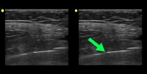
Show Answer
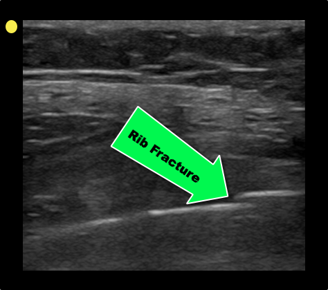
Show References
Metoprolol Usage Cardioprotective
- Intravenous (IV) metoprolol is sparingly used in STEMI given concern about precipitation of cardiogenic shock (COMMIT/CCS-2 Trial)
- A recent study (n=220) looked at usage of IV metoprolol versus controls in patients with STEMI and a killip class II or less prior to primary PCI
- MRI was preformed 5-7 days after STEMI revealing reduced infarct size and increased left ventricular ejection fraction in the IV metoprolol group
- IV beta-blockade appears cardioprotective in those with a low killip score and should be considered prior to primary PCI in certain subgroups
Show References
JNC8 (the Eigth Joint National Commission) released their recommendations for blood pressure management this week. The full article as published in JAMA can be found at http://jama.jamanetwork.com/article.aspx?articleid=1791497
Highlights from this report are
- Older adults do not need to be placed on antihypertensive medications unless their SBP > 150 or DBP > 90.
- Younger patients should still be started if their SBP > 140 or DBP > 90.
- Firstline drug treatment recommendations are:
- Non-black patients: start with thiazide diuretics, calcium channel blockers, angiotension converting enzyme (ACE) inhibitors, or angiotension-receptor blockers (ARBs).
- For black patients start with thiazide diuretics or calcium channel blockers.
- Patients with chronic kidney disease should be on an ACE or ARB.
General Pearl: Remember to be cautious in acutely lowering the blood pressure in asymptomatic patients. Acute lowerings can cause watershed ischemia leading to strokes.
A new drug is coming onto the drug scene with some case reports beginning to build. The internet appears to have been a major driver or mode of distribution for this particular drug.
One study of users showed that this ketamine analog has more vivid hallucinations that would liken it to LSD. It has been theorized that this drug has the dissociative effects of ketamine but also has prominent serotninergic effects making additions more likely and hallucinations possible.
If you see a case in your ED, you can say you heard it here first!
Show References
In November 2013, the CDC issued multiple Alerts on various polio outbreaks in Asia and Africa. Countries currently with the heaviest burden are Syria, Pakistan, Somalia, Kenya, and Cameroon. Nigeria and Afghanistan have also had persistent epidemics.
General Information:
- 95% of Polio cases are asymptomatic. (Not important clinically, but important for transmission)
- 4-8% present with non-specific flu-like symptoms +/- nuchal rigidity
- Only 1% have the classic syndrome of flaccid limb paralysis with decreased limb reflexes
- Paralysis may affect respiratory muscles leading to respiratory failure and death
- Treatment is supportive, but immunization of contacts is important
Relevance to the EM Physician:
The diagnosis can be made by detecting:
- Virus in stool sample or a nasopharyngeal swap is sensitive and specific in all patients.
- Polio antibodies in the patient’s serum is sensitive and specific in symptomatic patients.
The CSF analysis results will resemble that of aseptic meningitis.
Bottom Line:
Have a high suspicion for travellers to affected regions and recognize the high prevalence of asymptomatic infection (and thus importable epidemic potential). Pre-travel vaccination is essential.
University of Maryland Section of Global Emergency Health
Author: Walid Hammad, MB ChB
Show References
Hepatic Encephalopathy (HE)
Pathogenesis: Several theories exist that include accumulation of ammonia from the gut because of impaired hepatic clearance that can lead to accumulation of glutamine in brain astrocytes leading to swelling in patients with hepatic insufficiency from acute liver failure or cirrhosis.
Clinical Features:
- Impaired mental status
- impaired neuromotor function (hyperreflexia, hypertonicity, asterixis)
- Subtle signs include personality changes, decreased energy level, and impaired sleep-wake cycle
Diagnostic tests: Ammonia levels are routinely drawn but must be drawn correctly without the use of a tourniquet, transported on ice, and analyzed within 20 minutes to get an accurate result. Severity of HE does not correlate with increasing levels.
Management:
1. Airway protection as needed
2. Correct precipitating factors (GI bleed, infection-SBP, hypovolemia, renal failure)
3. Consider neuro-imaging if new focal neurologic findings are found on exam
4. Correct electrolyte imbalances
5. Lactulose by mouth (PO/Naso-gastric tube or Rectally)
a. 10-30 g every 1-2 hours until bowel movement or lactulose enema (300 mL in 1 L water)
b. Facilitates conversion of NH3 to NH4+, decreases survival of urease-producing bacteria in the gut
6. Rifaximin 550 mg by mouth BID (minimally absorbed antibiotic with broad-spectrum activity)
7. Do not limit protein intake acutely
8. TIPS reduction in certain patients with recurrent HE
9. Transplant referral as needed
10. Consider other causes if patient does not improve within 24-48hrs.
Show References
Question
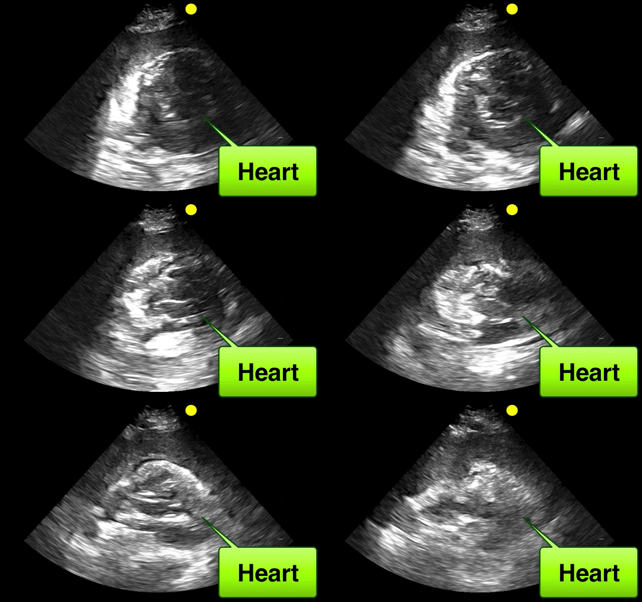
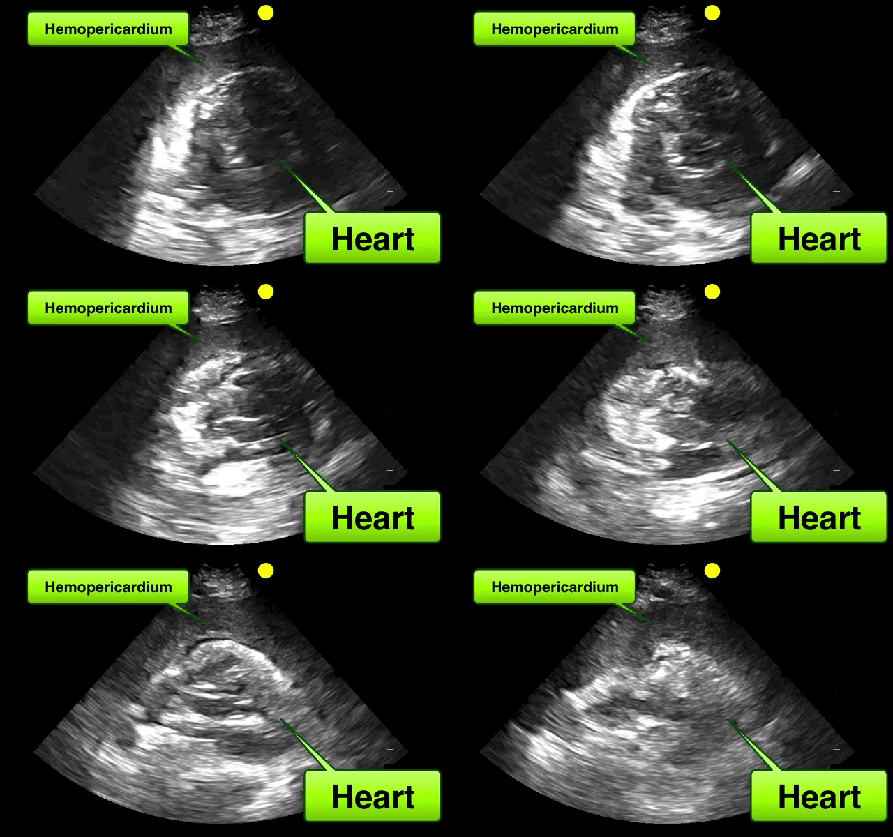
46 year-old female found unresponsive at a party. EMS transports the patient in cardiac arrest. A parasternal-long axis view of the heart is obtained during the pulse check. What's the diagnosis?
Show Answer
Show References
Treating knee osteoarthritis - from the American College of Rheumatology
Exercise whether it be aquatic, aerobic (land -based) or resistance can decrease pain and improve functional capacity. Exercise should be performed 3 to 5 times a week. Effects are usually noted after 3 to 6 months.
Weight loss of 5% or greater body weight is associated with a small improvement in pain and physical function. The main benefit of weight loss has more to do to effects on co-morbid conditions.
Walking aids: A single crutch or cane should be held on the side contralateral to the affected knee and should be advanced with the affected limb when walking to reduce the load on the affected joint.
Cane sizing: The distance from the floor to the patient's greater trochanter (brings the elbow to 15º to 20º of flexion.
Show References
- Significant morbidity and mortality has been consistently documented in pediatric sickle cell patients due to overwhelming sepsis from encapsulated organisms, especially S. pneumoniae
- All pediatric sickle cell patients presenting with fevers greater than 101.5F (38.6C) should receive antibiotics within 60 minutes of triage.
- Historically, and still in many pediatric sickle cell centers, ceftriaxone (75mg/kg/dose) is administered
- However, reported cases of deadly intravascular hemolysis in pediatric sickle cell patients whom had recieved multiple doses of ceftriaxone has led to new recommendations for antibiotic coverage to include cefuroxime (200mg/kg/day) or ampicillin/sulbactam (200mg/kg/day)