University of Maryland School of Medicine logo
Emergency Medicine
All Departments
UMSOM Assistant

UMEM Educational Pearls

  • Home
  • Departments
  • Emergency Medicine
  • CME
  • UMEM Educational Pearls
  • CME
  • Courses and Conferences
  • Free Open Access Medical Education
  • UMEM Educational Pearls
Previous  |  1 |  ... |  123 |  124 |  125 |  126 |  127 |  128 |  129 |  130 |  131 |  132 |  133 |  ... |  239 |  Next

Title: What's the diagnosis?

Category: International EM

Keywords: HIV, global health, infectious disease, rash, puritis (PubMed Search)

Posted: 4/2/2014 by Andrea Tenner, MD (Updated: 4/2/2014)

Question

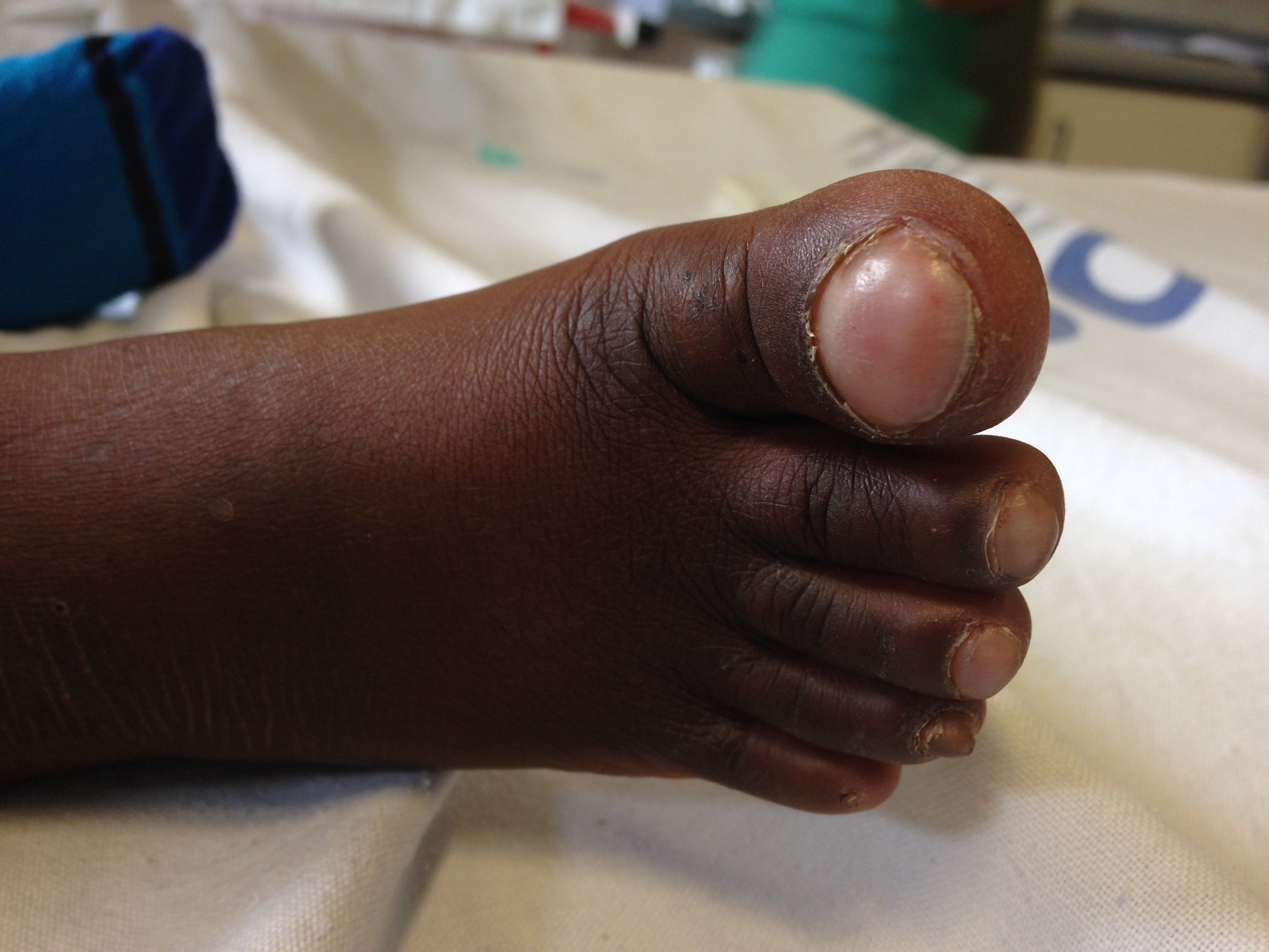
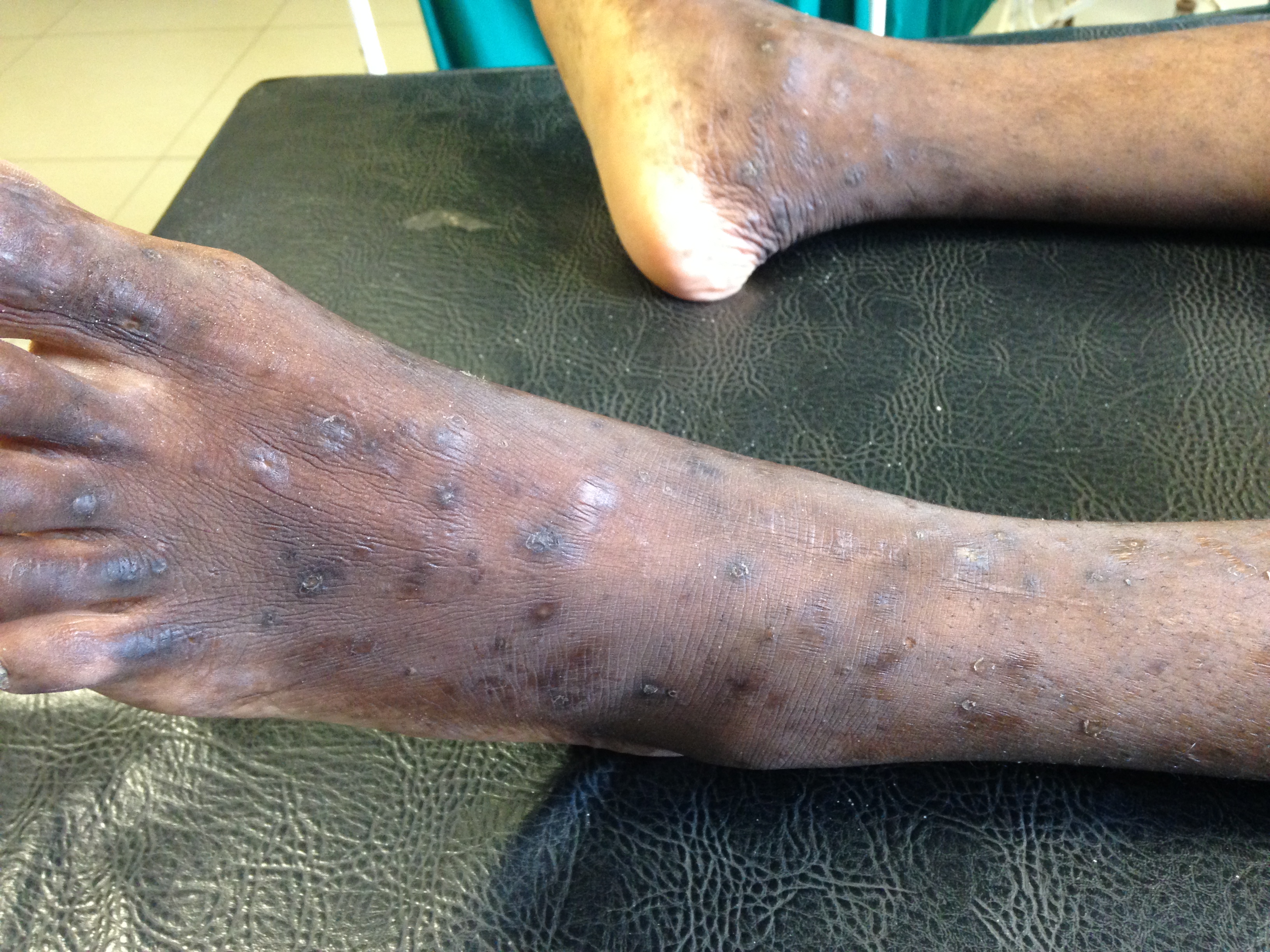
You are working in a clinic in Tanzania (or Baltimore, for that matter) when a 24 year old presents with this itchy rash on his feet.  What's the diagnosis and what underlying systemic condition does it indicate?


Show Answer

Pruritic Papular Eruptions in HIV

  • The most common cutaneous manifestation of HIV.  
  • The presenting symptom of HIV in 25-75% of patients and can be the initial indicator of advanced immunosuppression (the rash typically occurs once a patients CD4 is < 200)  

Presentation:

  • Multiple, discrete red bumps, which are puritic, symmetric and diffusely distributed.
  • Typically seen on the extremities and trunk with sparing of the mucous membranes, palms and webspaces.  
Treatment:
  • Topical steroids, emollients, and antihistamines are first line therapies.
  • If this fails, a trial of phototherapy is appropriate.  However, PPE has been found to be resistant to most of these treatments.

University of Maryland Section for Global Emergency Health

Authors: Van Pham, MD and Colleen Holley, MD

Show References

http://dermnetnz.org/viral/papulopruritic-hiv.html

Attachments

  • 1404012249_HIV-Associated_Puritic_Papular_Eruptions.JPG (2,920 Kb)


Title: Disseminated Intravascular Coagulation

Category: Critical Care

Posted: 4/1/2014 by Mike Winters, MBA, MD (Updated: 9/17/2026)

Coagulopathies in Critical Illness - DIC

  • Disseminated intravascular coagulation (DIC) is an acquired syndrome of intravascular coagulation and is commonly encountered in critically ill patients.
  • Think about DIC in the critically ill patient with oozing at vascular sites (or wounds) and the following lab abnormalities:
    • Thrombocytopenia
    • Prolonged PT and aPTT
    • Decreased fibrinogen
    • Elevated fibrin split products and D-dimer
  • Guidelines for the management of DIC are primarily based on expert opinion and include:
    • Treat the underlying condition (i.e., sepsis)
    • Transfuse platelets if < 50,000 per mm3
    • Transfuse FFP to maintain PT and aPTT < 1.5 times normal control
    • Transfuse cryoprecipitate to maintain fibrinogen levels > 1.5 g/L
  • The use of heparin remains controversial and cannot be routinely recommended.

Show References

Hunt B. Bleeding and coagulopathies in critical care. NEJM 2014;370:847-59.


Title: What's the Diagnosis? Image by Dr. Ashley Strobel

Category: Visual Diagnosis

Posted: 3/31/2014 by Haney Mallemat, MD (Updated: 4/1/2014)

Question

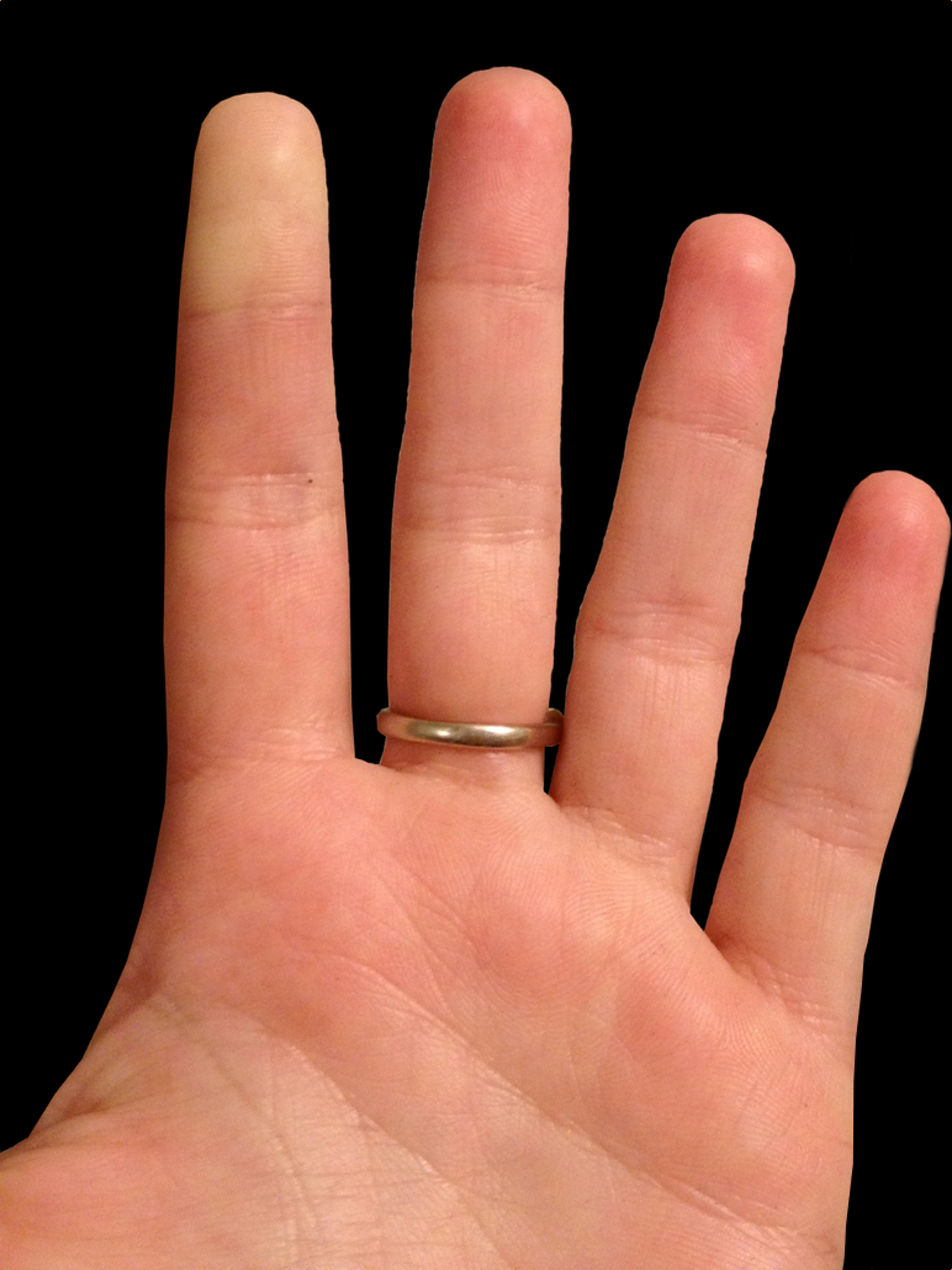
25 year-old female presents with the following. It seems to have occurred spontaneously and spontaneously resolves during her ED evaluation.
 

Show Answer
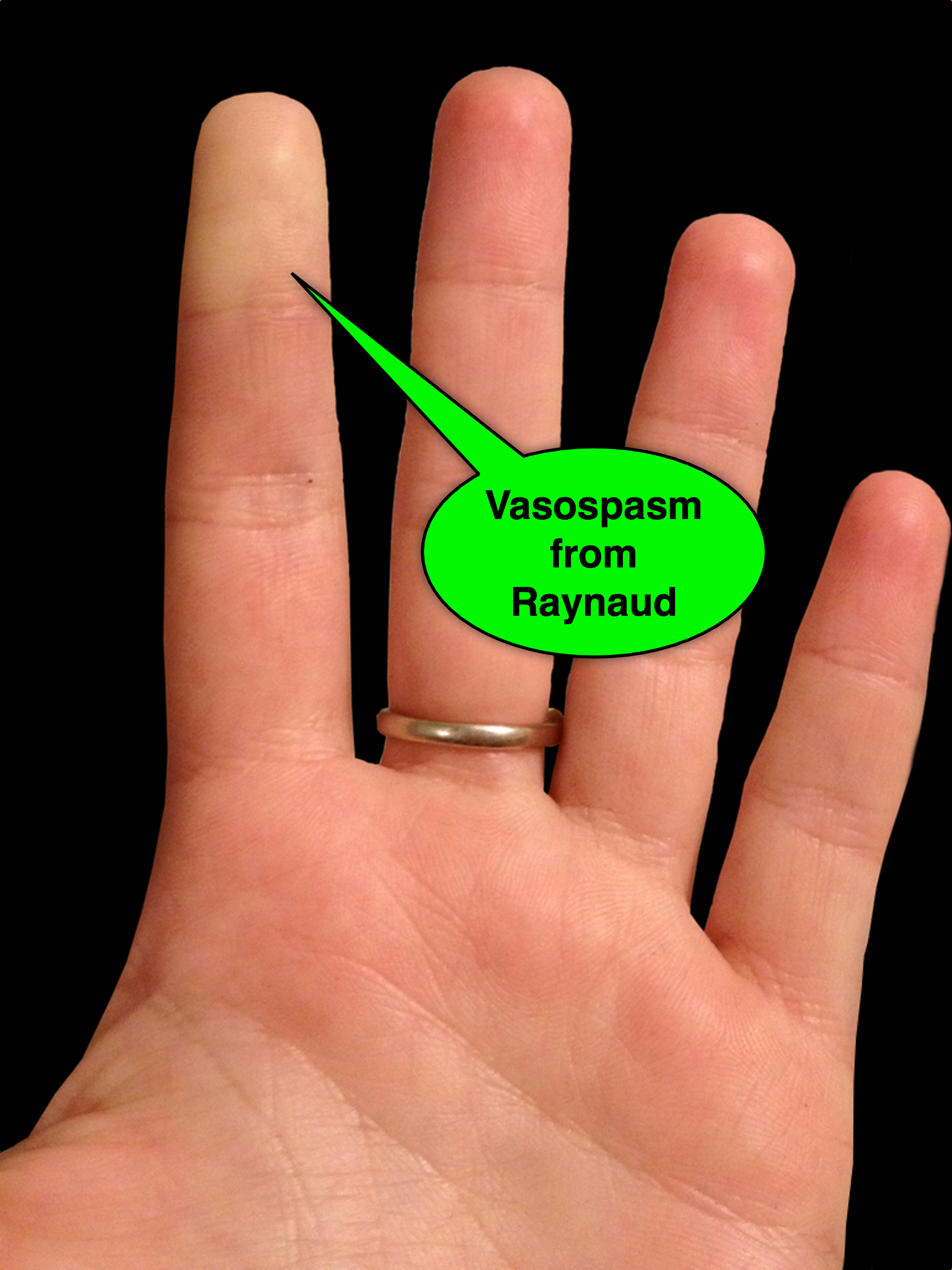
Answer: Raynaud phenomenon

  • Raynaud phenomenon is recurrent vasospasm of fingers or toes
  • Causes
    • Primary (e.g., external factors; such as cold temperature or stress)
    • Secondary to underlying illness, typically autoimmune disease (e.g., progressive systemic sclerosis or SLE)
  • Treatment
    • Avoid known triggers
    • Low-dose calcium channel blockers (e.g., nifedipine)
    • Topical nitroglycerine applied to areas of vasoconstriction
    • Identify and treat secondary disorders

Show References

Follow me on Twitter (@criticalcarenow) or Google+ (+criticalcarenow)


Title: DeQuervains versus Intersection Syndromes

Category: Orthopedics

Keywords: DeQuervain, Intersection, Syndrome, Tenosynovitis (PubMed Search)

Posted: 3/30/2014 by Michael Bond, MD (Updated: 9/17/2026)

DeQuervain and Intersection Syndromes:
 

  • DeQuervain's Syndrome (Tenosynovitis of the Abductor Pollicus Longus and Extensor Pollicus Brevis tendons) is a common disorder that has received a lot of press lately as BlackBerry Thumb or Gamer's Thumb.
    • This condition can be diagnosised by the Finklestein test [Have the patient bend their thumb into the palm of their hand, and then make a fist.  They should then ulnar deviate their wrist.  Pain along the tendons secures the diagnosis.]
    • The pain of DeQuervain's syndrome is typically along the distal end of the radius at the base of the thumb.
  • Intersection syndrome is a less common disorder though closely related to DeQuervain's Syndrome
    • The pain is usually felt on the top of the forearm about three inches proximal to the wrist. 
    • The pain from this condition is due to tenosynovitis of the Extensor carpi radialis longus and Extensor Carpi radialis brevis muscles/tendons caused by the intersection of them with the Extensor pollicus brevis and Abductor pollicus longus tendons.
    • Occurs due to excessive wrist movements.
    • Intersection syndrome can be seen in weight lifters, skiers, and can be seen in homeowners in the fall and winter when they rake a lot of leaves or shovel snow.
  • Treatment is the similar for both conditions and consists of:
    • NSAIDS
    • Cortisone injections can be effective
    • Thumb and wrist immobilization with a Thumb Spica Splint or Cock Up Wrist Splint


Title: Prescription Drug Abuse - What are Risk Factors for OD Death?

Category: Toxicology

Keywords: opioids (PubMed Search)

Posted: 3/27/2014 by Fermin Barrueto (Updated: 9/17/2026)

What are characteristics that increase the chance a patient is at risk for opioid-related death? A recent JAMA article begins to tackle this very issues. Baumblatt et al. found the following:

1) Patient with 4 or more prescribers had adjusted odds ratio 6.5 for opioid-related death

2) Patient with 4 or more pharmacies where they get their prescriptions aOR - 6.0

3) Patient with more than 100 mg of morphine equivalents mean per day aOR - 11.2

With the new Maryland Prescription Drug Monitoring program (PDMP)  we can start looking at a patient's prescription drug use pattern. The recent JAMA article can help you identify patients at high risk to die an opioid-related death. Use the PDMP and be wary if a patient has more than 4 prescribers or pharmacies or has >100mg of morphine equivalents per day.

Show References

High-Risk Use by Patients Prescribed Opioids for Pain and Its Role in Overdose Deaths.

Baumblatt JA, Wiedeman C, Dunn JR, Schaffner W, Paulozzi LJ, Jones TF.

JAMA Intern Med. 2014 Mar 3. doi: 10.1001/jamainternmed.2013.12711. [


Title: What's the diagnosis?

Category: International EM

Keywords: multiple myeloma, x-ray, global, neoplasm (PubMed Search)

Posted: 3/26/2014 by Andrea Tenner, MD

Question

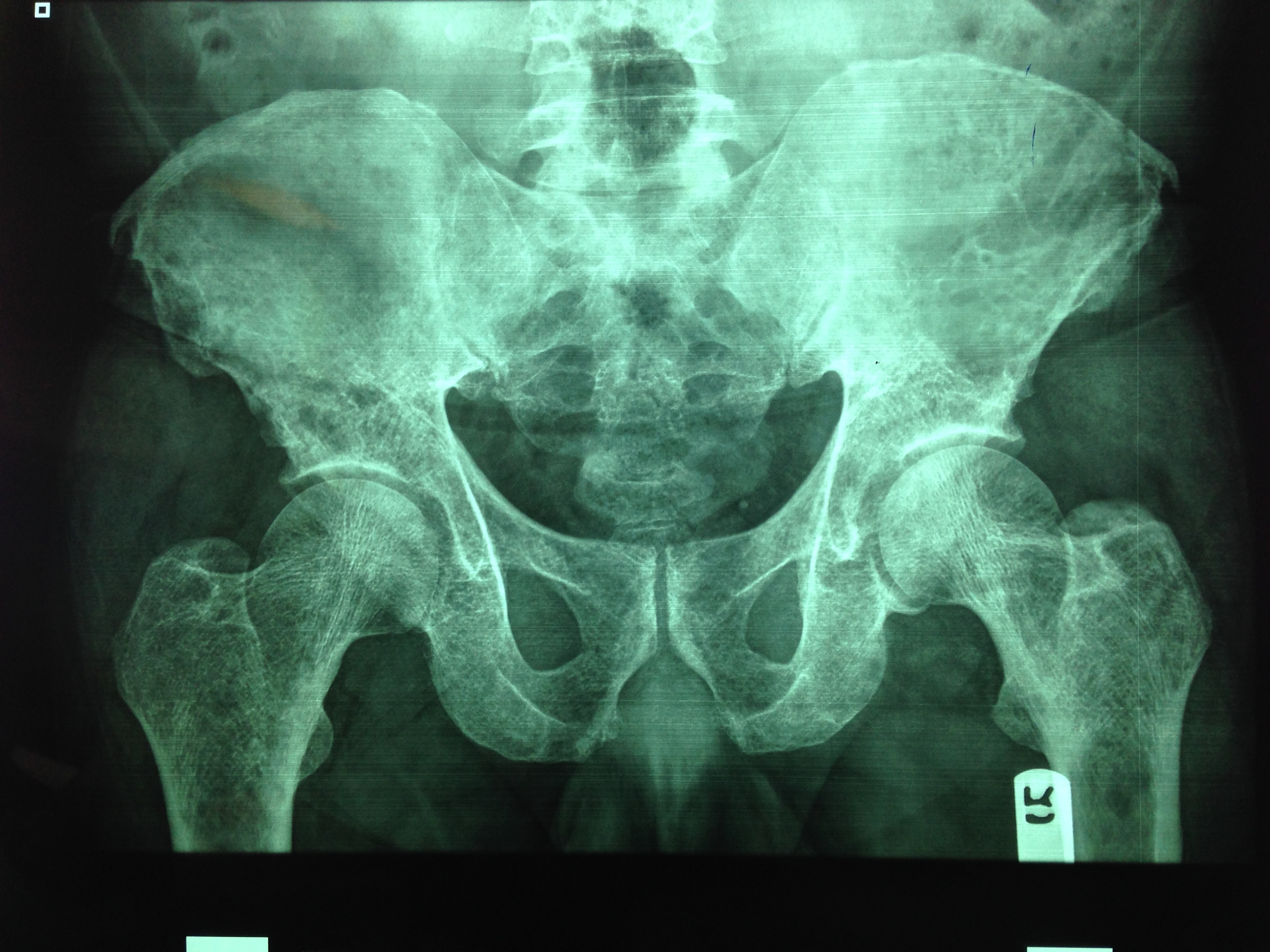
You are evaluating a 40 year old trauma victim and see this on pelvic xray. What are you worried about?
Show Answer

The patient likely has multiple myeloma. 
 
Multiple myeloma is a malignancy of plasma cells affecting the skeletal system. It has an osteoblastic effect on bones causing punched out lytic lesions in the skull, spine and pelvis. Skeletal survey is the initial diagnostic imaging of choice. It usually includes at least 3 of the following: a lateral radiograph of the skull, AP and lateral views of the spine, and AP views of the humeri, ribs, pelvis, and femora. It is also used as a staging tool to assess the extension of the disease.
 
Stage IA – Normal skeletal survey or single lesion
Stage IB - Five focal lesions or mild diffuse spine disease
Stage IIA/B - Five to 20 focal lesions or moderately diffuse spine disease
Stage IIIA/B - More than 20 focal lesions or severe diffuse spine disease
 
Treatment options are chemotherapy, radiation, steroids and stem cell transplant. Unfortunately, in patients with stage II these therapies are rarely curative.


University of Maryland Section for Global Emergency Health

Authors: Colleen Holley, MD and Van Pham, MD


Show References

Mulligan, Michael. Multiple Myeloma Imaging. available: http://emedicine.medscape.com/article/391742-overview#a19. accessed 24 March 2014.

Attachments

  • 1403261757_IMG_0312.jpg (2,427 Kb)


Title: There appears to be NO role for iNO in ARDS

Category: Critical Care

Keywords: ARDS, Nitric Oxide, acute respiratory failure, mechanical ventilation (PubMed Search)

Posted: 3/25/2014 by John Greenwood, MD (Updated: 3/26/2014)


Nitric Oxide appears to have NO role in ARDS

Background: The use of inhaled nitric oxide (iNO) in acute respiratory distress syndrome (ARDS) & severe hypoxemic respiratory failure has been thought to potentially improve oxygenation and clinical outcomes.  It is estimated that iNO is used in up to 14% of patients, despite a lack of evidence to show improved outcomes. 

Mechanism: Inhaled NO works as a selective pulmonary vasodilator which has been found to improve PaO2/FiO2 by 5-13%, but is costly ($1,500 - $3,000 per day) and increases risk of renal failure in the critically ill.

Study: A recent systematic review analyzed 9 different RCTs (N=1142) and compared mortality between those with severe (PaO2/FiO2 < 100) and less severe (PaO2/FiO2 > 100) ARDS and found that iNO does not reduce mortality in patients with ARDS, regardless of the severity of hypoxemia.


Bottom Line: Inhaled NO is an intriguing option for the treatment of refractory hypoxemic respiratory failure, however there does not appear to be a mortality benefit to justify it's high cost and potentially negative side effects.  In the ED, it is important to focus on appropriate lung protective ventilation strategies (TV: 6-8 cc/kg IBW) and maintaining plateau pressures < 30 cm H2O in the initial stages of ARDS to prevent ventilator induced lung injury while awaiting ICU admission.

Show References

Reference

Adhikari NK, Dellinger RP, Lundin S, et al. Inhaled nitric oxide does not reduce mortality in patients with acute respiratory distress syndrome regardless of severity: systematic review and meta-analysis. Crit Care Med. 2014;42(2):404-12. [PMID: 24132038]

Follow me on Twitter (@JohnGreenwoodMD)


Title: Are chest compressions safe in arresting LVAD patients?

Category: Cardiology

Keywords: Cardiac arrest, LVAD, CPR, Chest compressions (PubMed Search)

Posted: 3/23/2014 by Ali Farzad, MD

The number of patients with left ventricular assist devices (LVADs) is increasing and development of optimal resuscitative strategies is becoming increasingly important. Despite a lack of evidence, many device manufacturers and hospitals have recommended against performing chest compressions because of fear of cannula dislodgment or damage to the outflow conduit.

A recent retrospective analysis of outcomes in LVAD patients who received chest compressions for cardiac arrest did not support the theory that LVADs would be harmed by conventional resuscitation algorithms.

The study was a limited case series of only 8 LVAD patients over a 4 year period. All patients received compressions and device integrity was subsequently assessed by blood flow data from the LVAD control monitor or by examination on autopsy. Although more research is necessary to determine the utility and effectiveness of compressions in this population, none of the patients in this study had cannula dislodgment and half of the patients had return of neurologic function.

Show References

Shinar Z, Bellezzo J, Stahovich M, et al. Chest compressions may be safe in arresting patients with left ventricular assist devices (LVADs). Resuscitation. 2014. doi:10.1016/j.resuscitation.2014.01.003.


Want more emergency cardiology pearls?
Follow me on Twitter @alifarzadmd


Title: Ankle Syndesmosis Injuries

Category: Orthopedics

Keywords: ankle sprain (PubMed Search)

Posted: 3/22/2014 by Brian Corwell, MD

Ankle Syndesmosis Injuries are also called high ankle sprains as they involve trauma to the ligaments above the ankle joint

Most ankle sprains are lateral ankle sprains. High ankle sprains are relatively uncommon.

Usual mechanism: External rotation injuries

Exam: Tenderness at the syndesmosis and compression of the tib/fib at the mid calf level causing syndesmosis pain (squeeze test)

Median recovery time is almost 4 times as long as a lateral ankle sprain 62days vs. 15days

Emergency department care is similar tto that of other ankle sprains but the added benefit of patient education and advice may improve overall care and follow-up.


Title: Isolated vomiting in pediatric head injuries

Category: Pediatrics

Keywords: Head injury, vomiting, PECARN (PubMed Search)

Posted: 3/21/2014 by Jenny Guyther, MD


Parents will often bring children to the ED for evaluation after a minor head injury.  Vomiting has been considered a risk factor for traumatic brain injury (TBI).  Is isolated vomiting clinically significant?
 
A PECARN study looked at children < 18 years.
 
Isolated vomiting with minor head trauma was defined as: No history of LOC, GCS of 15, no altered consciousness (ie sleepiness, agitation), no palpable skull fracture or signs of basilar skull fracture, acting
normally per parent/guardian, no scalp hematoma or other traumatic scalp finding (ie abrasion or laceration), no headache (for patients 2-18 y), no seizure after the head trauma, no neurological deficits
(eg, motor or sensory abnormalities) and no amnesia (for patients 2-18 y).
 
42,112 children were enrolled.
5,557 (13.2%) had a history of vomiting, of whom 815 of 5,392 (15.1%) with complete data had isolated vomiting.
Clinically important TBI (death, neurosurgical procedure, intubation for at least 24 hours for TBI, or hospitalization for 2 or more nights because of the head trauma in association with TBI on cranial CT) occurred in 2 of 815 patients with isolated vomiting compared with 114 of 4,577 with non isolated vomiting.
Of patients with isolated vomiting for whom CT was performed, TBI on CT occurred in 5 of 298 compared with 211 of 3,284 with non isolated vomiting
 
There was no association found with timing of onset or time since the last episode of vomiting.
 
Bottom line: TBI on CT is uncommon and clinically important traumatic brain injury is very uncommon in children with minor blunt head trauma when vomiting is their only sign or symptom. Observation in the emergency department before determining the need for CT appears appropriate for these children to observe for deterioration.

Show References


Dayan et al.  Association of Traumatic Brain Injuries With Vomiting in Children With Blunt Head Trauma.  Annals of Emergency Medicine. Article in press.  Available for view Feb 14, 2014.


Title: Visual Diagnosis

Category: International EM

Keywords: echocardiography, rheumatic heart disease, endocarditis, international (PubMed Search)

Posted: 3/19/2014 by Andrea Tenner, MD

Question

35yo M with history of rheumatic heart disease presents with fever.  What disease process is suggested by the echo?

Show Answer

Answer: Mitral valve vegetation/Endocarditis
 
Any prior defects to cardiac valves increases your risk of endocarditis.  Rheumatic heart disease classically causes damage to either the mitral or aortic valve.   Of these patients, around 60%  will have mitral stenosis  and 20% will have pure mitral insufficiency.  The bacteria most commonly involved is Group A hemolytic strep.  Recommended antibiotics include penicillin and ceftriaxone.  
 
In a patient with known cardiac valvular disease, including rheumatic heart disease, keep endocarditis in your differential!
 
Univeristy of Maryland Section for Global Emergency Health
Authors: Colleen Holley and Van Pham

Show References

Burke AP.  Pathology of rheumatic heart disease.  Medscape.  2013.  Accessed 19 Mar 2013 at http://emedicine.medscape.com/article/1962779-overview#aw2aab6b4.

Attachments

  • 1403192137_IMG_0320_(1).JPG (2,033 Kb)


Title: A New Era in Management of the Septic Patient?

Category: Critical Care

Posted: 3/19/2014 by Haney Mallemat, MD

In 2001, Rivers et al. published a landmark article demonstrating an early-goal directed protocol of resuscitation that reduced mortality in septic Emergency Department patients.

Many questions have arisen throughout the years with respect to that trial; critics have complained about the overwhelming change in clinical practice based on this one single-center randomized trial.

Challenging Rivers data are the ProCESS (Protocolized Care for Early Septic Shock) investigators, who released the results from a multi-center randomized control trial of 1351 septic Emergency Department patients; the primary end-point was 60-day mortality. Click here for NEJM article.

Patients in this trial were randomized to one of three groups:

  • Protocol-based EGDT

  • Protocol-based standard (did not require central lines, inotropes, or blood transfusions

  • Usual care (no specific protocol; care was left to the bedside clinicians)

Bottom-line: The investigators did not find any difference in mortality between patients in the three groups and comment that the most important aspects of managing the septic patient may be prompt recognition and early treatment with IV fluids and antibiotics.

Show References

Follow me on Twitter (@criticalcarenow) or Google+ (+criticalcarenow)


Title: What's the Diagnosis? Case by Dr. Ali Farzad

Category: Visual Diagnosis

Posted: 3/17/2014 by Haney Mallemat, MD

Question

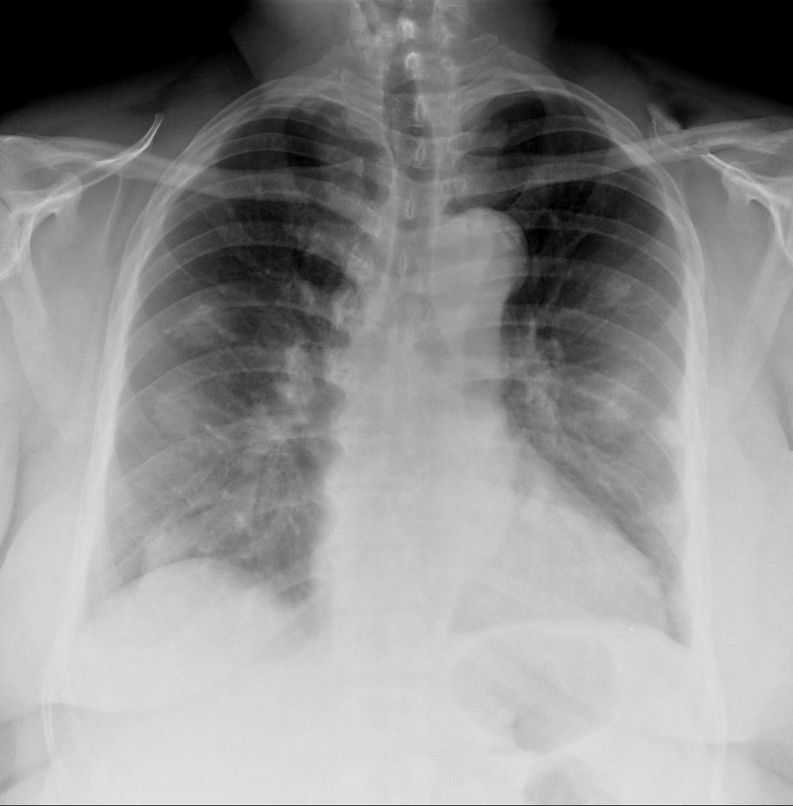
62 year-old male presents with weakness, chills, cough, and malaise. Recently, he had four teeth extracted but felt fine immediately after the extraction. Past medical history includes diabetes and hypertension; CXR is below. What’s the diagnosis?
 

Show Answer
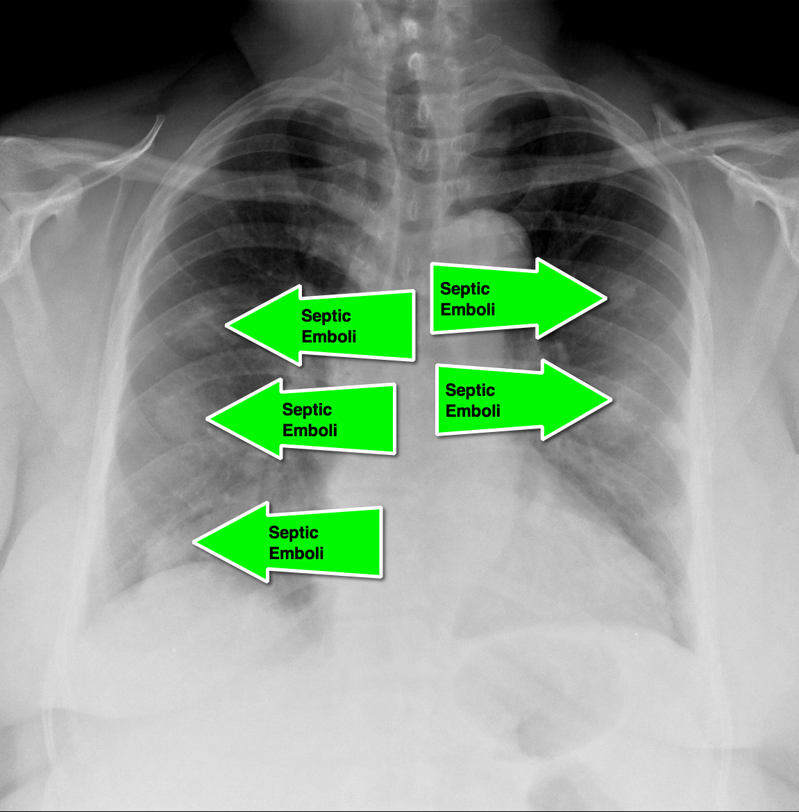
Answer: Septic pulmonary emboli (a.k.a. pulmonary puffballs); he was eventually diagnosed with bacterial endocarditis.

  • This history with the presence of "puffballs" on CXR, should clue the practitioner to the diagnosis of septic pulmonary emboli secondary to endocarditis
  • CT of the chest will confirm the diagnosis and possibly demonstrate cavitary lesions.
  • Blood cultures should be promptly obtained and the patient should be started on broad-spectrum antibiotics.

Show References

Follow me on Twitter (@criticalcarenow) or Google+ (+criticalcarenow)


Title: The HEART Score

Category: Cardiology

Posted: 3/16/2014 by Semhar Tewelde, MD

The HEART Score

Acute coronary syndrome defines a spectrum of diseases (unstable angina, NSTEMI, STEMI), without clear ECG abnormalities the diagnosis and disposition can be challenging

Several scoring systems have attempted to risk stratify patients: TIMI, PURSUIT, and GRACE

The TIMI & PURSUIT scores were designed to identify higher-risk patients and long-term mortality

A pilot/observational study has utilized a novel scoring system to risk stratify low to intermediate risk patients

The HEART (History, ECG, Age, Risk factors and Troponin) score: 

  • 0-3 points ~ 2.5% risk (data supporting discharge)
  • 4-6 points ~20.3% risk (data supporting observation)
  • ≥7points ~ 72.7% risk (data supporting early invasive strategies)

This scoring system is limited given the small study size and requires further study/validation, but may be an easy, quick, and reliable predictor of outcome in chest pain patients

Show References

Six AJ, Backus BE, Kelder JC. Chest pain in the emergency room: value of the HEART score. Neth Heart J. Jun 2008; 16(6): 191–196.


Title: Carbon Monoxide Poisoning from Kinjal Sethuraman

Category: Toxicology

Keywords: Carbon Monoxide, (PubMed Search)

Posted: 3/15/2014 by Michael Bond, MD

Carbon Monoxide is a odorless but deadly gas.  It is important to note that CO has an elimination half-life and it varies under different conditions.
 
When evaluating a patient, we can calculate backwards to determine the COHb level at time of exposure in an acute event.   

Carbon Monoxide Half-Life:

  • Average elimination on room air: 5-6 hours
  • 100% Oxygen: 70-130 minutes
  • 100% Oxygen under hyperbaric conditions at 3 ATA: 23 minutes
There is NO need to recheck COHb level again after initial level because it will be lower- (except in the case of Methylene Chloride exposure).

Show References

Thom, Stephen R.  "Carbon Monoxide Pathophysiology and Treatment."   Physiology and Medicine of Hyperbaric Oxygen Therapy.   Ed. Tom S. Newman and Stephen R. Thom, Saunders Elsevier, Philadelphia, PA 2008. Pp: 321-348.


Title: What's the Diagnosis? Image by Dr. Kami Hu

Category: Visual Diagnosis

Posted: 3/14/2014 by Haney Mallemat, MD

Question

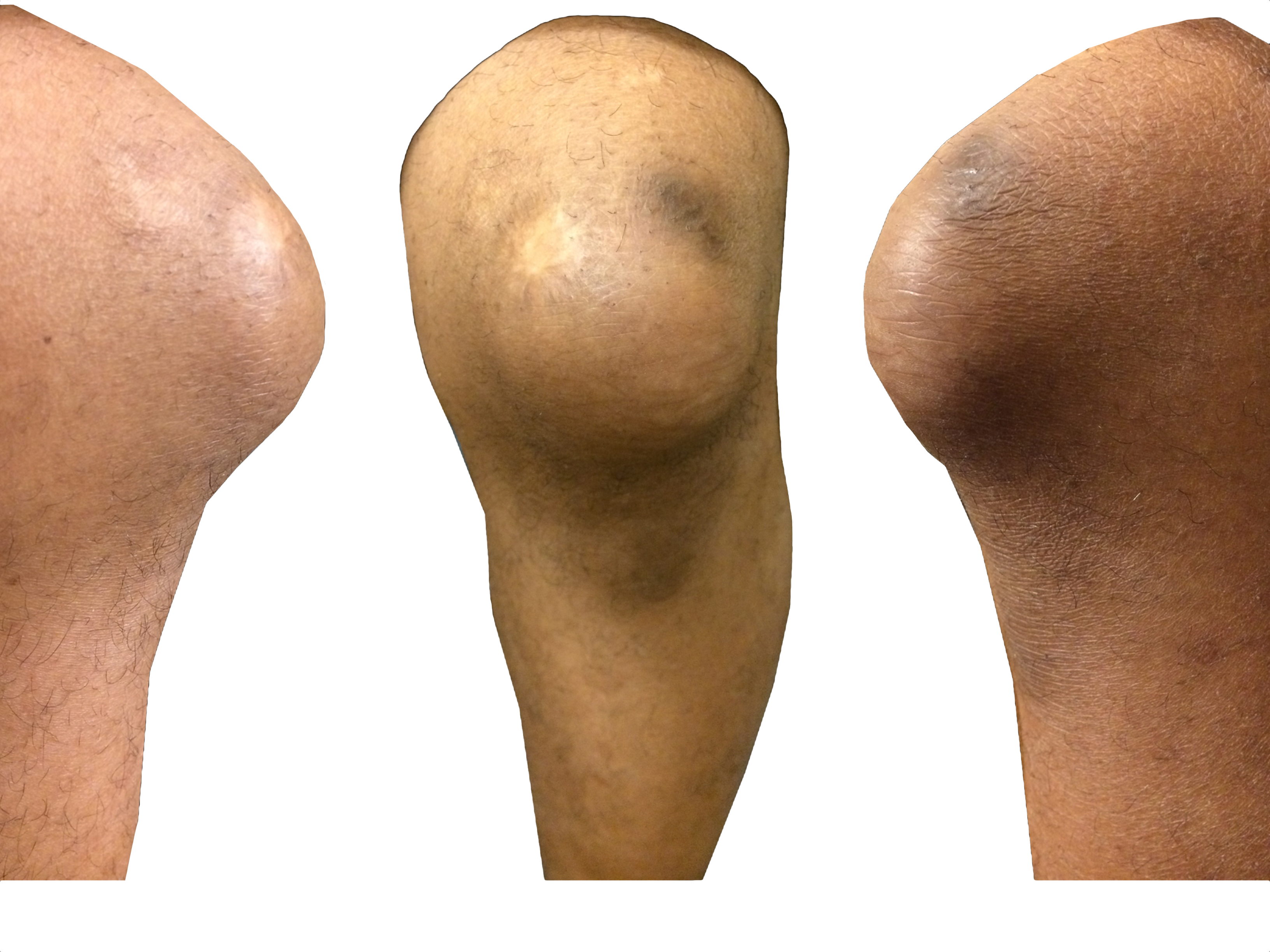
35 year-old carpet-layer presents with swelling of the superior portion of his knee that has progressively gotten worse over one week. He has no fever and has full range of motion (although pain is worse with movement). The knee is not tender to touch and the area is not erythematous or warm. What's the diagnosis?
 

Show Answer

Answer: Pre-patellar bursitis

  • The pre-patellar bursa is a superficial and synovial-lined space that reduces friction at the knee, separating the patella from the skin and the patellar tendon.
  • Inflammation to the bursa occurs secondary to direct knee trauma (either acute or chronic) and is typically benign.
  • It is associated with occupations that have a high incidence of trauma to the knee or repeated kneeling; hence the condition’s many names (carpet-layer's knee, coal-miner's knee, housemaid's knee, nun's knee)
  • Caution must be taken not to miss patients with septic bursitis (e.g., bacterial), especially in immunocompromised patients. If septic bursitis is suspected, fluid should be aspirated from the bursa and sent for analysis
  • Treatment for non-septic bursitis includes rest, (i.e., reduce trauma), NSAIDs, and physical therapy. If it remains persistent, incision and drainage by a qualified clinicians may be performed.

Show References

Follow me on Twitter (@criticalcarenow) or Google+ (+criticalcarenow)


Title: Do Poison Centers Reduce Length of Stay and Hospital Charges?

Category: Toxicology

Keywords: poison center, length of stay, hospital, charges (PubMed Search)

Posted: 3/13/2014 by Bryan Hayes, PharmD (Updated: 3/13/2014)

In a collaborative effort between the Illinois Poison Center and the Illinois Hospital Association, a new study sought to determine a poison center's effect on hospital length of stay (LOS) and hospital charges.

While the methodology was understandably complex, the authors compared ~5,000 toxicology inpatients with poison center assistance to 5,000 toxicology inpatients without poison center assistance.

After adjusting for confounders, the LOS among patients with posion center assistance was 0.58 days shorter compared to that of patients without poison center assistance (CI 95%: -0.66, -0.51, p<0.001). Though hospital charges for poison center-assisted patients in the lower quintiles were significantly higher than patients without poison center-assistance (+$953; p<0.001), they were substantially lower in the most costly quintile of patients (-$4852; p<0.001).

Poison center assistance was associated with lower total charges only among the most expensive to treat. However, this outlier group is very important when discussing medical costs.

Show References
Friedman LS, et al. The association between U.S. Poison Center assistance and length of stay and hospital charges. Clin Toxicol 2014;52:198-206. [PMID 24580060]

Follow me on Twitter (@PharmERToxGuy) or Google Plus (+bryanhayes13)


Title: Visual Diagnosis

Category: Visual Diagnosis

Keywords: international, global, hypoxia, clubbing (PubMed Search)

Posted: 3/12/2014 by Andrea Tenner, MD

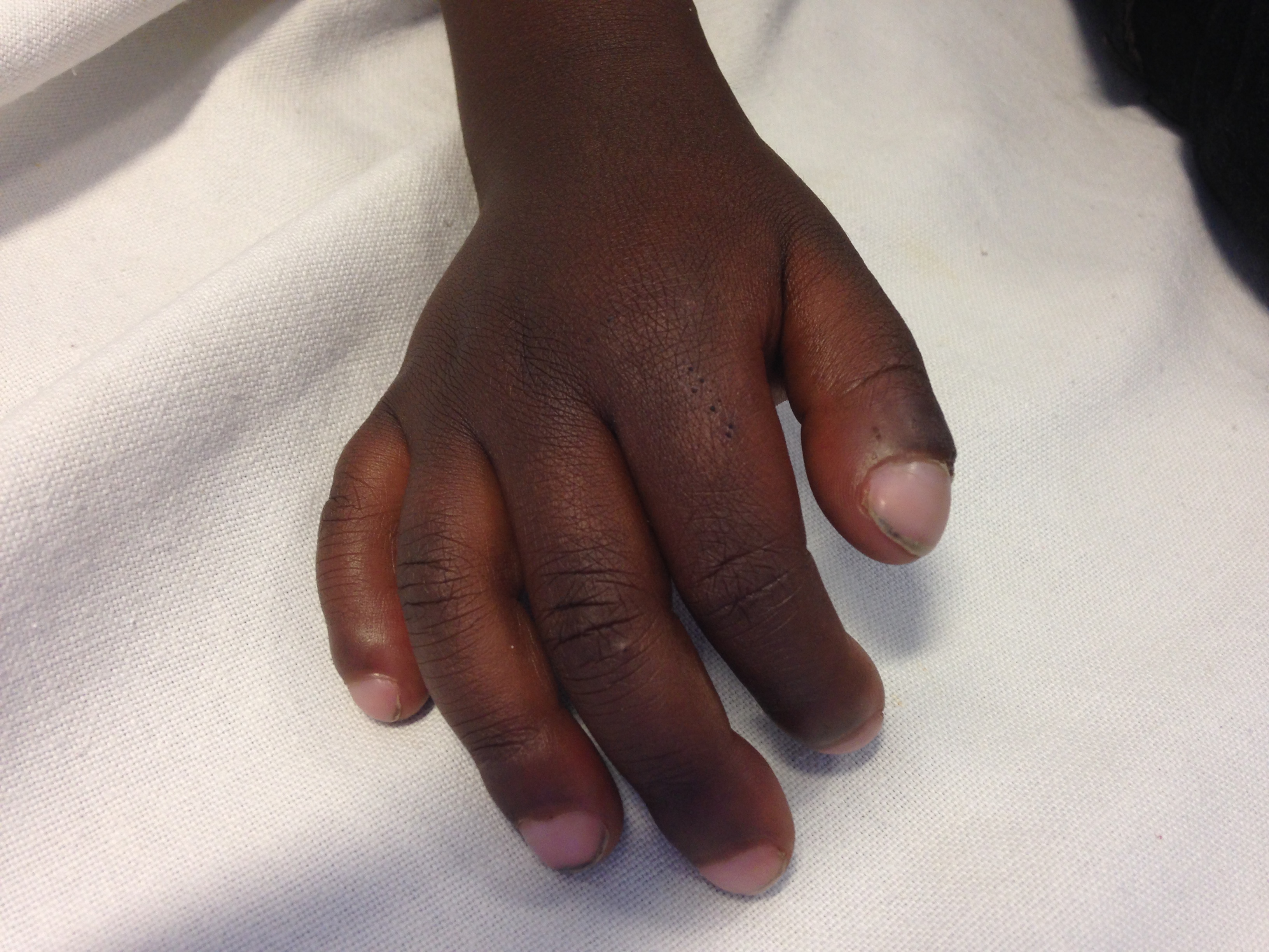
Question

What is this physical finding?

Show Answer

Answer: Clubbing

Differential Diagnosis of Clubbing in Children:

  • Cystic Fibrosis
  • TB
  • Sarcoid
  • Cyanotic congenital heart disease
  • Thyrotoxicosis
  • HIV
  • Malnutrition
  • Empyema
  • Hepatobiliary Disease

Clubbing was first documented by Hippocrates in a patient with empyema. It has been associated with cardiopulmonary disease, but also found in other disease processes such as hepatobiliary, infectious, and endocrine diseases. It can also occur without an underlying pathology. The pathophysiology behind these physical findings remain unknown. The workup and treatment should be aimed at managing the underlying cause.

University of Maryland Section for Global Emergency Health

Author: Van Pham, MD


Show References

Schwartz, Robert. Clubbing of the Nails. 3.12.14. http://emedicine.medscape.com/article/1105946-overview#showall


Title: Lung Ultrasound in Pulmonary Edema

Category: Critical Care

Keywords: lung ultrasound, pulmonary edema, B-lines (PubMed Search)

Posted: 3/11/2014 by Feras Khan, MD

  • “B-Lines” can be seen in patients with pulmonary edema (see attached image below)
  • A “B-line” is a reverberation artifact defined by Lichtenstein as having several properties:

1.     A comet-tail artifact

2.     Arising from the pleural line

3.     Well defined

4.     Hyperechoic

5.     Long (does not fade)

6.     Erases A lines

7.     Moves with lung sliding


  • A large amount of B-lines is pathologic
  • These artifacts are also called “comet-tails” due to their appearance
  • One or two B-lines can be seen in dependent lung zones in normal lungs
  • AIS (Alveolar interstitial syndrome) describes a group of conditions including pulmonary edema, interstitial pneumonia, and pulmonary fibrosis that show similar findings on lung ultrasonography
  • The most common presentation of this syndrome is from cardiogenic pulmonary edema and is characterized by B-lines in multiple lung zones
  •  B lines correspond with interlobular septal thickening on CT scans, which represent pulmonary vascular congestion 

Technique

  • B-mode is used with the micro-convex (cardiac) probe scanning in at least 8 lung zones
  • Quantify the number of B-lines in each zone
  • A lung zone is considered to be “positive” when three or more B-lines are present in a longitudinal plane between two ribs
  • Two or more regions bilaterally are required to be defined as AIS
  • Bilateral diffuse B-lines have a specificity of 95% and a sensitivity of 97% for the diagnosis of pulmonary edema

Show References

1.     Lichtenstein D, Mezie re G, Biderman P, et al. The comet-tail artifact. An ultra- sound sign of alveolar-interstitial syndrome. Am J Respir Crit Care Med 1997; 156(5):1640–6. 

Attachments

  • 1403111625_Fig6.1BLINESFINAL.jpg (55 Kb)


Title: Dexamethasone for acute asthma exacerbations

Category: Pediatrics

Keywords: asthma, pediatrics, dexamethasone, prednisone (PubMed Search)

Posted: 3/10/2014 by Danielle Devereaux, MD

Hot off the press! Pediatrics March 2014 just published results of a meta-analysis that compared 1 or 2 dose regimens of Dexamethasone versus 5 day course of Prednisone/Prednisolone for management of acute asthma exacerbations in pediatric patients. The results showed that Dexamethasone was as efficacious as the longer course of Prednisone. End points used were return trips to the emergency department and hospital admissions. On further review of the literature, parents tend to prefer the shorter duration of therapy with Dexamethasone. Also, there is less vomiting associated with Dexamethasone. There have been several articles published that show Dexamethasone is more cost-effective than Prednisone. Bottom line: consider giving single dose of Dexamethasone in the ER and then sending patient home with 1 additional dose.

Show References

Keeney G, Gray M, Morrison A, et al. Dexamethasone for Acute Asthma Exacerbations in Children: A Meta-analysis, Pediatrics March 2014, pp 493-499.

Williams K, Andrews A, Heine D, et al. Parental Preference for Short versus Long Course Corticosteroid Therapy in Children With Asthma Presenting to the Pediatric Emergency Department, Clinical Pediatrics January 2012, pp 30-34.

Andrews A, Wong K, Heine D, et al. A Cost-effectiveness Analysis of Dexamethasone versus Prednisone in Pediatric Acute Asthma Exacerbations, Annals of Emergency Medicine July 2012, pp 943-949


Previous  |  1 |  ... |  123 |  124 |  125 |  126 |  127 |  128 |  129 |  130 |  131 |  132 |  133 |  ... |  239 |  Next
University of Maryland School of Medicine logo
  • Departments
  • Programs
  • Research Centers
  • Institutes
  • Graduate Education
  • CIBR Cores
  • Offices of the Dean
    • University of Maryland, Baltimore
    • University of Maryland Medical Center
    • Medical Alumni Association of the University of Maryland
    • Health Sciences and Human Services Library
University of Maryland School of Medicine
655 W. Baltimore Street
Baltimore, MD 21201
Contact Us

© University of Maryland School of Medicine

    • Webmaster
    • Web Accessibility
    • Site Index
    • UMB Hotline
    • Employment
    • Map