
High-Flow Nasal Cannula for Apneic Oxygenation
- In recent years, much has been written about the use of apneic oxygenation for patients who require endotracheal intubation (ETI).
- Critically ill patients often have little cardiopulmonary reserve and can rapidly desaturate during ETI.
- High-flow nasal cannula (HFNC) devices can deliver heated, humidified O2 up to 60 L/min and can provide a modest amount of positive pressure.
- A recent study evaluated the use of a HFNC device for apneic oxygenation in ICU patients requiring ETI:
- Prospective, quasi-experimental, before-after study
- 101 patients in a single ICU in France
- Compared NRB + nasal cannula to HFNC for preoxygenation/apneic oxygenation
- Prevelance of severe hypoxemia (SpO2 < 80%) was significantly lower in the HFNC group
- Clinical Application: Consider using HFNC for apneic oxygenation in critically ill patients with mild-to-moderate hypoxemia who require ETI.
Show References
Question
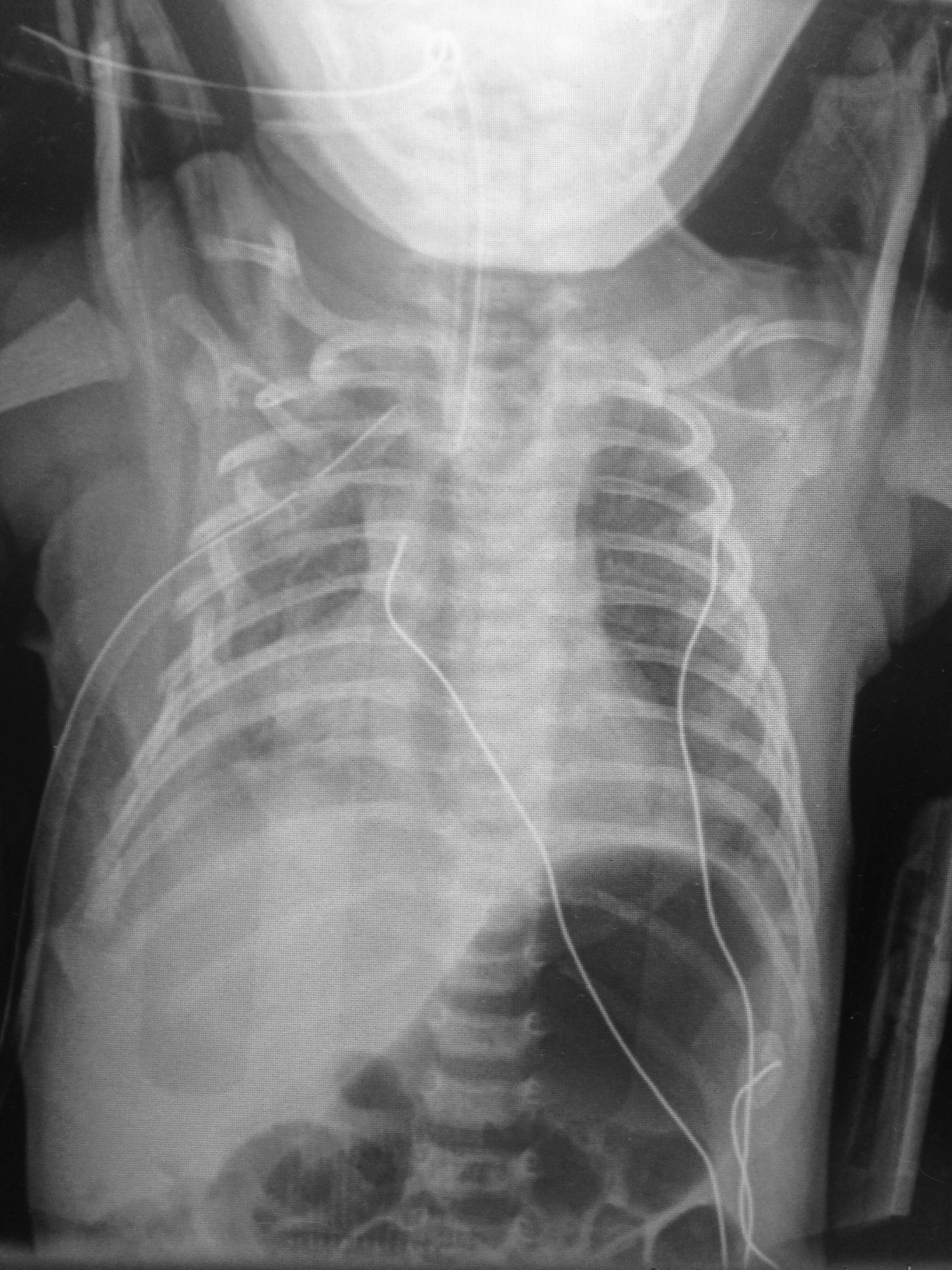
6 day-old child is brought in by parents with 1 day of reduced oral intake and 4 hours of rapid breathing. The child has no fever and no significant birth history. The child is tachycardic, hypotensive, and hypoxic. What’s the diagnosis?
Show Answer
Show References
Safety Risk? Digoxin in Atrial Fibrillation
- Digoxin is commonly utilize for atrial fibrillation/flutter with rapid ventricular response, though beta blockers and/or calcium channel blockers are a better 1st line therapy given digoxin’s narrow therapeutic index and lack of mortality benefit.
- Digoxin in the acute setting is often favored given its ability to reduce the heart rate while maintaining or slightly augmenting blood pressure.
- 2014 AHA/ACC guidelines recommend digoxin, specifically for rate control in patients with heart failure and/or reduced ejection fraction.
- There have been 2 post hoc studies from the AFFIRM trial which showed conflicting results w/regards to digoxin and risk of mortality.
1. Increased risk of mortality associated w/digoxin (on-treatment analytic strategy)
2. No association w/mortality (intent-to-treat analytic strategy)
- A recent retrospective cohort examination of newly diagnosed afib patients without heart failure & no prior use of digoxin; digoxin was independently associated with a 71% higher risk of death & a 63% higher risk of hospitalization.
- Consistent and substantial increase in mortality and hospitalization risk was seen using both on-treatment and intent-to-treat analytic methods.
- Given other available rate control options, digoxin should be used with caution.
Show References
Sesamoid Injuries
The first MTP joint contains the 2 sesamoid bones. They play a significant part in the proper functioning of the great toe. 30% of individuals have a bipartite medial or lateral sesamoid.
http://www.coreconcepts.com.sg/mcr/wp-content/uploads/2008/05/sesamoid_foot.jpg
Injury can occur from trauma, stress fracture or sprain of the sesamoid articulation or of the sesamoid metatarsal articulation. Overuse injuries tend to occur in sports with a great deal of forefoot loading (basketball/tennis).
SXs: Pain with weight bearing, pain with movement of first MTP, ambulation on lateral part of foot.
PE: Tenderness and swelling over medial or lateral sesamoid. Resisted plantar flexion (flexor hallucis) reveals pain and weakness.
Imaging: plain film with sesamoid view to assess for a sesamoid fracture. Stress fractures may take 3-4 weeks to show on plain film.
http://www.agoodgroup.com/running/Fracture002.jpg
Treatment for fractures and suspected stress fractures involve 4 to 6 weeks of non weight bearing.
Orthopedic Causes of Chest Pain
The first thing that pops into everybody’s mind when they hear a patient state they have chest pain radiating to the left arm is Acute Coronary Syndrome and specifically a Myocardial Infarction. However, there are a lot of orthopedic causes of chest pain that can also radiate to the left arm. It is estimate that up to 20% of patients with pectoral symptoms have an underlying orthopedic problem.
Some of them are:
- Herniated Disc
- Cervicothoracic tension syndrome
- Blockage of intervertebral or rib joints
- intercostal neuralgia
Some other less common causes are
- Arthritis of the shoulder
- Spondylocystitis
- Osteoporotic fractures
- Bone tumors
So instead of just ordering some troponin and admitting to medicine, consider that the cause can be orthopedic in origin.
Show References
Question
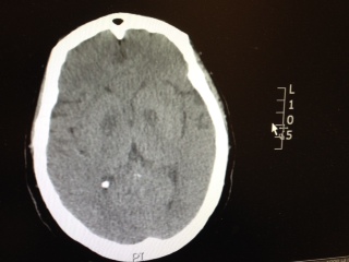
Patient has the following Head CT, what is your differential diagnosis? There are only a few characteristic toxins that can cause this type of finding on CT.
Show Answer
Neurologic causes of cardiac arrest have not been well described. Two recent retrospective studies looked at the epidemiology and clinical features of these patients.
Hubner P. et al.
- Over 20 years, 154 patients suffered cardiac arrest from neurologic causes.
- Diagnoses were made by CT in 123 patients (80%), autopsy in 28 patients (18%), and by history and clinical presentation in 4 patients (3%).
- PEA was the presenting rhythm in 77 patients (50%). Whereas 61 patients (40%) presented in asystole.
- Neurologic causes included subarachnoid hemorrhage in 74 patients (48%), intracerebral hemorrhage in 33 patients (21%), seizures in 23 patients (15%), and ischemic stroke in 11 patients (7%).
Arnaout M. et al.
- Over 13 years, 86 patients suffered out-of-hospital cardiac arrest from neurologic causes (2.3%).
- PEA was the presenting rhythm in 16 patients (19%). Whereas 66 patients (77%) presented in asystole.
- After ROSC, 64% of cases had ECGs with possible ischemic abnormalities.
- Neurologic causes included subarachnoid hemorrhage in 73 patients (85%), intracerebral hemorrage in 5 patients (6%), ischemic strokes in 5 patients (6%).
Neurologic causes of cardiac arrest are uncommon presentations that may be difficult to distinguish from cardiac etiology of cardiac arrest. If history and clinical presentation suggests a neurologic cause, obtain a non-contrast head CT for evaluation.
Show References
The Role of the CVP in a Post- “7 Mares” Era
The role for using central venous pressure (CVP) as a measure of volume responsiveness has largely fallen out of favor over the years.1 There are certainly better indices for fluid responsiveness, but don’t be fooled – the CVP isn’t a one trick pony. In fact, a high or rapidly rising CVP should raise a significant concern for impending cardiovascular collapse.
Consider the following differential diagnosis in the patient with an abnormally high or rising CVP ( >10 cm H2O).
- Excessive pressures outside of the heart or impediments to venous return (juxta-cardiac pressures)
- Cardiac tamponade
- Auto PEEP or breath stacking during mechanical ventilation
- Tension pneumothorax
- Venous return that’s more than the right ventricle can handle
- RV failure
- Severe tricuspid valve disease
- Massive increase in pulmonary vascular resistance (massive PE, pulmonary hypertension, ARDS, LV failure)
Bottom Line: In a time where the utility of the CVP has been largely dismissed, remember that an abnormal CVP offers great deal of information beyond a simple measure of volume status.
References
- Marik PE, Baram M, Vahid B. Does central venous pressure predict fluid responsiveness? A systematic review of the literature and the tale of seven mares. Chest. 2008;134(1):172-8.
- Berlin DA, Bakker J. Starling curves and central venous pressure. Critical Care. 2015;19(1):55.
Follow me on Twitter: @JohnGreenwoodMD
Question
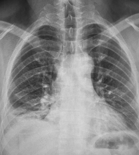
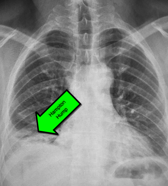
45 year-old male complains of pleuritic chest pain following a "long" flight. What's the diagnosis and what's this sign called?
Show Answer
Show References
This study is a case control study of the association of congenital heart disease (CHD) and stroke using a base population of 2.5 million Kaiser patients in California. 412 cases of stroke were identified and compared to 1236 controls. Of these stroke patients, 11/216 ischemic strokes and 4/196 hemorrhagic strokes were attributed to CHD (both cyanotic and acyanotic lesions). CHD was found in 7/1236 controls.
Children with CHD and history of cardiac surgery had the strongest risk of stroke (31 fold over the control group). Many of these children had strokes years after their surgery. Children with CHD who did not have cardiac surgery had a trend towards elevated stroke risk, but the confidence intervals included the null. More children without CHD history presented with headache.
Bottom line: Stroke risk (both hemorrhagic and ischemic) extend past the immediate postoperative period in patients with CHD.
Show References
The Centers for Disease Control continues to report increased numbers of measles patients in the US. From January 1 to February 13, 2015 there have been 141 cases. It has spread to 17 states and the District of Columbia, with 80% linked to the multistate outbreak from Disneyland.
Measles is not a benign disease!
Per the World Health Organization, there were 146,700 measles deaths globally in 2013. Most of these deaths occur in lower- and middle-income countries,
Even in the US, measles can cause serious complications and death. Complications from measles can be seen in any age group, but particularly in children <5 years of age and in adults >20 years of age.
Measles Complications:
Common:
- Ear infections (about 1 in 10 children)
- Can result in permanent hearing loss
- Diarrhea (about 1 in 10 people with measles)
Severe:
- Pneumonia (as many as 1 in 20 children)
- Most common cause of death
- Encephalitis (about 1 in 1,000 children)
- Can lead to seizures, hearing loss, intellectual disability
- Death (1 to 2 per 1,000 children)
- Measles in pregnancy can cause premature birth and low-birth-weight babies
Long-term:
- Subacute sclerosing panencephalitis (SSPE)
- Aside from the long-term complications above, an estimate 4 to 11 out of every 100,000 will develop this fatal disease of the central nervous system 7 to 10 years after infection.
Bottom Line:
Per Dr. Anne Schuchat of the CDC: “This is not a problem with the measles vaccine not working. This is a problem of the measles vaccine not being used.”
Show References
As the cold and snow rips through the United States, hypothermia is a major concern because each year approximately 1,300 Americans die of hypothermia.
Classification of hypothermia:
- Mild (32-35 Celsius): shivering, hyperventilation, tachycardia, but patients are usually hemodynamically stable.
- Moderate (28-32 Celsius): CNS depression, hypoventilation, loss of shivering, risk of arrhythmias, and paradoxical undressing
- Severe (<28 degrees Celsius): increased risk of ventricular tachycardia/fibrillation, pulmonary edema, and coma
The risk of cardiac arrest increases when the core temperature is less than 32 Celsius and significantly rises when the temperature is less than 28 Celsius. Rapid rewarming is required as part of resuscitation should cardiac arrest occur.
A rescue therapy to consider (when available) is extra corporeal membrane oxygenation (ECMO). ECMO not only provides circulatory support for patients in cardiac arrest, but allows re-warming of patients by 8-12 Celsius per hour.
Some studies quote survival rates of 50% with hypothermic cardiac arrest patients receiving ECMO versus 10% in similar patients who do not receive ECMO.
As winter lingers in the United States, consider speaking to your cardiac surgeons now to plan an Emergency Department protocol for hypothermic patients that may require ECMO.
Show References
Question
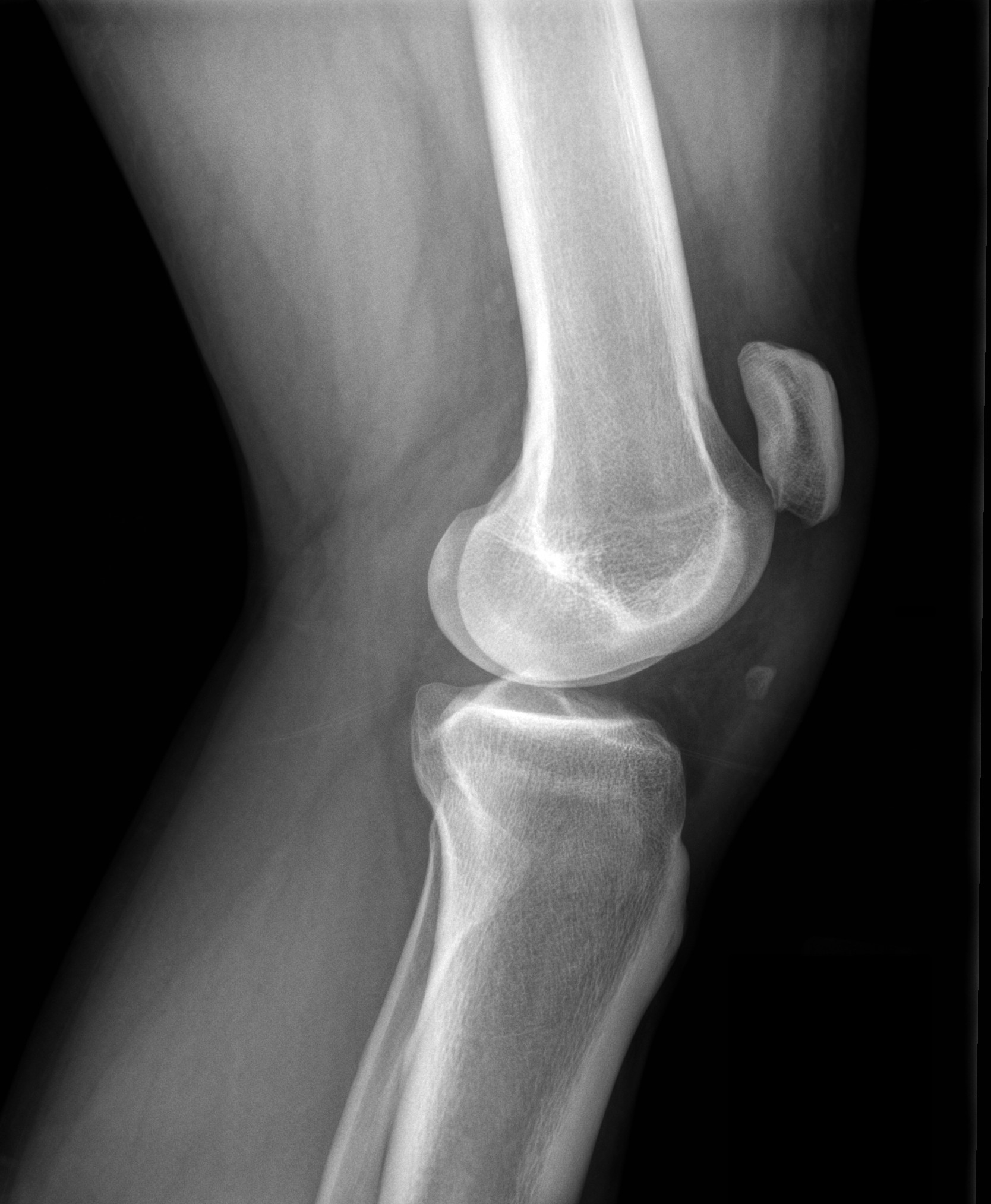
Show Answer
Show References
The Unforgotten: ECG Utilization to Differentiate Athletic Heart vs. Brugada
- Highly trained athletes develop ECG changes as a physiologic consequence of increased vagal tone; The ECG manifestations of early repolarization (ER) can range from simple J–point elevation to anterior (V1 to V3) "domed" ST-segment elevation and negative T wave.
- The former raises problems of differential some forms of ER with the “ coved-type” pattern seen in Brugada Syndrome (BS).
- A recent study compared the ECG tracings of 61 athletes w/a “domed” ST-segment elevation & negative T wave and 92 age/sex-matched BS patients w/a “ coved-type” pattern to identify an ECG criteria for distinguishing benign athletic changes seen in ER from BS.
- ECG analysis focused on ST-segment elevation at J-point (STJ ) and at 80 milliseconds after J-point (ST80 ).
- Athletes had a lower maximum amplitude of STJ (p < 0.001) & lower STJ /ST80 (p < 0.001)
- All patients (100%) with BS showed a downsloping ST-segment configuration (STJ/ST80 > 1) versus only 2 (3%) athletes (p < 0.001)
- An upsloping ST-segment configuration (STJ /ST80 < 1) showed a sensitivity of 97%, a specificity of 100%, and a diagnostic accuracy of 98.7% for the diagnosis of ER.
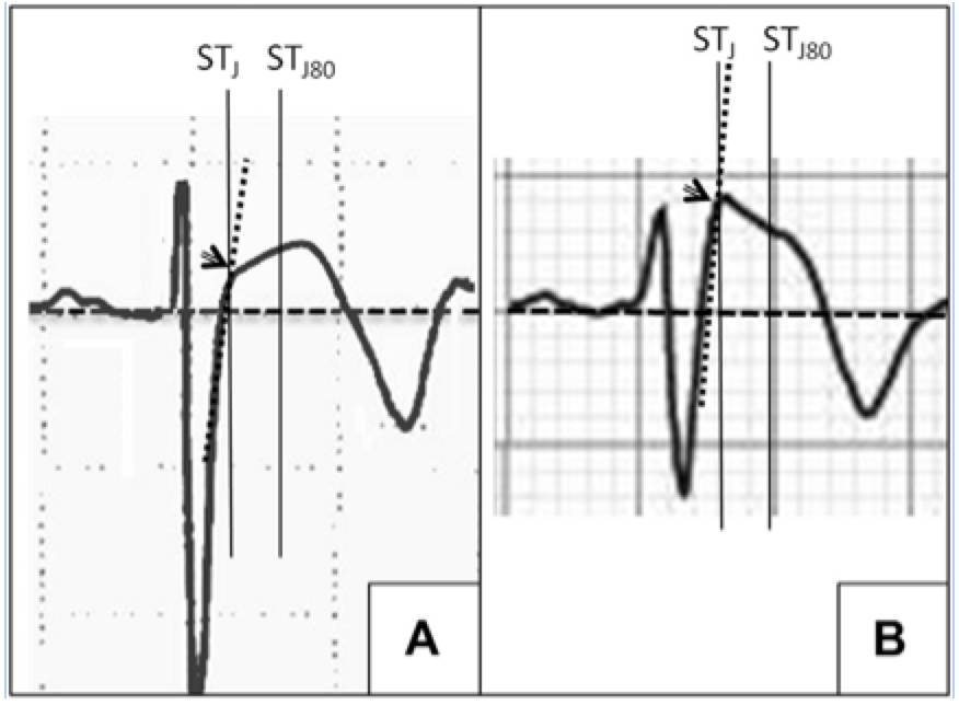
A: ER
B: Brugada
Show References
LATERAL ANKLE TENDINOPATHY
Hx: subacute onset (weeks) of the pain seen in athletes esp. runners (banked or uneven surfaces).
PE: Tenderness to palpation posterior to the lateral malleolus or over the course of the tendon. Pain worse with resisted ankle eversion from a dorsiflexed postion. Examine for subluxation of tendon.
The diagnosis is made from the above and does not require imaging.
Tx: Rest, conservative care, physical therapy (eccentric exercise focus), ankle taping or lace up brace. Severe cases may even require a walking boot.
http://www.epainassist.com/images/Article-Images/Peroneal_Tendonitis.jpg
Pediatric Caffeine Overdose
As the in-service draws closer and the hours to study wind down, I find myself becoming more and more of a caffeine enthusiast. While a No-Doz or Diet Mt. Dew may put a little more pep in my step, the caffeine found in energy drinks, caffeine pills, and diet supplements can quickly result in an dangerous overdose in a young child.
Caffeine Overdose Presentation- Sympathomimetic Toxidrome
- Tachycardia, dysrhythmia, hypertension
- Diaphoresis, piloerection
- Nausea, vomiting
- Hyperthermia
- Dilated pupils
- Agitation, delusions, paranoia
- Seizures, coma
- Sometimes: Metabolic acidosis, hypokalemia
- Rhabdo- muscle breakdown by Ca++ sequestration in the sarcoplasmic reticulum
Available Sources of Caffeine-
- NoDoz- 200mg/tab
- Excedrin 65mg/tab
- Starbuck Double Shot 130mg/6.5oz
- Monster Energy Drink 160mg/16oz
- Caffeine Solution for Neonates with Apnea of Prematurity
- So many more!
Toxic Doses
- 15 mg/L- tachycardia, arrhythmia, HTN, seizure, vomiting, irritable, delusions, hallucinations (approx 1500 mg for an adult)
- >80 mg/L- Coma or Death
Management- treat the symptoms (metabolic, cardiovascular, and neurologic)
- IV Fluids
- Anti-emetics
- Sodium Bicarb if refractory metabolic acidosis
- Benzos for severe agitation or seizure
- PALS protocols for cardiac arrhythmias
Good Luck on the In-Service!

Show References
A new review summarized published adverse effects when IV lipid emulsion is used along with venous-arterial extracorporeal membrane oxygenation (VA-ECMO) in patients with cardiotoxic drug poisoning.
Not surprisingly, running fat through the ECMO circuit can cause some issues. Here's what's been published:
- cracking of stopcocks
- fat emulsion agglutination
- clogging and associated malfunction of the membrane oxygenator
- increased blood clot formation in the circuit
It's unclear how these findings should change management if using both treatment modalities, but at the very least, be aware that fat depostion in the VA-ECMO circuits and increased blood clot formation can occur.
Show References
Magnesium, another failed neuroprotectant?
Stroke is a leading cause of adult disability and the second leading cause of death worldwide. Currently available therapies for acute ischemic stroke are based on restoring perfusion to the ischemic penumbra. However, they are only moderately effective.
A series of pathological cascades leading to neuronal death are triggered in acute ischemia. Thus it may be logical to suggest that if one can interrupt the propagation of these cascades, perhaps part of the brain tissue can be protected and salvaged.
Magnesium has been shown in various animal models to have pluripotent neuroprotective properties. It is also widely available, simple to administer, and has a favorable risk profile. A prior study of magnesium in acute ischemic stroke (IMAGES) did not show a benefit when the agent was administered a median 7.4 hours after symptom onset. However, a subgroup of patients treated within 3 hours of symptom onset showed possible benefit.
The Field Administration of Stroke Therapy - Magnesium (FAST-MAG) trial, funded by the NIH, looked at magnesium administered within 2 hours after symptom onset on the degree of disability at 90 days after stroke as measured by the modified Rankin scale.
- A total of 1700 patients were included in the study.
- 73.3% of patients had a final diagnosis of ischemic stroke, compared with 22.8% with intracranial hemorrhage and 3.9% with stroke-mimicking condition.
- Of the patients with ischemic stroke, 52.4% were treated with tPA.
Magnesium was not found to have any benefit in functional outcome at 90 days.
This study was unique in several ways:
- It examined the use of a neuroprotective agent in the hyperacute window of 2 hours, as a common criticism of prior neuroprotective studies is that those agents may not have been administered within the optimal therapeutic window
- Administration of the neuroprotective agent began in the prehospital setting. Logistically, this was done with a pre-randomized study kit containing the initial bolus dose to be administered by EMS and the maintenance dose to be given to the receiving hospital for administration in the ED.
- Despite the short window for enrollment and drug administration, patients were screened using a previously validated prehospital stroke scale and 98.7% of patients were enrolled after explicit written informed consent from the patient or a legally authorized representative.
However, despite this study being very well executed, demonstrating the feasibility of conducting a phase 3 trial with targeted intervention within the hyperacute window, it is another neuroprotective agent that failed to translate from the laboratory bench to the clinical realm.
Potential explanations for the discrepancies between preclinical and clinical outcomes of neuroprotective agents thus far include discrepancies on outcome measures, functional assessments, pre-morbid conditions, therapeutic windows, and drug-dosing schedules between animal studies and clinical trials.
Take Home Point: Magnesium does not have any clear benefit in acute ischemic stroke at this time.
Show References
Updates in preventative strategies in the ICU
Preventing Ventilator Associated Pneumonia (VAP)
- Traditionally ICUs use techniques such as head of bed elevation> 30 degrees, chlorhexidine mouth rinses, reduced sedation, and controlling cuff pressure between 20-30 cm H2O to reduce VAP
- A new trial confirms that subglottic suctioning also reduces VAP
- Endotracheal tubes are made with a suction line along the edge with fenestrations below the vocal cords and above the cuff
- This is hooked to wall suction removing secretions before they are aspirated
- VAP rates are very low in the US (most likely due to under-reporting)
- It is reported at around 15 VAPs/ 1000 ventilator days in Europe
The trial
- 5 ICUs in Belgium; 352 total patients with suctioning vs control were randomized
- Reduced incidence of confirmed VAP 9% vs 18%, decrease ventilator days 10 vs 20 and antibiotic use 7% absolute reduction
Bottom Line
- More expensive around $20 or more vs $1 for a regular ETT
- NNT around 11 to prevent one VAP: it is cost efficient
- Use them in patients who will remain intubated for > 48hrs (not elective surgical patients)
Daily bathing with chlorhexidine does not reduce health care associated infections
- It is believed that daily bathing with chlorhexidine antibiotic washes decrease rates of infection in the ICU; this is debatable
The trial
- One center, 5 ICUs, 9340 patients
- 10 week cleaning period followed by a two week washout then crossover to the alternate treatment (non-antibiotic washes)
- Looking for CLABSIs, CAUTIs, VAP and C. diff infections
- 55 infections occurred in the chlorhexidine group; 60 in the control goup.
- 2.86 per 1000 patient days (chlorhexidine group) vs 2.9 per 1000 patient days (control)
Bottom Line
- Does not appear to be helpful (perhaps specific patient groups such as bone marrow units may benefit)
- More expensive to use these washes and can lead to resistance
- Very well designed study with a variety of ICUs used (although one center)
Show References
We know vancomycin should be dosed based on weight rather than the default 1 gm dose so many patients receive. A past Academic Life in EM post explores the nuances of proper vancomycin dosing. But do higher loading doses in the ED actually lead to more therapeutic trough levels?
New Data
A new randomized trial compared ED patients receiving 30 mg/kg initial doses vs. 15 mg/kg. [1] There was a significantly greater proportion of patients reaching target trough levels of 15 mg/L at 12 hours among the patients who received a 30 mg/kg loading dose as compared with a traditional 15 mg/kg dose (34% vs 3%, P < 0.01). This study did not use a max dose of 2 gm. They included patients up to 120 kg who received 3.6 gm loading doses! Patients with creatinine clearance < 50 mL/min were excluded. There was no difference in incidence of nephrotoxicity between the groups.
Application to Clinical Practice
- Advocate for change in your ED's order sets to weight-based dosing of vancomycin and remove 1 gm as a default option. [2, 3]
- While 34% attaininment of adequate trough levels still isn't great, properly loading vancomycin with up to 30 mg/kg is a step in the right direction. It also takes longer than one dose to reach steady-state levels.
- This study did not evaluate clinical cure rates, just trough levels.