Search
Subscribe to daily UMEM Pearls (FREE!) via:
281-300 of 889 results with category "Critical Care"
This week we anticipate treating more COVID19 cases as they progress to ARDS. The World Health Organization issued guidelines on 3/13/20 for treating Severe Acute Respiratory Infection (SARI) due to COVID19.
How to identify ARDS?
No different than before COVID. Order a CXR, ABG, and perform bedside ultrasound evaluation of cardiac function and volume status. If there are bilateral opacifications you cannot explain entirely with volume overload, nodules, or lobar collapse, AND if the ratio of PaO2/FiO2 is < 300 (mild), < 200 (moderate), or < 100 (severe), then treat for ARDS.
***While you are waiting for your blood gas, SpO2/FiO2 <315 suggests ARDS.
What is the oxygen goal?
During resuscitation: > 93%
Once stabilized: > 89%
What is the expected clinical course?
Patients experience RAPID deterioration to respiratory failure. You should expect to intubate. This should be performed with N95 protection and should be done by the person with greatest first pass success.
Be CONSERVATIVE with fluids. Do not give a 30mL/kg bolus. Give 250-500mL bolus and re-evaluate. Excess fluid results in prolonged hypoxia and mechanical ventilation.
Should empiric treatments change?
No. Co-infection with influenza, bacterial pneumonia, and all other pathogens is possible, so you should continue to cover all suspected pathogens and de-escalate as microbiology labs result.
Should ventilator settings change?
No. Use lung protective volumes and permissive hypercapnia. The volume is based on the patient's height, not weight. A quick way to do this? Measure the height in cm. Subtract 100 for a man and subtract 110 for a woman and this is the ideal body weight. Provide 6mL/kg of tidal volume with a goal plateau pressure < 30. Use the high PEEP strategy from the ARDSnet trial and even consider clamping the ET tube when transitioning from machine to bag for transport in order to preserve PEEP.
Do patients benefit from proning?
Yes. 12-16 hours/day for severe ARDS. Not true in pregnancy as a whole, though early pregnancy may still benefit.
Is ECMO beneficial in refractory cases?
Unknown. In the case of MERS-CoV, ECMO reduced mortality.
Are corrticosteroids useful?
No. Do not administer steroids routinely to these patients. You may give steroids where indicated, including cases of refractory shock following pressors.
Show Additional Information
Show References
(*It is important to note that many of the percentages in these early studies will change as more asymptomatic or minimally symptomatic patients are identified with increased testing)
Epidemiology
Among more than 44,000 confirmed cases of COVID-19 in China as of Feb 11, 2020:
- 30–69 years: ~78%
- severely or critically ill: ~19%
Case-fatality proportion:
-60-69 years: 3.6%
-70-79 years: 8%
-≥80 years: 14.8%.
-With no underlying medical conditions: overall case fatality of 0.9%
-With comorbidities:
-cardiovascular disease (10.5%), diabetes (7%)
-chronic respiratory disease, hypertension, and cancer (6% each)
Presentation
For patients admitted to the hospital, many non-specific signs and symptoms:
- fever (77–98%) and cough (46%–82%) were most common
- of note, gastrointestinal symptoms (~10%) such as diarrhea and nausea present prior to developing fever and lower respiratory tract signs and symptoms.
Diagnosis
No general lab tests have great sensitivity or specificity
A normal CT scan does NOT rule out COVID-19 infection
-In an early study, 20/36 (56%) of patients imaged 0-2 days (‘early’) after symptom onset had a normal CT with complete absence of ground-glass opacities and consolidation
Treatment-
Mainstay of treatment will be management of hypoxemia including early intubation if necessary. However, specifically:
-Steroid therapy is controversial and the WHO is currently recommending against it unless it is being administered for another reason
-has not been associated with any benefit
-associated with possible harm in previous smaller studies with SARS and MERS
-associated with prolonged viremia
-intravenous remdesivir (a nucleotide analogue prodrug with promising in-vitro results against SARS-CoV and MERS-CoV) is available for compassionate use
-lopinavir-ritonavir has been used without any associated benefit
Show References
Settings: Multicenter randomized controlled trial
Patients: 710 patients
Intervention: 345 patients. no sedative but only boluses of morphine as clinically indicated (Sedation group)
Comparison: 356 patients. light sedation with daily interruption (Nonsedation group)
Outcome: all-cause mortality at 90 days after randomization
Study Results:
42.4% of nonsedation group died vs 37% of sedation group (95% confidence interval [CI], −2.2 to 12.2; P = 0.65).
Number of ventilator-free days for nonsedation group was 27 days vs. 26 for sedation group.
Discussion:
This study did not agree with previous studies that lighter sedation was associated with shorter length of stay on mechanical ventilation , ICU or hospital. The authors attributed to the findings that RASS score was not significantly different between the 2 groups.
Conclusion:
Critically ill adult patients receiving mechanical ventilation, there was no difference in 90-day mortality between patients receiving light sedation or no sedation.
Show References
With ED-boarding of critically-ill patients becoming more common, it is likely that ED physicians may find themselves caring for a patient who develops ACS – that is, abdominal compartment syndrome. While intraabdominal hypertension (IAH) is common and is defined as intraabdominal pressure > 12 mmHg, ACS is defined as a sustained intraabdominal pressure > 20mmHg with associated organ injury.
WHY you need to know it:
ACS → Increased mortality & recognition is key to appropriate management
WHO is at risk:
- Decreased abdominal wall compliance (obese, post-surgical)
- Increased intrabadominal contents (hemoperitoneum, ascites, tumor)
- Increased intraluminal contents (gastroparesis, ileus)
- Capillary leak / aggressive fluid resuscitation (sepsis, burns)
HOW it kills:
- Decreased blood flow to organs due to extraluminal pressure (mesenteric, renal, hepatic ischemia)
- Decreased diaphragmatic mobility, hypoventilation/oxygenation
- Decreased venous return, decreased cardiac output
→ Lactic acidosis, respiratory acidosis, multisystem organ failure, cardiovascular collapse & death
WHEN to consider it:
- Most patients who develop ACS are already intubated or altered – but consider in responsive patients c/o severe abdominal pain, marked distension, and SOB with tachypnea
- Intubated patients – recurrent, ongoing high pressure alarms with relatively low lung volumes, tachypnea
- Abdomen distended and minimally ballotable
- New / worsening oliguria / anuria
- Labs demonstrate increased creatinine, LFTs, lactate elevated “out of proportion” to patient presentation prior to decompensation
- Imaging may reveal underlying etiology or sequelae of ACS but cannot rule it out
WHAT to do:
- Confirm diagnosis with bladder pressure (via urinary catheter) *see cited paper for how-to in the ED*
- Emergent surgical consultation (emergent laparotomy → improved hemodynamics, organ function, & survival.
- Optimize abdominal perfusion pressure (MAP - intraabdominal pressure; recommended > 60mmHg) as much as possible:
- Adequate analgeisia and sedation, if needed, to avoid agitation
- Avoid intubation if able, to avoid the positive pressure. In intubated patients, aim for low PEEPs and plateau pressures and consider short-term paralytic
- Lower the head of bed (supine to 30mmHg) to minimize abdominal "crunch"
- Aim for intravascular euvolemia. If volume overload is a contributing factor then IVF for hypotension will worsen the ACS -- start vasopressor instaed
- Evacuate intraluminal contents if able (NGT/rectal tube for decompression, consider erythromycin/reglan, or neostigmine for colonic pseudoobstruction)
- Evacuate intraabdominal extraluminal contents if able (therapeutic paracentesis for ascites(
- Burn patients with restrictive abdominal eschar should get escharotomy
Bottom Line: Abdominal compartment syndrome is an affliction of the critically ill, is assosciated with worsened mortality, and requires aggressive measures to lower the intraabdominal pressure while obtaining emergent surgical consultation for potential emergent laparotomy.
Show References
Don't forget cerebral fat embolism syndrome (FES) on the differential for altered trauma patients. FES is typically associated with long bone fractures, but has been reported with other fractures, orthopedic reaming (i.e. aggressive orthopedic procedures), and in rare cases even with non-fracture (soft-tissue) trauma. Typically symptoms occur between 24 and 72 hours after injury, but there have been cases both earlier and later. Diagnosis is clinical, but MRI may be helpful, and will often show multiple cerebral white matter lesions. It is debated whether FES is truly an embolic phenomena (i.e fat molecules traveling to and blocking blood supply of organs), or rather an inflammatory response to free fatty acids in the blood stream (i.e. more of a vasculitis type pathology). Management is supportive care, but give these patients time as there can be favorable outcomes, even after prolonged coma.
Show References
Dr. Bryan Hayes wrote a Pearl 10/4/2013 to remind us autoimmune encephalitis can present like neuroleptic malignant syndrome.
Dr. Danya Khouja wrote a Pearl 6/28/2017 to inform us autoimmune encephalitis is associated with tumors and can be investigated with serum and CSF antibody panels.
Since those publications, the number of validated autoimmune biomarkers in these panels has increased dramatically. In 2020 we now know, autoimmune encephalitis is at least as common as infectious encephalitis.
Here is how to diagnose it
1. Suspect the diagnosis in patients with subacute/rapidly progressive altered mental status, memory loss, or psychiatric symptoms. It can be mistaken for a new diagnosis of schizophrenia or bipolar disorder.
2. Look for one or more additional findings: new seizures, focal CNS findings, CSF pleocytosis, MRI findings
3. Exclude other likely etiologies (but try not to get hung up on a positive drug test, especially if drug use was not recent).
Why is this important?
Early treatment with steroids and plasmapheresis can prevent progression of disease (prevent seizures, prevent months-long hospitalizations).
Young girls are especially likely to have teratomas as a cause for the disease. Finding and resecting those tumors is life-saving.
Show Additional Information
Show References
Mechanical Ventilation Pearls for Acute Ischemic Stroke
- Patients with an acute ischemic stroke (AIS) may require intubation for various reasons.
- Two main goals of mechanical ventilation in patients with an AIS are to maintain appropriate oxygen levels and tight control of PaCO2.
- In terms of oxygenation:
- Target normoxia
- Administer O2 if the SpO2 is < 94%
- Supplemental O2 is not recommended in non-hypoxic patients
- In terms of CO2:
- Target normocapnia
- Hypercapnia increases the risk of intracranial hypertension
- Hypocapnia can worsen cerebral perfusion
Show References
Settings: multicenter, double-blind, phase 3 trial (apparently vitamin D worked in phase 2 trials).
- Patients:
- 1059 patients were enrolled within 12 hours of ICU admission. The patients had to have risk factors warranted ICU admisions (pneumonia, sepsis, mechanical ventilation, shock, pancreatitis, etc.).
- Vitamin D deficiency was defined as plasma level < 20 ng/ml
- Intervention:
- 531 patients received a single oral dose of 540,000 IU of vitamin D3 within 2 hours after randomization
- Comparison
- 528 patients received placebo
- Outcome
- 90-day all-cause mortality
Study Results:
- Total SOFA score was similar in both groups (5.6 vs. 5.4).
- On day 3, mean plasma vitamin D was higher (47 ng/ml) in treatment group vs 11 ng/ml in placebo group
- 90-day all cause mortality was similar. Treatment group was 23.5% vs. 20.6% for placebo (95% CI, −2.1 to 7.9; P = 0.26).
- Vitamin D-related adverse events were similar in both groups.
Discussion:
- This trial enrolled patients early in their critical illness compared to phase 2 trial which enrolled patients after 3 days in the ICU.
- This phase 3 trial also enrolled mostly medical-related illness, whereas 75% of patients in phase 2 had either surgical or neurology-related illnesses.
Conclusion:
Early administration of high dose vitamin D did not improve 90-day all cause mortality.
Show References
The arrival of a critically ill pregnant patient to the ED can be anxiety-provoking for emergency physicians as two lives and outcomes must be considered.
Some basic tenets of care, regardless of underlying issue, include:
- Obtain IV access above the diaphragm to avoid delay/prevention of administered products reaching central circulation due to compression of the IVC by the gravid uterus.
- Provide supplemental oxygen as needed to maintain a saturation of >95% which corresponds to a PaO2 >70 mmHg. A PaO2 <60 mmHg is associated with fetal hypoxemia which will quickly lead to fetal acidosis and bradycardia.
- Goal maternal PaCO2 is 28-32 mmHg; this respiratory alkalosis maintains a CO2 gradient to help shift offload fetal CO2 into the maternal circulation for clearance.
- Hypotensive pregnant patients with a large uterus (20+ weeks) should be turned to the left lateral decubitus position or tilted leftward by at least 15 degrees to offload aortocaval compression and minimize secondary decrease in venous return) by the gravid uterus.
- In cases of maternal cardiac arrest, the patient should be kept supine for chest compressions with the gravid uterus manually displaced to the left.
- Keeping the mother alive is the best way to keep the fetus alive. Standard sedatives, vasopressors, and inotropes are okay if they are needed. Exception for ketamine, which has mixed effects in existing studies and while low doses are probably safe if needed, use as a firstline agent is not recommended. Notify the NICU team of medications given to mother if there is a precipitous delivery.
- Fetal tococardiometry monitoring if available, or regular POCUS assessment of FHR, in all viable pregnancies.
Finally, once critical illness is identified the OB and NICU teams should be consulted immediately. Fetal distress in a viable pregnancy may be an indication for delivery, and initiation of the transfer process should occur if the supportive specialties are not in-house.
Show References
There are few conditions that can be as dramatic or difficult to control as variceal GI bleeding in a cirrhotic patient. It is important to be familiar with all options in these cases, from Blakemore/Minnesota tube placement to massive transfusion to when and which consultants to get involved. In cases that are refractory or not amenable to endoscopic intervention, emergent interventional radiology consultation for Transjugular Intrahepatic Portosystemic Shunt (TIPS) may be a consideration. In high risk cases, think about getting IR on the phone at the same time as you engage GI, in case endoscopic management fails. Variceal bleed patients can decompensate rapidly, get your consultants involved early!
Generally accepted indications for emergent TIPS (both of the following should be true):
-GI bleeding not amenable or not controllable by endoscopy
-Cause is felt to be variceal. May also consider in portal hypertensive gastropathy
Contraindications:
-Right heart failure or pulmonary hypertension
-Severe liver failure (MELD > 22, T Bili > 3 or Child-Pugh C. In these cases TIPS may not confer a significant survival benefit)
-Hepatic encephalopathy (relative contradindication. HE may be worsened by TIPS).
-Polycystic liver disease (makes TIPS technically challenging)
-Chronic portal vein thrombus (makes TIPS technically challenging. Acute PV thrombus is NOT considered a contraindication)
Bottom Line: In cases of variceal GI bleeding from portal hypertension, consider getting IR on the phone early to discuss emergent TIPS.
Show References
Pearl: consider desmopressin (DDAVP) for patients with an intracranial hemorrhage who are taking an antiplatelet. Caution, this is not for patients with an ischemic stroke with hemorrhagic conversion and it was not specifically evaluated for patients on anticoagulation or going to the OR with neurosurgery.
How strong is this evidence? International guidelines already give cautious approval for this practice, and now there is a retrospective review to support it. Though there were only 124 patients in the trial, the rate of hemorrhage expansion was much lower in the DDAVP group (10.9% vs 36.2%, P = .002) and there was no increased risk of hyponatremia (no events reported).
Show Additional Information
Show References
Interventions Shown to Reduce Mortality in RCTs
- Santacruz and colleagues recently performed a systematic review to determine which multicenter RCTs in critically ill patients have shown that an intervention was associated with a reduction in mortality.
- Approximately 13% of the 212 trials included in this review reported a statistically significant reduction in mortality. Unfortunately, many of the interventions were not associated with reduced mortality in subsequent studies.
- Interventions consistently shown to reduce mortality in multicenter RCTs in critically ill patients were limited tidal volume in patients with ARDS, noninvasive ventilation in acute hypercapnic respiratory failure, and noninvasive ventilation following extubation in complex cases.
- Corticosteroids in septic shock, selective digestive decontamination, and prone positioning in ARDS remain controversial.
Show References
Settings
- Patients: mechanical ventilation in the ICU. Randomization of 1000 patients.
- Intervention: conservative oxygen therapy, if spO2 reached 97%, then FiO2 was lowered to 0.21
- Comparison: no specific limits for FiO2 or SpO2.
- Outcome: number of ventilator-free days at 28 days after randomization.
Study Results:
- 484 conservative-oxygen group vs 481 to the usual oxygen group
- Comparing to the conservative-oxygen group had:
- more time at FiO2 21 (29 hours vs. 28 hours),
- less time with SpO2 > 97% (27 hours vs. 49 hours)
- Similar ventilator-free days: 21 days vs. 22 days.
Discussion:
This study’s results differed from previous single center study (Girardis JAMA 2016) or meta analysis (Chu DK, Lancer 2018), which showed mortality benefit in patients with conservative oxygen (Girardis & Chu) and more ventilator-free days (Girardis).
Conclusion: Conservative oxygen did not significantly affect the ventilator free days of mechanically ventilated patients.
Show References
When managing cardiac arrest, it is important to differentiate PEA, the presence of organized electrical activity without a pulse, from "pseudo-PEA,"where there is no pulse but there IS cardiac activity visualized on ultrasound.
Why:
- Pseudo-PEA is essentially a profound, low-flow shock state that often has reversible causes, such as hypovolemia, massive PE, tension pneumothorax, etcetera.
- Compared to PEA, with appropriate care patients with pseudo-PEA have a higher rate of ROSC as well as overall survival.
How:
- POCUS during rhythm check in cardiac arrest. Be careful not to prolong the pause in compressions; acquire the US, if needed, for review once hands are back on the chest.
What:
- In addition to searching for & addressing reversible causes of the pseudo-PEA, manage the profound shock state with pressors and/or inotropic support.
- In EDs where TEE is utilized during cardiac arrest resuscitations, strongly consider synchronization of external compressions with intrinsic cardiac activity to potentially improve ventricular filling and therefore coronary perfusion pressure.
Bottom Line: Pseudo-PEA is different from PEA. Utilize POCUS during your cardiac arrests to identify it and to help diagnose reversible causes, and treat it as a profound shock state with the appropriate supportive measures, i.e. pressors or inotropy.
Show References
Rapid Assessment of the RV on Bedside Echo
There are several causes of acute RV dysfunction resulting in a patient presenting to the ER with unstable hemodynamics. Some of these include acute cor pulmonale, acute right sided myocardial infarction and acute submassive or massive pulmonary embolism. While bedside assessment of the LV function is often performed by the ED physician, simultaneous evaluation of the RV can provide crucial information that can help guide therapeutic decisions to prevent worsening of the patient’s clinical condition. A rough guideline to determine RV size and function is below using the apical 4 chamber view.
Normal RV size : <2/3 the size of the LV
Mildly enlarged RV : >2/3 the size of the LV, but not equal in size
Moderately enlarged RV: RV size = LV size
Severely enlarged RV: RV size > LV size
Patients who are found to have RV dilation should be given fluids in a judicious fashion as the RV is not tolerant of fluid overload. Early diagnosis of the cause of acute RV failure should be sought to guide definitive therapy, but early institution of inotropic support should be considered. Frequent reassessments of biventricular function during resuscitation should be performed.
Show References
Attachments
Ever been in an acute rescucitation and found yourself unable to remember all of those famous ACLS Hs and Ts? I know I have. A few years ago Littman et al published an alternative approach to critically ill, hypotensive medical patients with non shockable rhythms. Unfortunately, it seems like some of the enthusiasm for this approach has died down, but I still think it's something you're more likely to recall in a pinch than the Hs and Ts and is a better way of getting started with a hypotensive non-trauma patient. And it's so simple you may actually remember it!
1) Look at the monitor. Is the rhythm narrow or wide?
2a) Narrow - more likely a mechanical problem (tamponade, tension PTX, autoPEEP, or PE). Give IVF and search for one of these causes (and correct it!). Keep in mind that ultrasound can help you differentiate a lot of these.
2b) Wide - more likely a metabolic problem (hyperK, sodium channel blockade, etc*). Give empiric calcium, bicarb, and other therapies targeted for these problems (if desired) and get stat labs.
Take a minute and either go to this REBEL EM post:
https://rebelem.com/a-new-pulseless-electrical-activity-algorithm/
To review this, or look at the attached diagrams.
*Dr. Mattu would want me to remind you that hyperkalemia IS a sodium channel poisoned state, so there's no need to think of these two separately
Show References
Attachments
- 1910292200_Narrow-Complex-PEA-Management-765x456.jpg (96 Kb)
- 1910292201_Wide-Complex-PEA-Management-765x444.jpg (83 Kb)
The Critically Ill Geriatric Patient with Sepsis
- Due to the age-related physiologic change of immunosenescence, geriatric patients have an increased susceptibility to infection, a decreased ability to mount a response to infection, and an increased likelihood of atypical presentations.
- Atypical presentations of sepsis in the geriatric patient include confusion, decreased functional status, generalized weakness, and failure to thrive.
- In fact, up to 33% of geriatric patients with bacteremia will be afebrile upon presentation.
- Consider sepsis in the differential diagnosis of geriatric patients with these nonspecific complaints.
Show References
Rationale: Data regarding temperature management in patients suffered from cardiac arrest with nonshockable rhythm was inconclusive.
Objective: whether moderate hypothermia at 33C, compared with normothermia at 37C would improve neurologic outcome in patients with coma after cardiac arrest with nonshockable rhythm.
Outcome: survival with favorable 90-day neurologic outcome (Cerebral Performance Category scale 1-2/5)
SummaryThere was higher percentage of patients achieving CPC 1-2 in the hypothermia group (10.2%) vs normothermia group (5.7%, Hazard Ratio 4.5, 95% CI 0.1-8.9, p=0.04)
This randomized multicenter trial involved 581 patients with cardiac arrest and nonshockable rhythm. Hypothermia group included 284 patients vs. 297 in the normothermia group. Median GCS at enrollment = 3.
Majority of patients was cooled with the use of a basic external cooling device: 37% for hypothermia and 50.8% for normothermia group.
There was higher percentage of patients achieving CPC 1-2 in the hypothermia group (10.2%) vs normothermia group (5.7%, Hazard Ratio 4.5, 95% CI 0.1-8.9, p=0.04)
Limitation:
A. The study used strict enrollment criteria:
- CPR initiation within 10 minutes;
- CPR to ROSC within 60 minutes;
- epinephrine or norepinephrine infusion at < 1 ug/kg/min;
- No Child-Pugh class C liver cirrhosis
B. normothermia group had higher proportion of patients with temperature at 38C.
C. Hypothermia group underwent temperature management of 56 hours vs. 48 hours for normothermia patients.
Take home points:
In a selected group of patients with cardiac arrest and nonshockable rhythm, moderate hypothermia at 33C may improve neurologic outcome.
Show References
Blood Transfusion Thresholds in Specific Populations
Sepsis - 7 g/dL
- non-inferior to 9 g/dL (which was previously recommended in early goal-directed therapy and early Surviving Sepsis guidelines)
Acute Coronary Syndrome - no current specific recommendations pending further studies
- recent MINT pilot study showed unexpected trend toward higher combined mortality and major cardiac events in restrictive transfusion arm (8 g/dL) vs. liberal arm (10 g/dL)
Stable Cardiovascular Disease - 8 g/dL
- no difference in 30-day mortality compared to 10 g/dL, excluding those who have undergone cardiac surgery
Gastrointestinal Bleeds
- UGIB - 7 g/dL (unless intravascularly volume depleted or h/o CAD)
- better 6 week-survival, less re-bleeding compared to 9 g/dL
- LGIB - 7 g/dL, limited evidence, but based on UGIB data
Acute Neurologic Injury - Traumatic Brain Injury - 7 g/dL
- no significant difference in neurologic recovery at 6 weeks or mortality vs. 10 g/dL, although there were more brain tissue hypoxia events in restrictive arm
- anemia and transfusions both associated with worse outcomes in TBI
Postpartum Hemorrhage - 1:1:1 ratio strategy
- FFP/RBC ratio ≥ 1 associated with improved patient outcomes
Show References
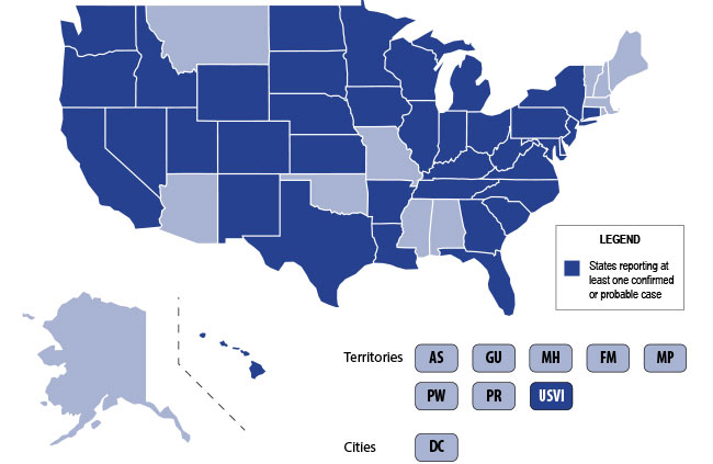
The U.S. is currently experiencing an epidemic of a severe lung disease termed Vaping-Associated Pulmonary Illness (VAPI), with over 500 cases and 7 deaths across 38 states and 1 U.S. territory since July 2019.
The clinical presentation of VAPI varies --
- Respiratory (SOB, cough, chest pain), constitutional (fever, tachycardia, headache, dizziness), and potentially GI symptoms (vomiting, diarrhea) after the use of vaping devices. GI symptoms may precede respiratory issues.
- Can take days or worsen over weeks and can present or end up with severe respiratory failure
Diagnostics --
- Labs nonspecific: Leukocytosis, elevated ESR, no specific infectious etiology
- Chest CT generally with bilateral infiltrates
- Bronchoscopy with BAL demonstrates PMNs and may have lipid-laden macrophages on Oil red O or Sudan staining
Treatment is supportive +/- steroids --
- Current recommendations to treat similarly to ARDS in intubated patients
- Potential benefit to steroids if not contraindicated
Bottom Line: Include vaping-associated pulmonary illness in your differential for patients presenting with acute lung disease.
- Ask patients about use of e-cigarette/vaping devices.
- Notify the CDC or your state health department of any suspected cases.
- Counsel your patients to avoid the use of these devices, at the very least until the specific causative agent is found.