
Search
Subscribe to daily UMEM Pearls (FREE!) via:
241-260 of 889 results with category "Critical Care"
Background:
The association between time intervals of ED antibiotic administration and outcome has been controversial. While single studies showed there was increased mortality associated with delayed antibiotic administration (1-3). A meta-analysis of 13 studies and 33000 patients showed that there was no mortality difference between septic patients receiving immediate Abx (< 1 hour) vs. those receiving early abx (1-3 hours) (4).
Since delay in recognition of sepsis (defined as ED triage to Abx order) and delay in antibiotics delivery (Abx order to administration) contribute to total delay of Abx administration, a new retrospective study (3) attempted to investigate the contributions of either factor to hospital mortality.
Results:
The study used generalized linear mixed models and involved 24000 patients.
For All patients and outcome of hospital mortality:
Recognition delay (ED triage to Abx order): OR 2.7 (95% CI 1.5-4.7)*
Administration delay at 2-2.5 hours (Abx order to administration): OR 1.5 (1.1-2.0)
These results was associated with non-statistical significance in patients with septic shocks.
Conclusion:
Delayed recognition of sepsis was associated with higher hospital mortality. Longer delay of abx administration was also associated with increased risk of hospital mortality.
Show References
Clinical Question:
- Does dexmedetomidine lead to better short-term and long-term outcomes than propofol in mechanically ventilated adults with sepsis?
Methods:
- Multicenter (13 US hospitals), double-blinded, 422 mechanically ventilated patients with sepsis
- Notable exclusion criteria: pregnant, 2nd- or 3rd-degree heart block or persistent bradycardia requiring intervention, indication for bzds, expected to have NMB > 48 hrs, already had received mechanical ventilation >96 hrs
- Pain was treated with opioid pushes or fentanyl gtt
- Primary end point: number of calendar days alive without delirium or coma during the 14-day intervention period
- Secondary efficacy end points included ventilator-free days at 28 days, death at 90 days, and global cognition at 6 months
Results:
- No sig difference in adjusted number of days alive without delirium or coma over the 14-day intervention period (dexmedetomidine: 10.7 days vs. propofol: 10.8 days; OR, 0.96; 95% CI, 0.74 to 1.26; P = 0.79
- No sig differences in the number of ventilator-free days at 28 days, in death at 90 days, or global cognitiion at 6 months
- Other notable findings:
- Fewer patients in the dexmedetomidine group had ARDS or signs of trial drug withdrawal
- Fewer patients in the propofol group extubated themselves
- Open-label propofol received by 13% in the dexmedetomidine group and 8% in the propofol group) and dexmedetomidine (4% in the dexmedetomidine group and 3% in the propofol group)
- Rescue midazolam was used in about half the patients, most often for procedural sedation or during NMB, 42% received antipsychotics
- Similar proportions of patients had organ dysfunction, hypotension, or severe lactic acidosis
- Symptomatic bradycardia requiring discontinuation of the trial drug was similar in the two groups
Take-home points:
- Dexmedetomidine or propofol are reasonable options for septic patients requiring mechanical ventilation without notable differences in delirium or mortality
Show References
Background: In respiratory failure due to COPD and cardiogenic pulmonary edema, noninvasive positive pressure ventilation decreases need for intubation and improves mortality,1 while its utility in other scenarios such as ARDS and pneumonia has yet to be proven.1,2 We know that patients on NIV with delays to needed intubation have a higher mortality,1,3 but intubation and mechanical ventilation come with risks that it is preferable to avoid if possible.
So how and when can we determine that NIV is not working?
The HACOR (Heart rate, Acidosis, Consciousness, Oxygenation, Respiratory rate) score at 1 hour after NIV initiation has been demonstrated to be highly predictive of NIV failure requiring intubation.4,5
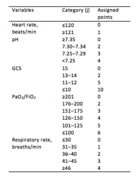
Initial development/validation: Score > 5 after 1 hour of NIV corresponds to >80% risk of NIV failure4
-
Earlier intubation (before 12 hours) in these patients = better survival
External validation: Score > 8 after 1 hour of NIV most predictive of eventual NIV failure 5
-
Average score @ 1-hour of patients with NIV success = 3.8
-
Score remained predictive at 6, 12, 24, 48 hours as well & mortality worsened as delay to intubation time increased
-
Baseline, pre-NIV score not predictive
-
Better predictive agreement in pneumonia and ARDS
Bottom Line:
-
Patients on NIV require close reassessment to prevent worsened survival due to intubation delay should invasive mechanical ventilation be indicated.
-
A HACOR score >8 after 1 hour of NIV should prompt intubation in most instances, with strong consideration given to a score >5.
*Note: ABGs were obtained for PaO2 assessment in the above studies -- the use of SpO2 was not evaluated -- but we are often not obtaining ABGs in our ED patients with acute respiratory failure. The following chart provides an estimated SpO2 to PaO2 conversion.
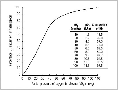
WHO 2001
Caveats:
- Pulse oximetry may be inaccurate in darker skin tones (overestimated by ~2%)6 and in certain disease processes (e.g. CO poisoning, profound shock states, etc.)
- The oxyhemoglobin dissociation curve shifts right with increasing pCO2/decreasing pH (lower saturation for a given PaO2).
Show References
Optimal oxygenation targets and the possible, theoretical, benefits of hyperoxygenating critically ill patients have long been points of controversy. Multiple studies have suggested harm in pursuing aggressive hyperoxygenation amongst critical patients with various conditions ranging from myocardial infarction to sepsis to neurologic conditions. In addition, oxygen toxicity is a known mechanism causing ARDS.
The HOT-ICU trial adds to the list of arguments against hyperoxygenation, by looking at 2928 ICU patients on high levels of supplemental oxygen and targeting a paO2 of 60 mm Hg (low oxygen group) vs paO2 of 90 mm Hg (high oxygen group). There was no difference in mortality, or other significant difference in outcomes.
Bottom Line: A lower paO2 goal of 60 (correlates to an O2 sat of 90%) is noninferior to a higher paO2 goal of 90 (O2 sat of approximately 96%). When titrating oxygen, targeting a pulse ox of 90-96% is reasonable in critically ill patients. Be sure to include an upper limit on the sat goal, beware an O2 sat of 100%, and titrate down supplemental oxygen when the spO2 is above goal, as the paO2 may be dangerously high.
Show References
Mechanical Ventilation in COPD
- Mechanical ventilation of the patient with obstructive lung disease can be challenging, primarily due to the presence of dynamic hyperinflation.
- In the initial phase of ventilation, it is important to prevent complications of hyperinflation and not to target normalization of blood gas values.
- Recommended initial ventilator settings include:
- Mode: Volume assist-control
- Inspiratory flow waveform: square
- Tidal volume: 6-8 ml/kg PBW
- RR: 12 bpm
- Inspiratory flow: 60-90 L/min
- The effect of PEEP is variable with each patient. When titrating PEEP, be sure to frequently measure plateau pressure and discontinue titration should Pplat increase.
Show References
| Ever wonder what to tell the families regarding prognosis of the sickest COVID-19 patients you intubate in the ED or ICU? Introduction: Case fatality rates (CFR) for COVID-19 patients requiring invasive mechanical ventilation have been widely variable. A study in the Blue Journal (AJRCCM) examined patients requiring intubation in a systemic review and meta-analysis. The case fatality risk of an infection is represented by the proportion of patients who die among all infected cases in a population over a period of time. Methods: 69 studies axross 23 countries for a total of 57,420 patients with COVID-19 who required mechanical ventilation were included in analysis. Results: Overall case fatality rate was 45%, or about 1 death for every two intubated patients.. Among studies that included age stratification, pooled estimates of CFR were 47.9% in patients <40 (CI 46.4-49.4%) and 84.4% in patients >80 (CI 83.3-85.4%). Overall heterogeneity is high (I 2 .90%), In early COVID epicenters, CFR was 70% among patients aged more than 60 years of age. CFR increased exponentially with increasing age. Specifically in the US across 21 studies (3,811 intubated patients), CFR was 47% (95% CI, 36–57%). Studies from New York reported a CFR of 54% (95% CI, 36–72%) whereas other regions in the United States reported a CFR of 41% (95% CI, 30–53%). Definitive hospital CFR (aka for those with hospital discharge outcomes, 13,120 patients) was 56% (CI 47-65%). Conclusion: Almost half of patients with COVID-19 receiving IMV died based on the reported CFR. The reported CFR was higher in older patients and in early pandemic epicenters, which may be influenced by limited ICU resources. Limitations: Overall CFR of 45% still included patients in the hospital. Definitive hospital outcomes were only for 13,120 patients (36.6%). Significant variation in CFR exists between studies. |
Show References
A single center (Nebraska, USA), retrospective analysis investigated the prevalence of intubation during hospital stay for 105 patients who had COVID-19 between March 24 to May 5, 2020 (1). 40 patients underwent awake proning vs. 60 patients did not undergo awake proning.
After adjusting for either SOFA or APACHE scores, patients with awake proning were associated with lower Hazard Ratios of intubation for SOFA (HR 0.30, 95% CI 0.09-0.96, p=0.043) and APACHE (HR 0.30, 95%CI 0.1-0.91, p=0.034).
Discussion
While this US study seemed promising, another Brazilian study being published earlier in July 2020 showed no difference in the prevalence of intubation between COVID-19 patients with proning or without proning (2).
These 2 studies highlighted the nature of this disease: high practice variability, uncertainty of therapeutic modalities. However, the complications from awake proning had been very low.
Conclusion:
Awake proning for hypoxic COVID-19 patients is a promising intervention but we will need more studies. In the meanwhile, we can try this therapeutic modality as the risk is low.
Show References
Study Question: What is the association of relative hypotension (degree and duration of MPP deficit) in patients with vasopressor-dependent shock with the incidence of new significant AKI and major adverse kidney events (MAKE)?
- Mean Perfusion Pressure (MPP) = MAP - CVP
- MAKE-14: composite measure of death, new initiation of RRT, or doubling of serum creatinine from the premorbid level at Day 14
- Basal MPP estimated using pre-illness BP readings in the chart, basal CVP estimated using prior echo findings or estimated mean values
Methods:
- Multicenter, prospective observational cohort study with 302 patients
- Notable exclusion criteria:
- age < 40, trauma as primary reason for ICU admission, active bleeding, unavailability of at least two preillness BP readings, pregnancy, "any condition specifically requiring a higher or a lower blood pressure target in the view of a treating clinician"
Results:
- for every percentage increase in the time-weighted average MPP deficit, the odds of developing new significant AKI and MAKE-14 increased by 5.6% (95% CI, 2.2–9.1; P = 0.001) and 5.9% (95% CI, 2.2–9.8; P = 0.002), respectively.
- Relationships between the risks of developing new significant AKI or MAKE-14 and the percentage of time spent with a MAP < 65 mm Hg were not statistically significant
Take-aways:
- Critically ill patients in shock who had higher and longer degrees of relative hypotension compared to their baseline BPs had a higher incidence of adverse kidney outcomes
- Sidenote: also consider venous congestion/volume overload when thinking about end-organ damage (e.g. MPP not just MAP)
Show References
PEEP in the Intubated Obese Patient
- Obesity has numerous adverse effects on the respiratory system, most notably a reduction in lung volumes.
- The reduction in lung volumes (i.e., FRC) often result in airway closure and atelectasis.
- The application of PEEP in the mechanically ventilated patient helps maintain alveolar patency by preventing derecruitment.
- Importantly, the typical initial PEEP setting of 5 cm H2O is insufficient for many ventilated obese patients.
- Pearl: In the ventilated obese patient start with an initial PEEP of 10-15 cm H2O.
Show References
Summary
Our group performed a meta-analysis to assess whether it is safe to infuse vasopressor through peripheral venous catheters. We identified 9 studies with a total of 1835 patients. The prevalence of complications among the pooled patient population was 9%. Up to 96% of the complications was extravasation and almost no complications required any treatment.
A few studies reported safe infusion of norepinephrine up to 0.1 mcg/kg/min for up to 24 hours.
In exploratory meta-regression, catheter size 20 or larger was negatively associated with the rate of complications.
We also observed that studies that were published within the past 5 years reported significantly lower rate of complications from older studies. This suggested that with careful planning and monitoring, it is safe to start vasopressor through peripheral IV.
Limitation
most of the included studies were observational. No studies had enough power to statistically analyze any variables that could predict complications.
Bottom line: we should start vasopressor as soon as indicated, if we have good, reliable IV access.
Show References
Show References
As the number of COVID-19 cases rises worldwide, prehospital and emergency department healthcare workers remain at high risk of exposure and infection during CPR for patients with cardiac arrest and potential SARS-CoV-2.
Existing evidence supports similar cardiac arrest outcomes in airways managed with a supraglottic airway (SGA) compared to endotracheal intubation (ETT).1 It is generally accepted that the best airway seal is provided with endotracheal intubation + viral filter, but how well do SGAs prevent spread of aerosols?
In CPR simulation studies:
- Cuffed endotracheal tube + viral filter provides effective seal to prevent aerosolization during CPR.2
- SGA + viral filter decreases AP spread compared to facemask and compared to bag-valve mask ventilation during CPR.3
- Notable aerosolization is seen with SGAs, with no difference between AuraGain, I-gel, LMA Proseal, LMA Supreme, Combitube, or LTS-D.2
- Ventilating through an SGA + viral filter is likely better to limit spread of aerosolized particles than bag-valve mask ventilation.
- SGAs allow egress of aerosolized particles, although the amount and area of distribution in clinical practice is unclear, and endotracheal intubation with a cuffed endotracheal tube remains the best way to avoid ongoing aerosolized particle spread with chest compressions.
- Appropriate PPE remains crucial to limiting healthcare workers' risk of infection and must be prioritized, even/especially in the management of patients in cardiac arrest.
Show References
N-acetylcysteine (NAC) is well known as the accepted antidote for acute acetaminophen (tylenol/paracetamol) overdose and is well studied for this indication. While the literature base is not nearly as strong in other causes of acute liver failure, NAC is increasingly used in these scenarios as well. In the emergency department in particular, the cause of fulminant hepatic failure is often not known. NAC may have some protective benefit in non-acetaminophen acute liver failure. Existing data do not show a mortality benefit to NAC in non-acetaminophen acute liver failure, but do show improvement in transplant-free survival. The AASLD guidelines (last revised in 2011) do not comment on NAC in non-acetaminophen acute liver failure. A common practice is to continue NAC until the INR is < 2 and AST/ALT have decreased at least 25% from their peak values.
Patients in fulminant liver failure should also be strongly considered for transfer to a center that does liver transplant, if presenting to a non-transplant center. The King's College criteria is the most commonly used prognostic score for determining need of transfer to a transplant center, but in addition to calculating a King's College score providers should generally consider consultation with a transplant hepatologist for any fulminant liver failure patient to discuss the risks/benefits of transfer for transplant evaluation.
Bottom Line: While not as strongly indicated as it is in acute acetaminophen induced liver failure, NAC should be considered in both non-acetaminophen liver failure and liver failure of unknown etiology. In addition, strongly consider consultation with a transplant hepatologist in any case of fulminant hepatic failure.
Show References
Blood Pressure Management in Acute Ischemic Stroke
- Blood pressure (BP) is elevated in many patients who present to the ED with an acute ischemic stroke (AIS).
- Severe elevations in BP are associated with hemorrhagic transformation, as well as cardiac and renal complications.
- As such, it is important to know the various BP goals for patients with an AIS.
- Permissive hypertension with a BP less than or equal 220/120 mm Hg is recommended for patients not receiving IV-tPA or endovascular therapy.
- BP should be lowered to less than or equal to 180/105 mm Hg for patients who have received IV-tPA.
- BP goals for patients who have received endovascular therapy remain controversial and should be individualized based on the degree of recanalization.
Show References
Historically, there has been debate on transporting outside hospital cardiac arrests, as well a trauma, with the question of whether to "scoop and run" or "stay and play".
Could hasty transportation of cardiac arrest patients put a damper on resuscitation quality?
A recent propensity-matched study in JAMA analyzed 192 EMS agencies across 10 N American sites.
Methods:
-Resuscitation Outcomes Consortium Cardiac Epidemiologic Registry, which counted 43,969 consecutive cases of nontraumatic adult EMS-treated OHCA (median age 67, 37% of whom were women) in 2011-2015.
-25% of these patients were transported to the hospital
-Matched 1:1 with patients in refractory arrest who were resuscitated on scene
-Primary outcome was survival to hospital discharge, secondary outcome survival to hospital discharge with a favorable neurological status
Results:
-Duration of out-of-hospital resuscitation was only 6 minutes longer in the intra-arrest transport group (29.1 and 22.9 minutes; not a statistically significant difference)
-Survival to hospital discharge was 3.8% for patients who underwent intra-arrest transport and 12.6% for those who received on-scene resuscitation
-In the propensity-matched cohort, which included 27,705 patients, survival to hospital discharge occurred in 4.0% of patients who underwent intra-arrest transport vs 8.5% who received on-scene resuscitation (risk difference, 4.6% [95% CI, 4.0- 5.1])
-Favorable neurological outcome occurred in 2.9% of patients who underwent intra-arrest transport vs 7.1% who received on-scene resuscitation (risk difference, 4.2% [95% CI, 3.5%-4.9%])
-Intra-arrest transport during resuscitation was associated with worse odds of survival to hospital discharge compared to on-scene resuscitation (4% vs 8.5%, RR 0.48, CI 0.43-0.54)
-Findings persisted across subgroups of initial shockable rhythm vs. non-shockable rhythms (most common initial rhythm was aystole), as well as EMS witness arrests vs. unwitnessed arrests
Conclusion:
-This study does not support the routine transportation of patients in cardiac arrest during rescuscitation.
-The neurologically intact survival benefit associated with on-scene resuscitation is both impressive and intriguing.
-However, what implications could this have on ECPR?
Limitations:
-Potential bias due to observational nature of study
-Duration of resuscitations very similar, unknown exactly how long transport times were or if this was in urban or rural populations
-External validity not generalizable due to heterogeneity of patient populations and EMS systems
-Further randomized clinical trials are required
Show Additional Information
Show References
A retrospective study analyzed data from 757 patients with spontaneous intraparenchymal hemorrhage.
Within the first 6 hours of admission, patients who had systolic blood pressure reduction between 40 – 60 mm Hg (OR 1.9, 95% CI 1.1-3.5) or reduction ≥ 60 mm Hg (OR 1.9, 95%CI 1.01-3.8) were associated with almost double likelihood of poor discharge functional outcome (defined as modified Rankin Scale 3-6).
Additionally, large systolic blood pressure reduction ≥ 60 mm Hg in patients with large hematoma (≥ 30.47 ml) was associated with higher likelihood of very poor functional outcome (mRS 5-6).
Take home points: while more studies are still needed to confirm these observations, perhaps we may not want to drop blood pressure in patients with spontaneous intraparenchymal hemorrhage too much and too fast.
Show References
Systematic review and meta-analysis of 5 studies with a total of 929 patients comparing early vs. late initiation of norepinephrine in patients with septic shock
- all were single-center studies
- included RCTs, prospective and retrospective cohort studies
Primary outcome:
- short-term mortality of the early group was lower than that of the late group ([OR] = 0.45; 95% CI, 0.34 to 0.61)
Secondary outcome:
- no difference in ICU LOS
- time to achieved target MAP of the early group was shorter than that of the late group (mean difference = − 1.39; 95% CI, −1.81 to −0.96)
- in the three studies that assessed the volume of intravenous fluids within 6 h, the volume of intravenous fluids within 6 h of the early group was less than that of the late group (mean difference = − 0.50L; 95% CI, −0.68 to −0.3)
Caveat:
- no clear definition of “early” initiation (ranged from within 1 to 6 hrs)
Take home point:
Early norepinephrine usage may improve mortality in septic shock
Show References
While the invasive monitoring of central venous pressure (CVP) in the critically ill septic patient has gone the way of also transfusing them to a hemoglobin of 10 mg/dL, it remains that an elevated CVP is associated with higher mortality1,2 and renal failure.2,3
Extrapolating from existing data looking at hepatic vein, portal vein, and renal vein pulsatility as measures of systemic venous hypertension and congestion,4,5,6 Beaubien-Souligny et al. developed the venous excess ultrasound (VExUS) grading system incorporating assessment of all 3, plus the IVC, using US to stage severity of venous congestion in post-cardiac surgery patients.7 They evaluated several variations, determining that the VExUS-C grading system was most predictive of subsequent renal dysfunction.
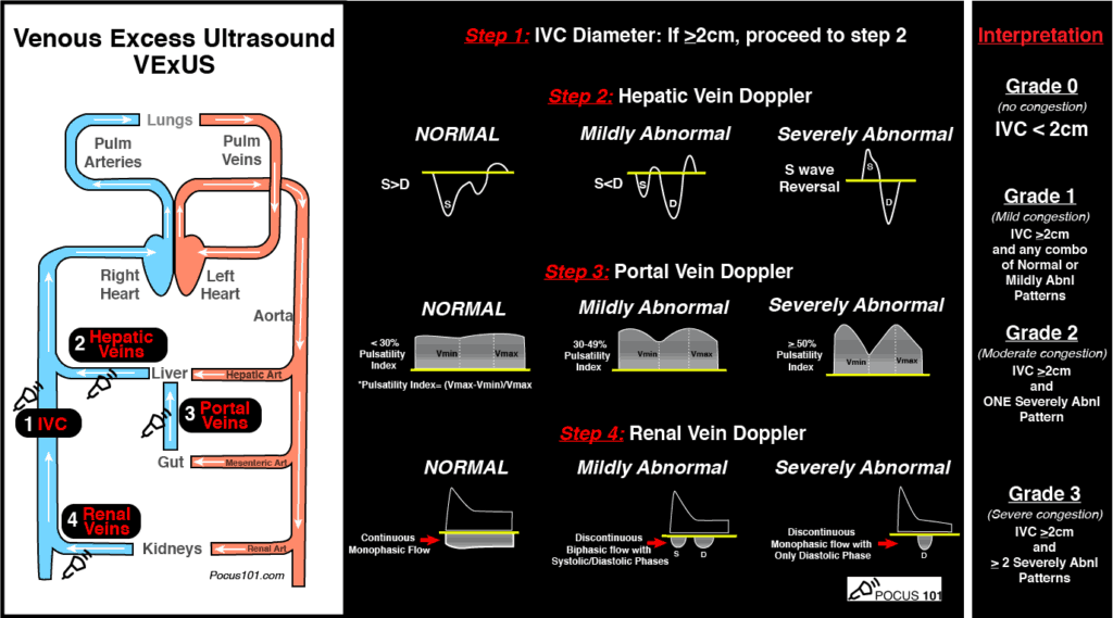
(Image from www.pocus101.com)
High Points
VExUS Grade 3 (severe) venous congestion:
- Correlated with higher CVP & NTproBNP levels, as well as overall fluid balance
- Had a 96% specificity for development of subsequent AKI
Caveats
- Evaluating all parameters yields the most benefit to avoid false positives
- Can be difficult to obtain all views (>25% of subjects excluded due to poor US image quality)
- Studied in a limited population, notably not primarily RV failure patients
Clinical Uses
- To limit harmful fluid administration in shock
- To help answer the prerenal vs cardiorenal AKI question in CHF
- To indicate when volume removal (diuresis) should be the strategy, even in patients with vasopressor-dependent shock
A great how-to can be found here:
https://www.pocus101.com/vexus-ultrasound-score-fluid-overload-and-venous-congestion-assessment/
Show References
STARRT-AKITrial
The Standard versus Accelerated initiation of Renal Replacement Therapy in Acute Kidney Injury
The development of acute kidney injury (AKI) in the critical care setting portends a greater morbidity and mortality for patients. Additionally, it places the patient at high risk of complications and requires a greater use of resources. Several studies in the past have examined if the timing of initiation of renal replacement therapy (RRT) would result in a mortality benefit, but have failed to demonstrate consistent outcomes.
The STARRT-AKI trial was a multinational, randomized controlled trial designed to determine if early initiation of RRT in critically ill adult patients with AKI lowered the risk of 90-day mortality. The Kidney Disease Improving Global Outcomes (KDIGO) classification was used to define AKI and over 2900 patients were randomly assigned to two groups over a 4 year period. Exclusion criteria included: recent RRT, a renal transplant within the preceding year, advanced CKD, an overdose necessitating RRT, or a strong suspicion of obstruction or autoimmune/vascular cause of their AKI.
Groups:
- The accelerated strategy group
- Initiation of RRT within 12 hours of meeting eligibility criteria (AKI based on KDIGO definition)
- The standard strategy group –
- General goal of withholding RRT unless the patient met the following specific parameters:
- K+ >6.0, pH <7.20, HCO3 <12mmol/L, moderate ARDS with clinical picture concerning for volume overload, or persistent AKI >72hr after randomization
Outcomes/Results:
- The study’s primary outcome measure was all cause mortality at 90 days
- There was no significant difference between the groups
- P=0.92 with RR 1.00
- Secondary outcomes evaluated several things including ventilator and vasoactive free days, hospital length of stay, number of days without RRT at 90 days as well as adverse events directly related to RRT
- Interestingly, at 90 days, the patients in the accelerated strategy group were more likely to have ongoing RRT needs at 10.4% compared to the standard strategy group at 6.0% (not statistically significant).
- Overall, no significant difference between the groups when assessed for death in the ICU, major adverse events, or with regard to hospital length of stay.
Take home points:
- This was a well done, well randomized trial from many countries and ICU settings
- No significant mortality benefit between groups at 90 days
- Interestingly, the patients in the accelerated group were more likely to have suffered adverse events related to RRT and were more likely to be dependent on RRT at 90 days
- It is unclear why this is, but suggestive that early initiation of RRT may compromise the intrinsic healing of the kidney
- Emphasizes a greater risk for adverse events without clear benefit
- Ultimately, the decision to initiate RRT should be based on the patient’s clinical picture, acid/base status, electrolyte abnormalities, and volume status and NOT on a general trend of their renal indices.
Show References
Ketamine In the Critically Ill Patient
- Ketamine has become a popular agent in the ED for both RSI and procedural sedation.
- Given the sedative, analgesic, dissociative, antidepressant, and anti-inflammatory properties, ketamine has also been used in a number of other critical illness conditions including:
- Acute pain management
- Status asthmaticus
- Alcohol withdrawal syndrome
- Status epilepticus
- Acute agitated delirium
- The authors of a recent review in Critical Care Medicine found that the evidence supporting the use of ketamine in the critically ill is most robust for adjunctive analgesia in the intubated patient. Surprisingly, the data is very limited to support the use of ketamine in these other conditions.
- Pearl: ketamine does have a myocardial depressant effect, which can be unmasked in states of catecholamine depletion and result in hypotension and bradycadia.