University of Maryland School of Medicine logo
Emergency Medicine
All Departments
UMSOM Assistant
UMEM Educational Pearls
  • Home
  • Departments
  • Emergency Medicine
  • CME
  • UMEM Educational Pearls
  • CME
  • Courses and Conferences
  • Free Open Access Medical Education
  • UMEM Educational Pearls

Search

41-60 of 321 results by Haney Mallemat

Previous  |  1 |  2 |  3 |  4 |  5 |  6 |  7 |  8 |  ... |  17 |  Next

Title: Long or Short?....That is the question

Category: Critical Care

Posted: 5/12/2015 by Haney Mallemat, MD

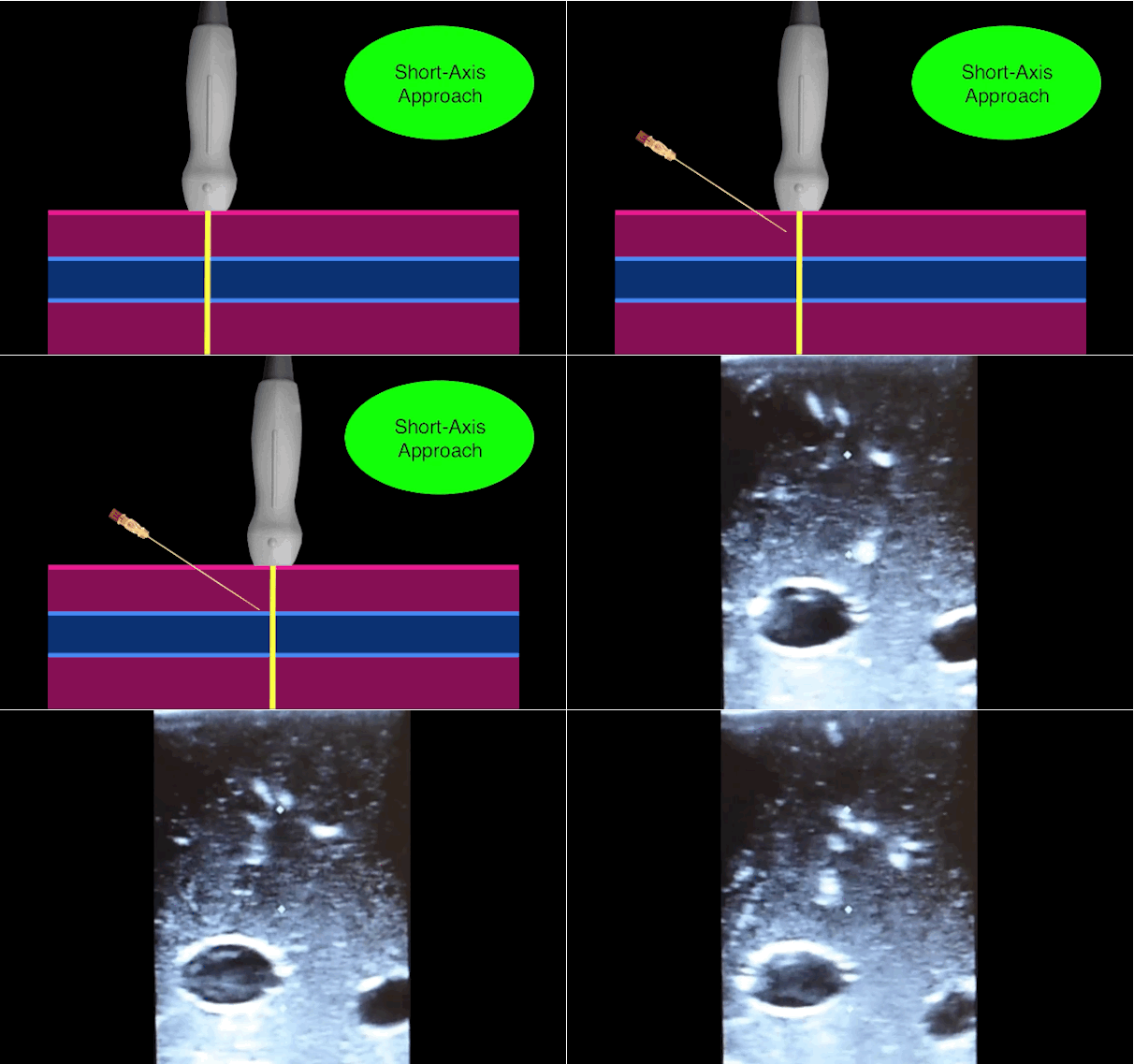
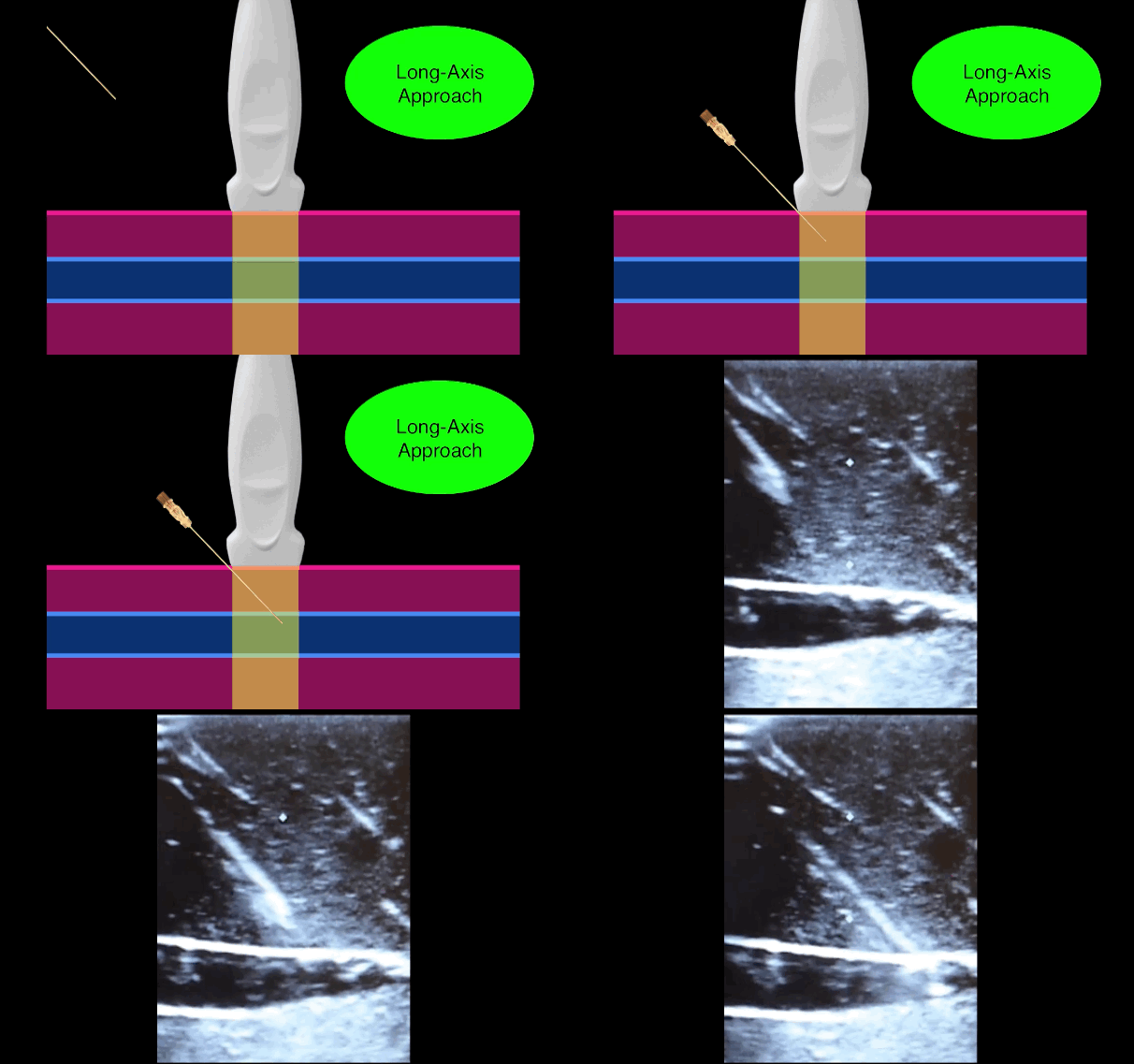
There is little debate that ultrasound-guided central lines are safer, faster, and more reliable compared to a landmark technique; there is some debate, however, as to whether the short axis (SA) or long axis (LA) approach is the best (see clips below).

The referenced study compared the SA and the LA technique for both the internal jugular (IJ) and subclavian (SC) venous approach. The authors measured number of skin breaks, number of needle redirections, and time to cannulation for each method.

This study demonstrated that the LA technique for subclavian placement had fewer redirections, decreased cannulation time, and fewer posterior wall punctures as compared to the SA. With respect to the IJ approach, the LA was also associated with fewer redirections than the SA view.

Bottom line: Consider the long-axis technique the next time you place an ultrasound guided central line.
Show References

Vogel, J. et al. Is long-axis view superior to short-axis view in ultrasound-guided central venous catheterization? Crit Care Med 2015 Apr;43(4):832-9.

Follow me on Twitter (@criticalcarenow) or Google+ (+criticalcarenow)


Title: What's the Diagnosis? Case by Dr. Nicholas Santavicca and Dr. Semhar Tewelde

Category: Visual Diagnosis

Posted: 5/11/2015 by Haney Mallemat, MD

Question

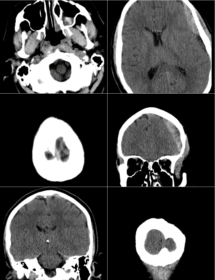
40 year-old male sustains a blunt force injury the left side of his lead. What's the diagnosis and what structure was injured?
Show Answer
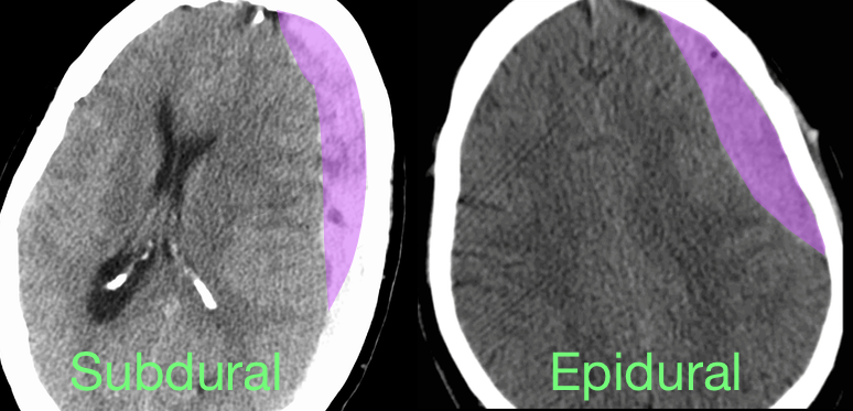
Epidural Hematoma with mid-line shift from middle meningeal artery injury; see image below for comparison of subdural and epidural

Epidural hematoma

  • Blood accumulates between dura and calvarium secondary to trauma; concurrent skull fractures are common
  • Middle menial artery is usually involved in the temporal region; other arteries may be involved depending on which area of the skull receives the impact
  • Up to one-third of cases present with the "classic" lucid interval from injury and loss of consciousness to a decline in mental status. Diagnosis should be suspected, however, based on mechanism
  • Treatment is surgical evacuation, however aggressive medical management of increased intracranial pressure should not be delayed:
    • Head of bed elevated and keep neck mid-line
    • Anti-emetics and analgesics
    • Osmotic agents such as mannitol or hypertonic saline
    • Avoid hypoxia, hypotension, and hyperthermia

Show References

Follow me on Twitter (@criticalcarenow) or Google+ (+criticalcarenow)


Title: What's the Diagnosis? Case by Dr. Ahmed Alrasheedi

Category: Visual Diagnosis

Posted: 5/4/2015 by Haney Mallemat, MD

Question

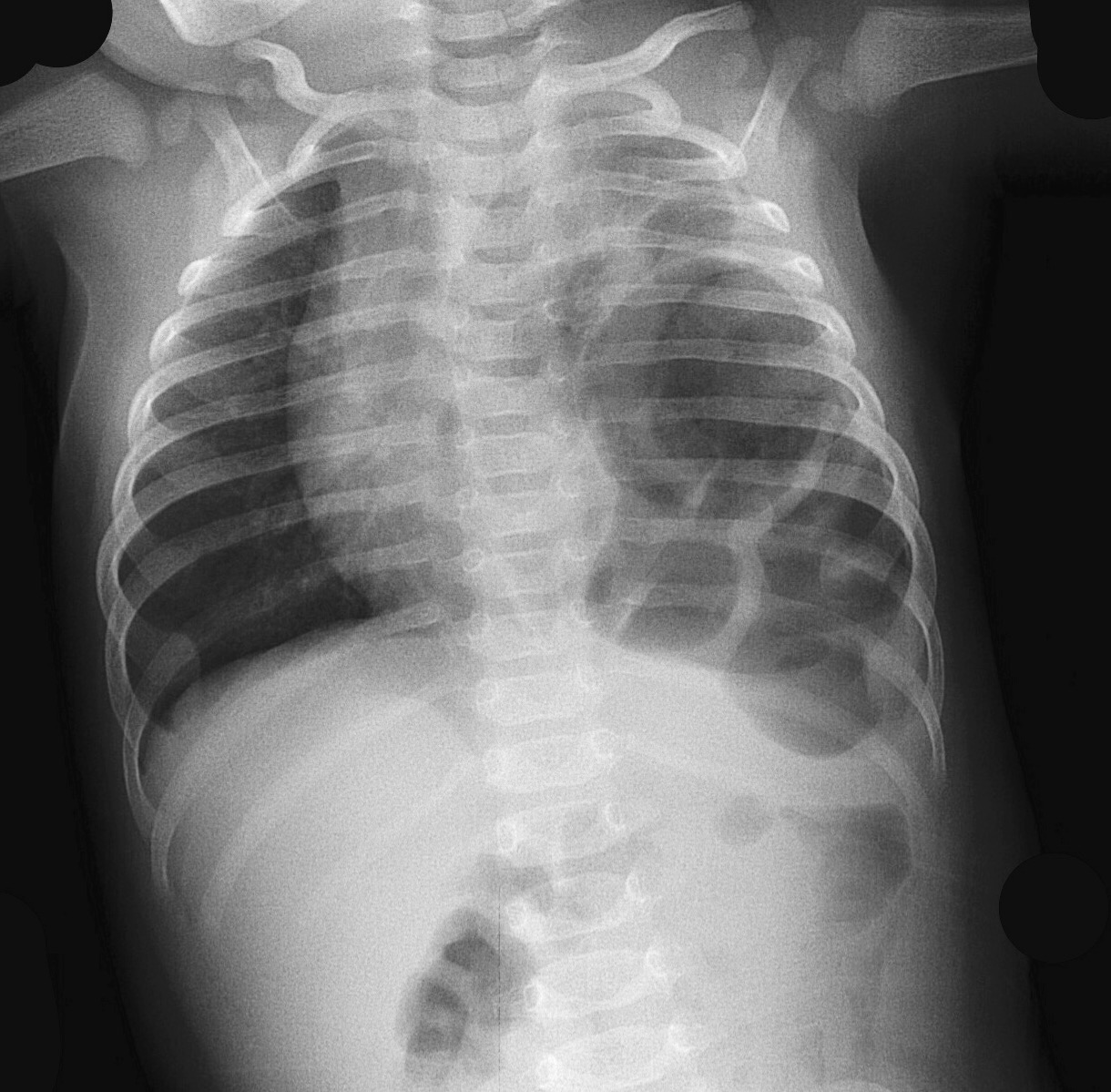
3 month-old male presents with severe respiratory distress; oxygen saturation is 81% (on room air), he is grunting, and there are no breath sounds on the left. What's the diagnosis?

Show Answer

Congenital diaphragmatic hernia
Management:
  • Aggressive fluid and vasopressor support
  • Early intubation and airway protection should be considered in severe cases; minimize BVM given the risk of gastric distention.
  • NG tube decompressing of the stomach will help the ventilation/oxygenation process.
  • ECMO may be considered if conventional therapy has failed and any one of the following criteria is met:
  1. Inability to maintain pre-ductal PaO2 saturations >85 percent or post-ductal PaO2 >30 mmHg
  2. Peak inspiratory pressure >28 cm H2O or mean airway pressure >15 cm H2O
  3. Hypotension that is resistant to fluid and inotropic support
  4. Inadequate oxygen delivery with persistent metabolic acidosis

Show References

Follow me on Twitter (@criticalcarenow) or Google+ (+criticalcarenow)


Title: What's the Diagnosis?

Category: Visual Diagnosis

Posted: 4/27/2015 by Haney Mallemat, MD

Question

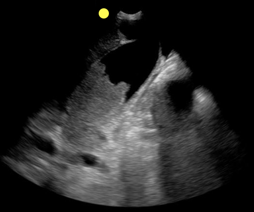
70 year-old female presents from a nursing home with fever and abdominal pain. A right upper quadrant ultrasound is shown, what's the diagnosis?
Show Answer
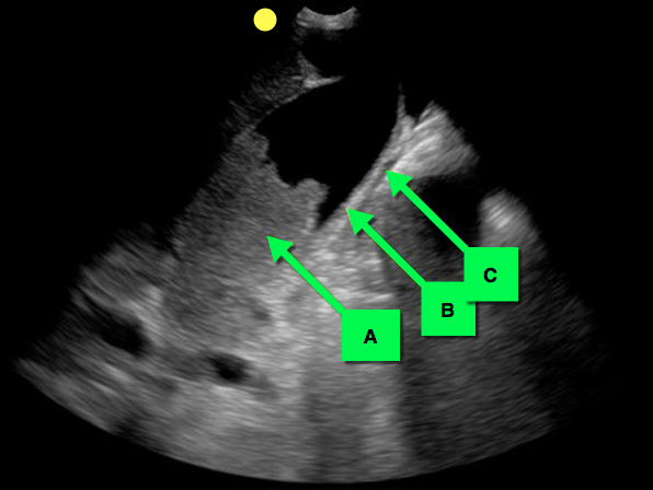
Acalculous cholecystitis

Acalculous Cholecystitis

  • Acalculous cholecystitis is due to gallbladder (GB) wall ischemia and GB stasis (i.e., from reduced GB contraction secondary to reduced PO intake).
  • The result is gallbladder "sludge" (A), thickened GB walls (B), and pericholecystic fluid (C).
  • Risk factors for acalculous cholecystitis include
    • critical illness (especially sepsis)
    • acute cholecystitis
    • chronic immobility
    • total parenteral nutrition
    • diabetes and immunosuppression
  • Treatment includes antibiotics covering enteric / biliary pathogens and cholecystectomy (if a surgical candidate) or percutaneous cholecystostomy if unsuitable for the OR.
  • Complications include perforation, GB gangrene, and extra-biliary abscess.

Show References

Follow me on Twitter (@criticalcarenow) or Google+ (+criticalcarenow)


Title: What's the Diagnosis?

Category: Visual Diagnosis

Posted: 4/20/2015 by Haney Mallemat, MD

Question

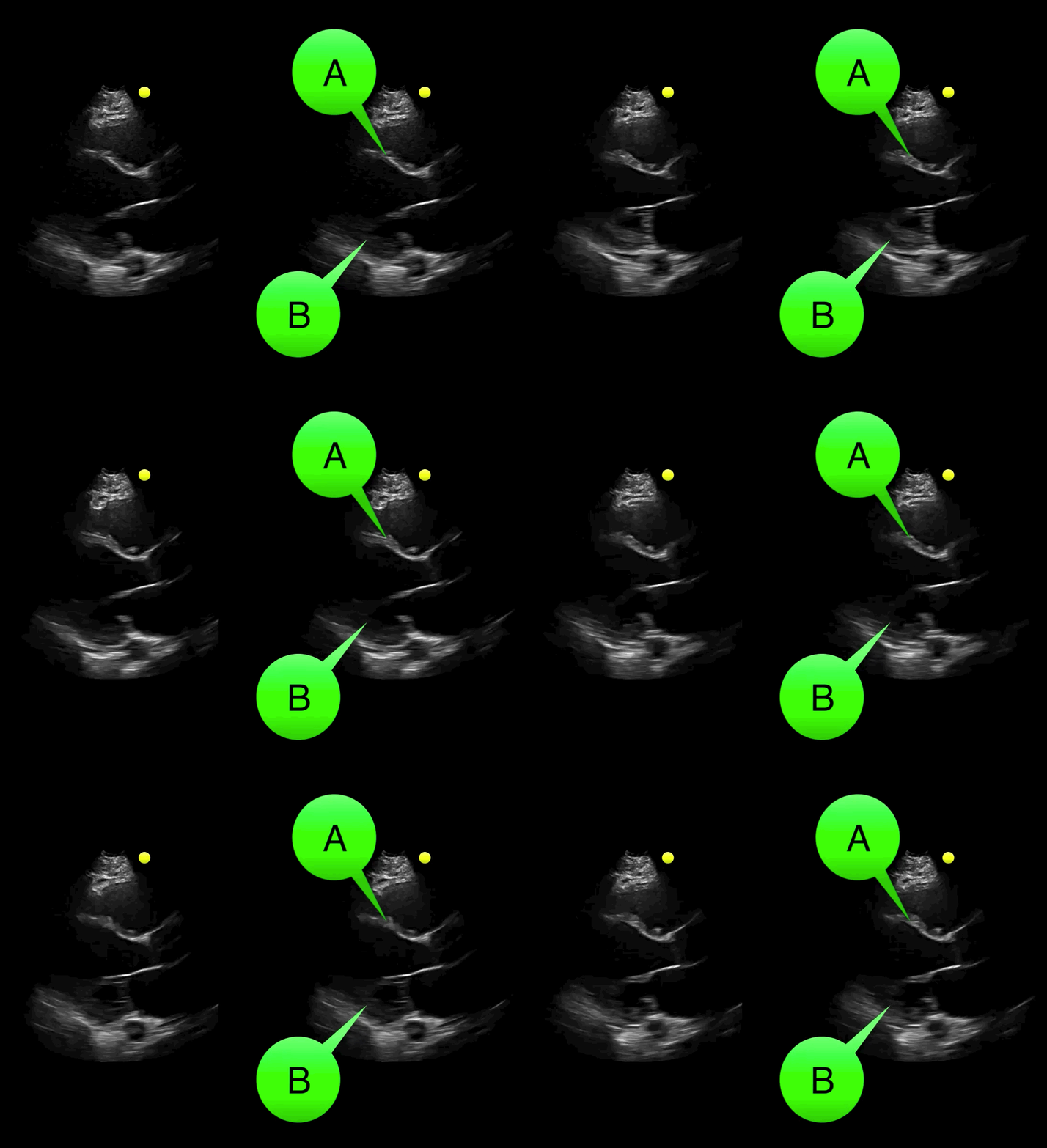
You see the following on a parasternal long-axis view; what's the diagnosis and what coronary distribution is involved?
Show Answer

Severe hypokinesis with anterioseptal wall motion abnormality.

  • A: Anteroseptal wall (perfused by left anterior descending)
  • B: Inferolateral wall (perfused by left circulmflex artery and variable contributions from posterior descending artery)
  • Click on the links to read more about cardiac wall segments and their coronary distribution
    • Parasternal view and Apical four chamber view

Show References

Follow me on Twitter (@criticalcarenow) or Google+ (+criticalcarenow)


Title: Back to Basics: Is that the RV or the LV?

Category: Critical Care

Posted: 4/14/2015 by Haney Mallemat, MD

Question

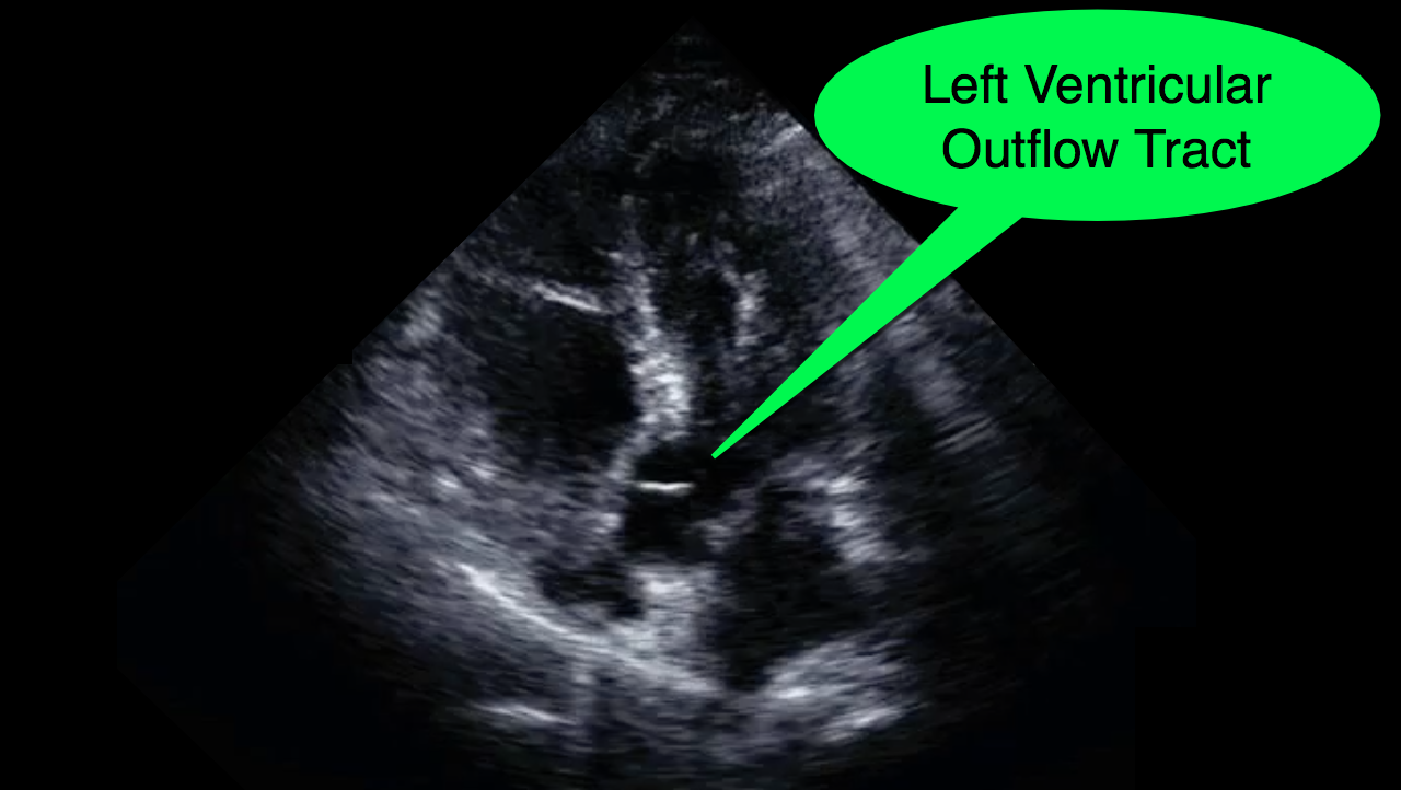
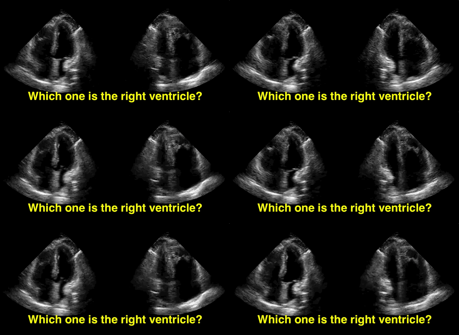
You decide to do a R.U.S.H. exam on your hypotensive patient and perform an apical four-chamber view.You see one of the two clips below; are there any tricks to figure out which is the left ventricle and which is the right ventricle?
Show Answer
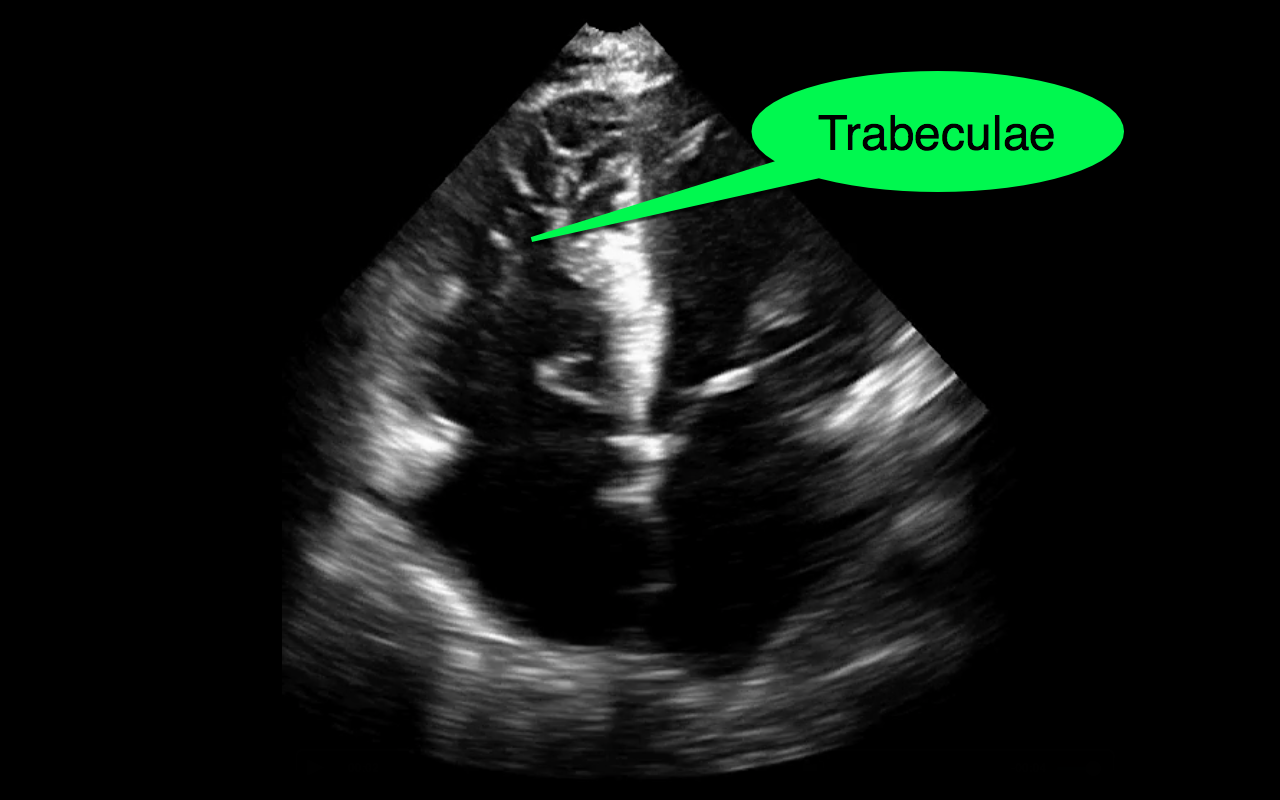
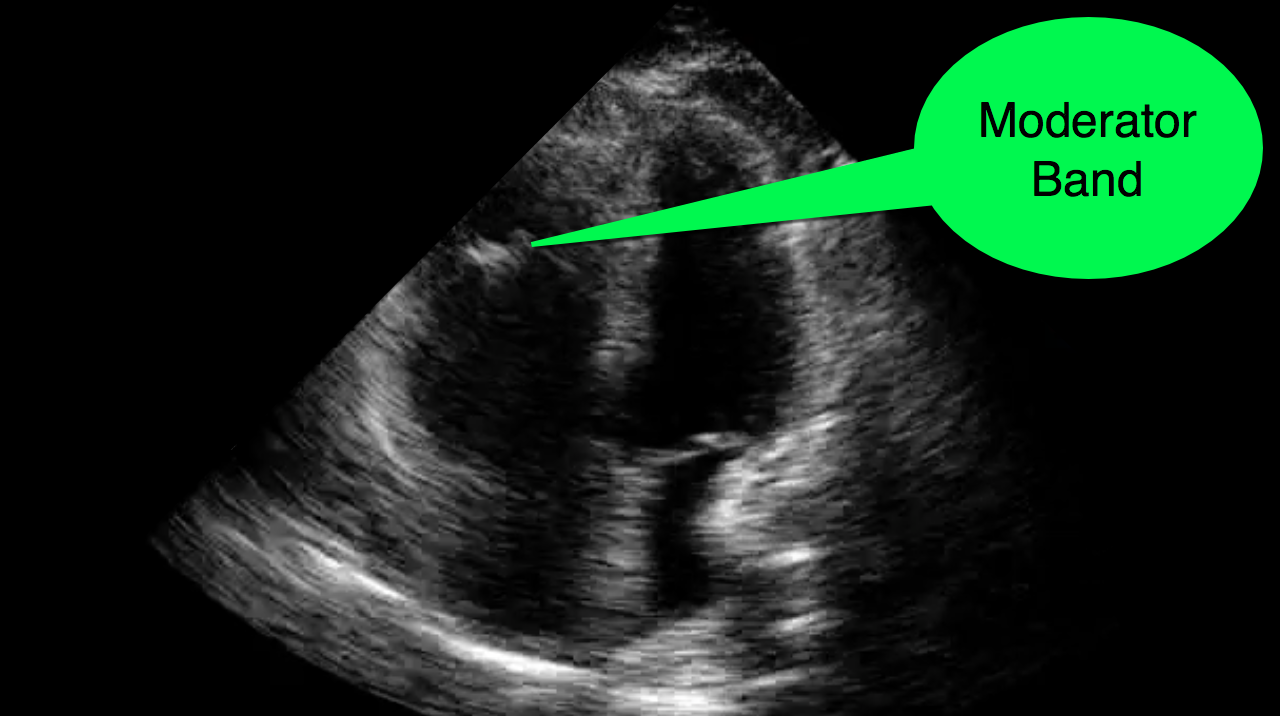
Tricks to distinguish the RV from LV in apical four-chamber view

  • The right ventricle (RV) is normally smaller in size (2/3 the size) as compared to the left ventricle (LV).
  • When scanning critically ill patients you may encounter patients with right ventricles that are dilated secondary to acute or chronic illness. This enlargement may make the RV appear similar to a typical LV on an apical four-chamber view.
  • If you are new to ultrasound or just get confused by the probe marker, screen orientation, or just unsure, there are some tricks that you can use to quickly identify the LV and the RV on the apical four-chamber view.
    • The tricuspid valve is more "apically" displaced when compared to the mitral valve
    • The RV is heavily trabeculated as compared to the LV
    • The RV has the moderator band, while the LV lacks such a structure
    • The LV has the "fifth" chamber (i.e., the left ventricular outflow tract); you can identify this by tilting the probe slightly anteriorly after finding the apical four-chamber view.

Show References

Follow me on Twitter (@criticalcarenow) or Google+ (+criticalcarenow)


Title: What's the Diagnosis? Case by Dr. Leen Alblaihed

Category: Visual Diagnosis

Posted: 4/13/2015 by Haney Mallemat, MD

Question

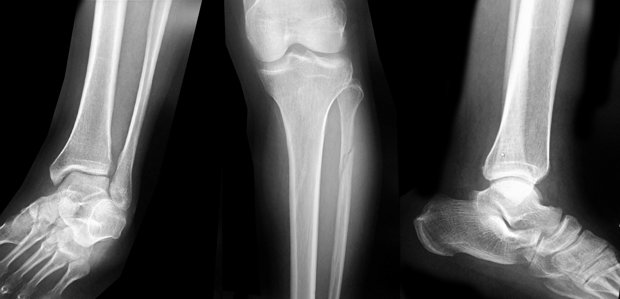
Patient presents with leg and ankle pain after a fall 3 weeks earlier. Initial ankle Xrays were negative. Patient presents today with persistent leg and ankle pain. What's the diagnosis and what other imaging would you perform and why?
 

Show Answer
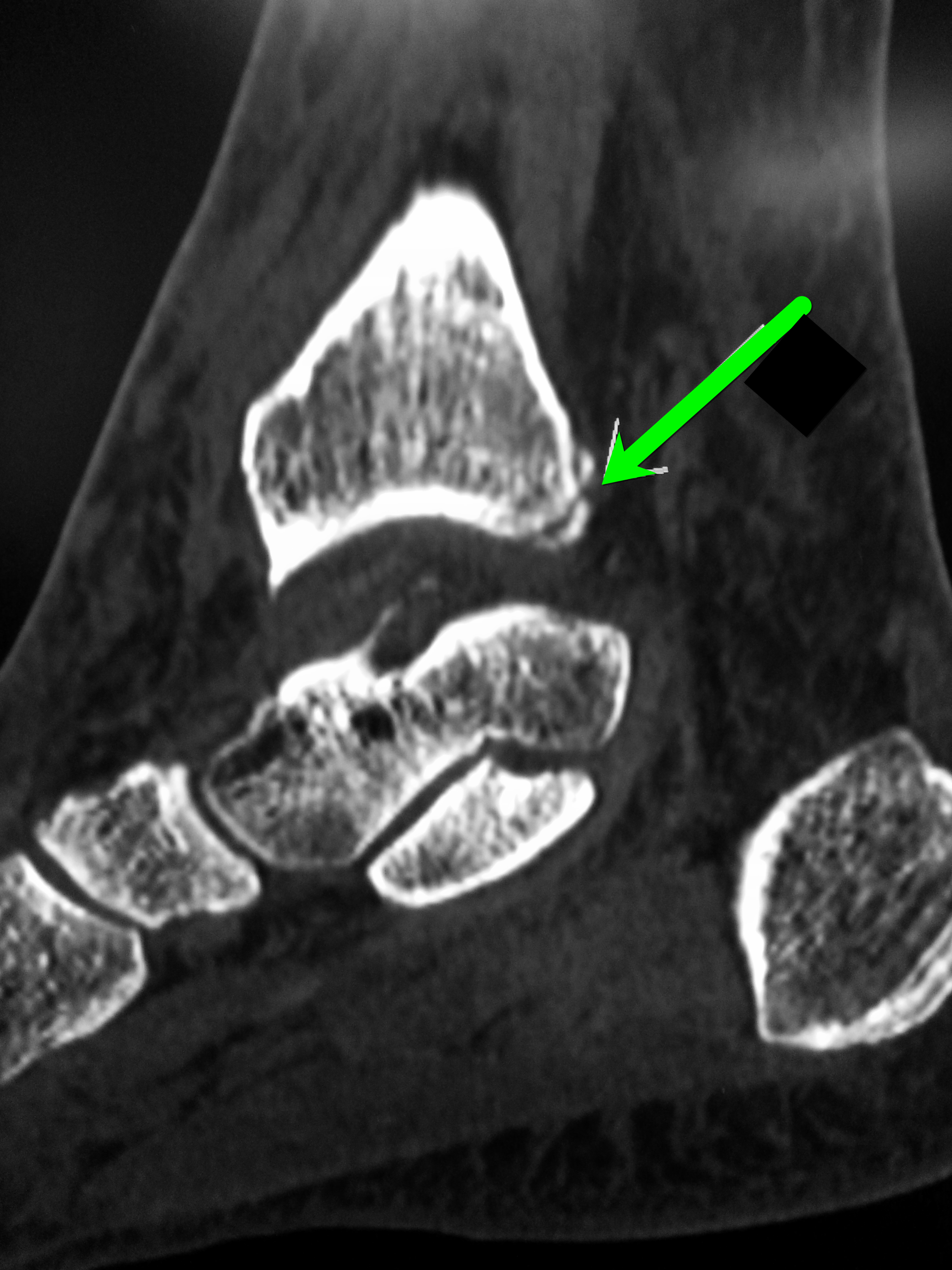
Proximal fibula fracture; have a high-clinical suspicion for a Maisonneuve fracture and consider CT scan of the ankle

Maisonneuve fracture

  • Fracture of proximal third of the fibula with associated injury of the tibiofibular syndesmosis and interosseous membrane; there is typically an associated fracture of the medial malleolus or ruptured deltoid ligament.
  • This patient had a normal ankle X-ray, but the proximal fibular fracture and the persistent ankle pain lead to an ankle CT scan demonstrating a non-displaced fracture at the medial aspect of the posterior malleolus, extending into the tibiotalar joint (see below). 
  • The take home point is to always look for an associated ankle injury when a proximal fibular fracture is discovered and Xray above and below the fracture. If Xrays are negative but there is still high clinical suspicion consider advanced imaging such as CT scan.

Show References

Follow me on Twitter (@criticalcarenow) or Google+ (+criticalcarenow)


Title: What's the Diagnosis?

Category: Visual Diagnosis

Posted: 4/6/2015 by Haney Mallemat, MD

Question

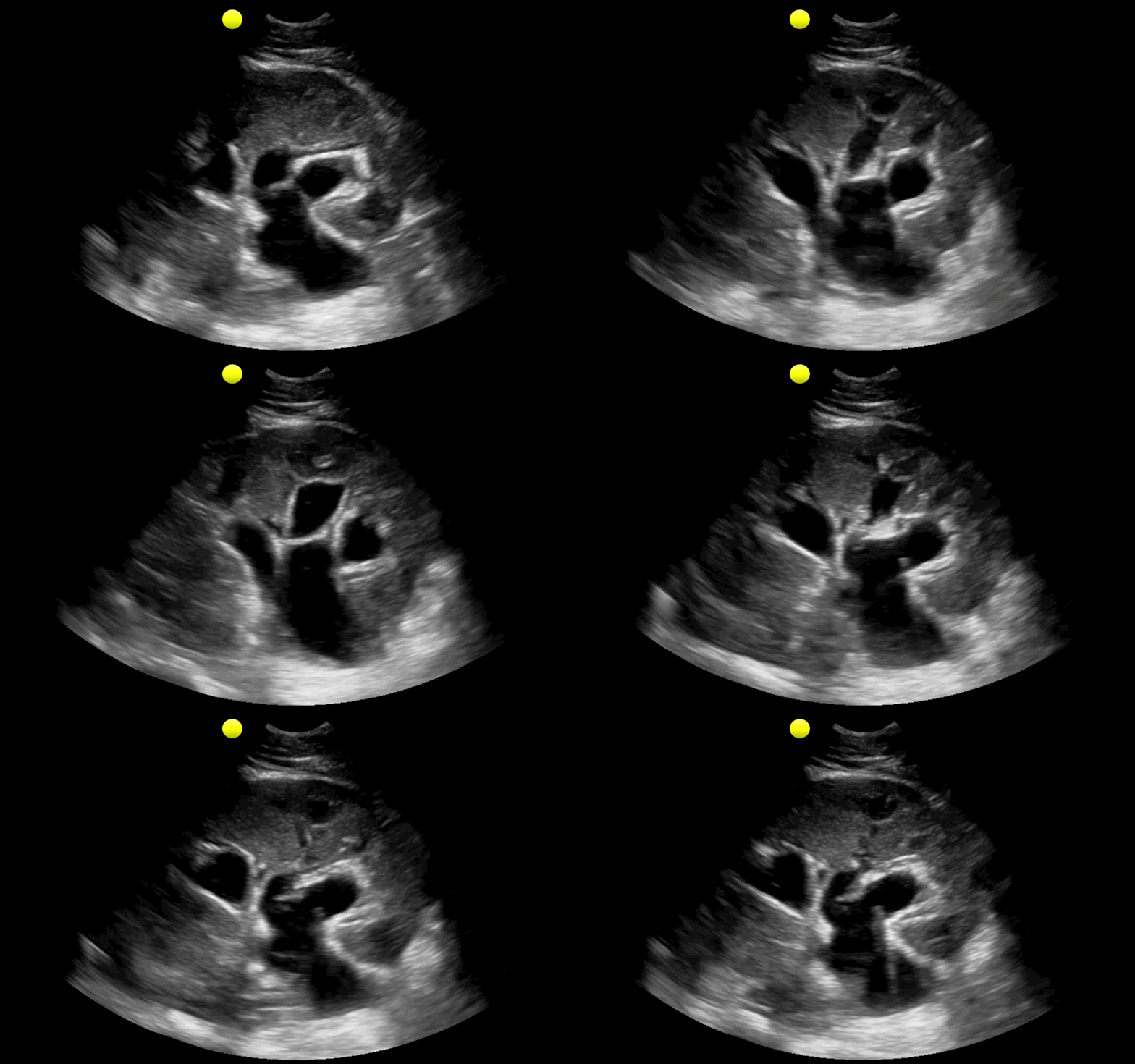
25 year-old male with the acute onset of right flank pain. Ultrasound of the right flank is shown. What's the diagnosis?
Show Answer

  • This is an ultrasound of the kidney demonstrating moderate to severe hydronephrosis seondary to an obstructing renal stone (not shown)
  • The areas that are anechoic (or black) are the blocked ureter, renal pelvis, and calyces

Show References

Follow me on Twitter (@criticalcarenow) or Google+ (+criticalcarenow)


Title: What's the Diagnosis? Image by Dr. Tu Carol Nguyen & Dr. R. Gentry Wilkerson

Category: Visual Diagnosis

Posted: 3/30/2015 by Haney Mallemat, MD

Question

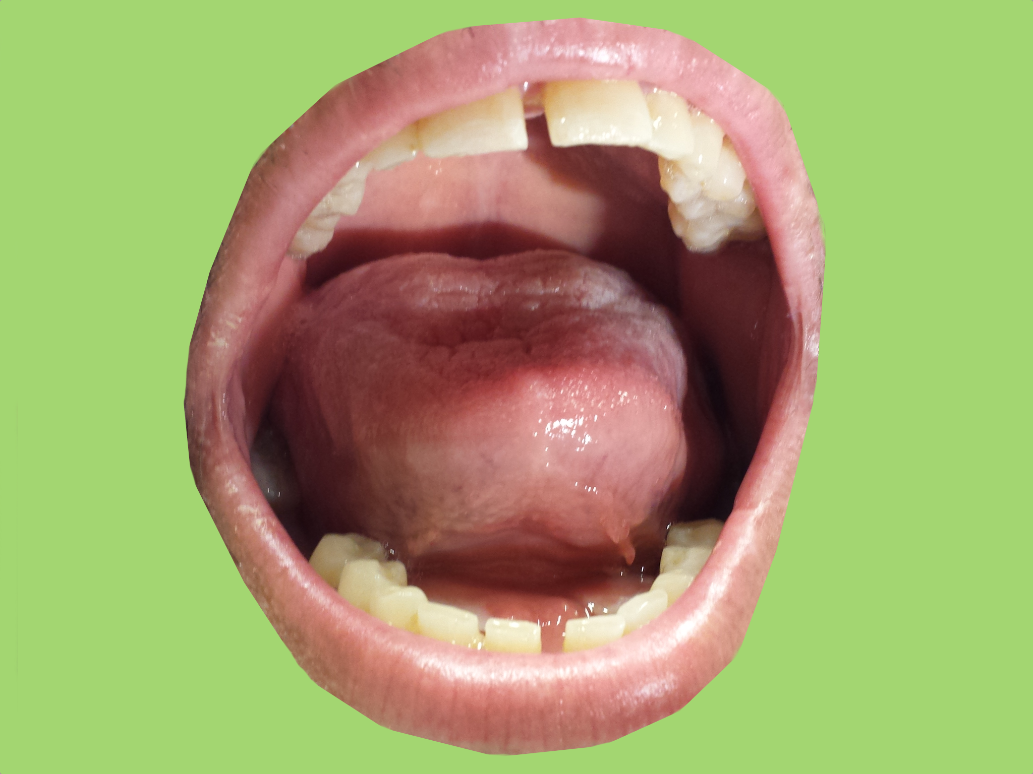
35 year-old male presents with increasing difficulty swallowing and tenderness in the floor of him mouth. What's the diagnosis?
Show Answer

Ludwig's angina

Show References

Follow me on Twitter (@criticalcarenow) or Google+ (+criticalcarenow)


Title: What's the Diagnosis? Case by Dr. David Bostick and Dr. Gentry Wilkerson

Category: Visual Diagnosis

Posted: 3/23/2015 by Haney Mallemat, MD

Question

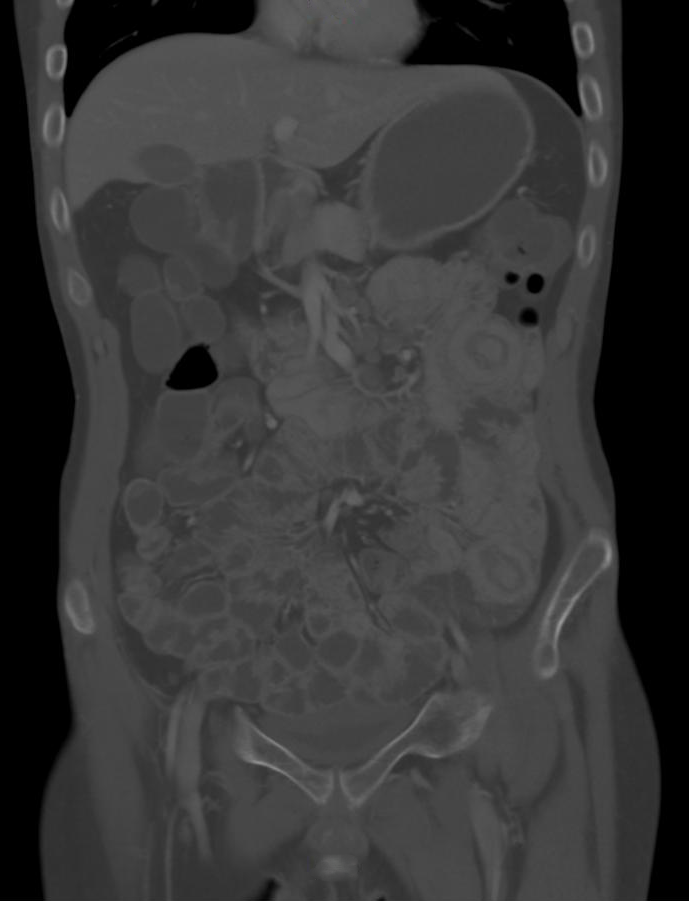
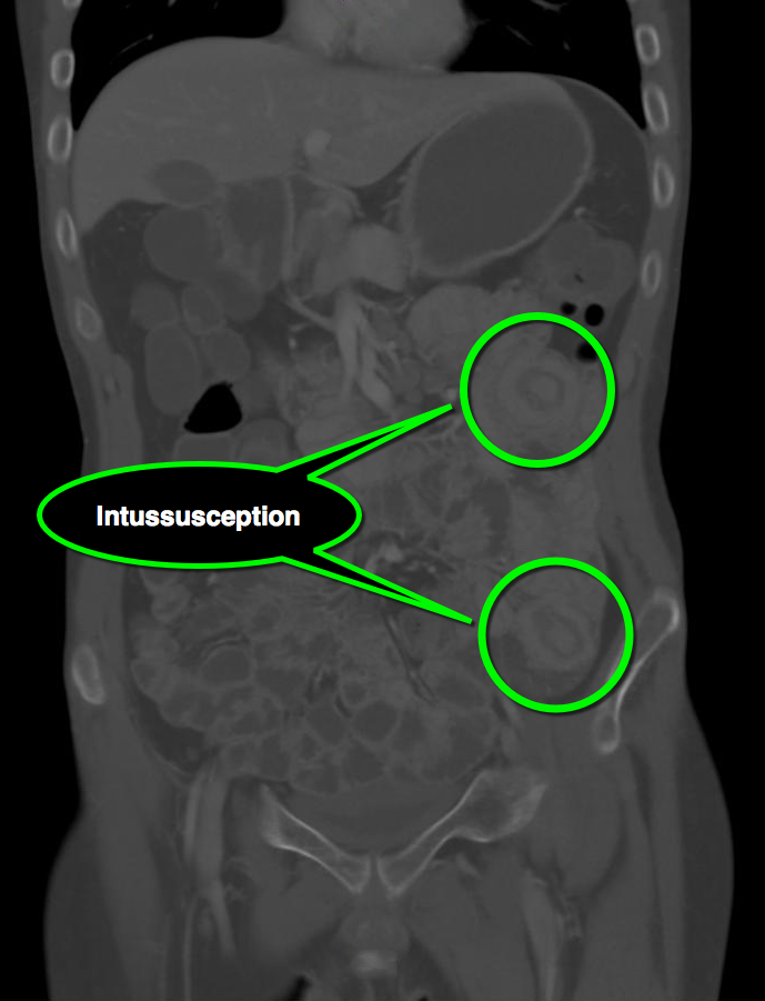
25 year-old male with autoimmune enteropathy presents with intractable vomiting and diarrhea for 7 days. What's the diagnosis?
Show Answer
Show References

Follow me on Twitter (@criticalcarenow) or Google+ (+criticalcarenow)


Title: The results are in! The ProMISe Trial

Category: Critical Care

Posted: 3/18/2015 by Haney Mallemat, MD (Updated: 3/18/2015)

The results of a multi-center trial from the UK, the ProMISe trial, were just released and it confirms what two prior studies (i.e., ProCESS and ARISE) have already shown; there does not appear to be any difference in mortality when septic patients are treated with a strategy of early-goal directed therapy as compared to usual care.

Patients were included in the ProMISe trial if they were in septic shock and were then randomized to either the EGDT group (630 patients) or the usual care group (630 patients); a total of 1,260.

The primary end-point was all cause mortality at 90 days and there was no difference shown in the primary outcome. There were no differences found in the measured secondary outcomes (e.g., serious adverse events)

This trial adds to the evidence that septic patients may not benefit from protocolized (i.e., EGDT) care versus usual care. One explaination why, is that our "usual care" in 2015 has significantly changed since the introduction of EGDT in 2001.

Show References
Follow me on Twitter (@criticalcarenow) or Google+ (+criticalcarenow)

http://www.nejm.org/doi/pdf/10.1056/NEJMoa1500896


Title: Take the #whatsthedx challenge! Image by Dr. Joseph Martinez

Category: Visual Diagnosis

Posted: 3/16/2015 by Haney Mallemat, MD

Question

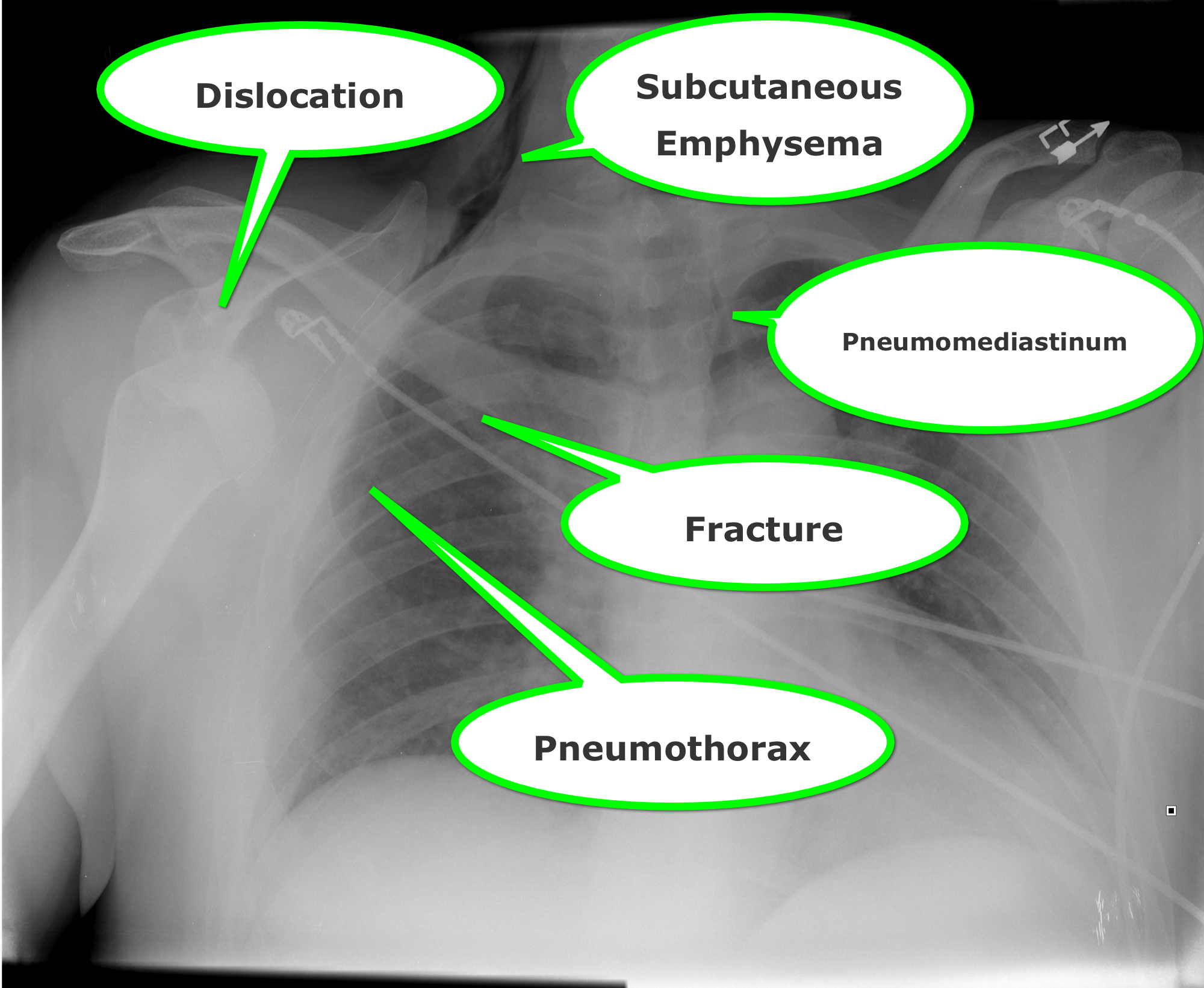
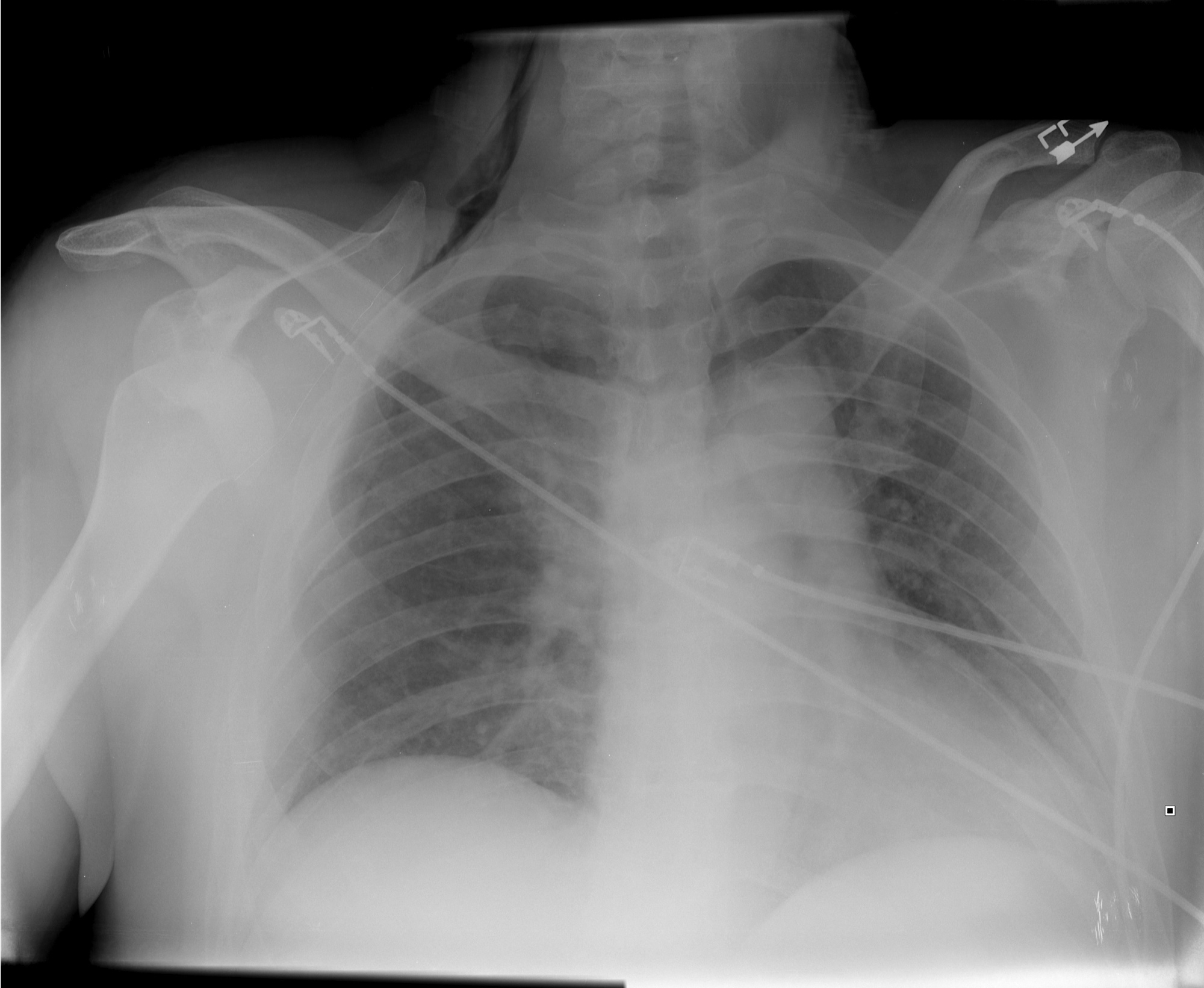
How many abnormalities can you find below?

Show Answer

Show References

Follow me on Twitter (@criticalcarenow) or Google+ (+criticalcarenow)


Title: What's the Diagnosis? Image by Dr. Connor Lundy

Category: Visual Diagnosis

Posted: 3/9/2015 by Haney Mallemat, MD

Question

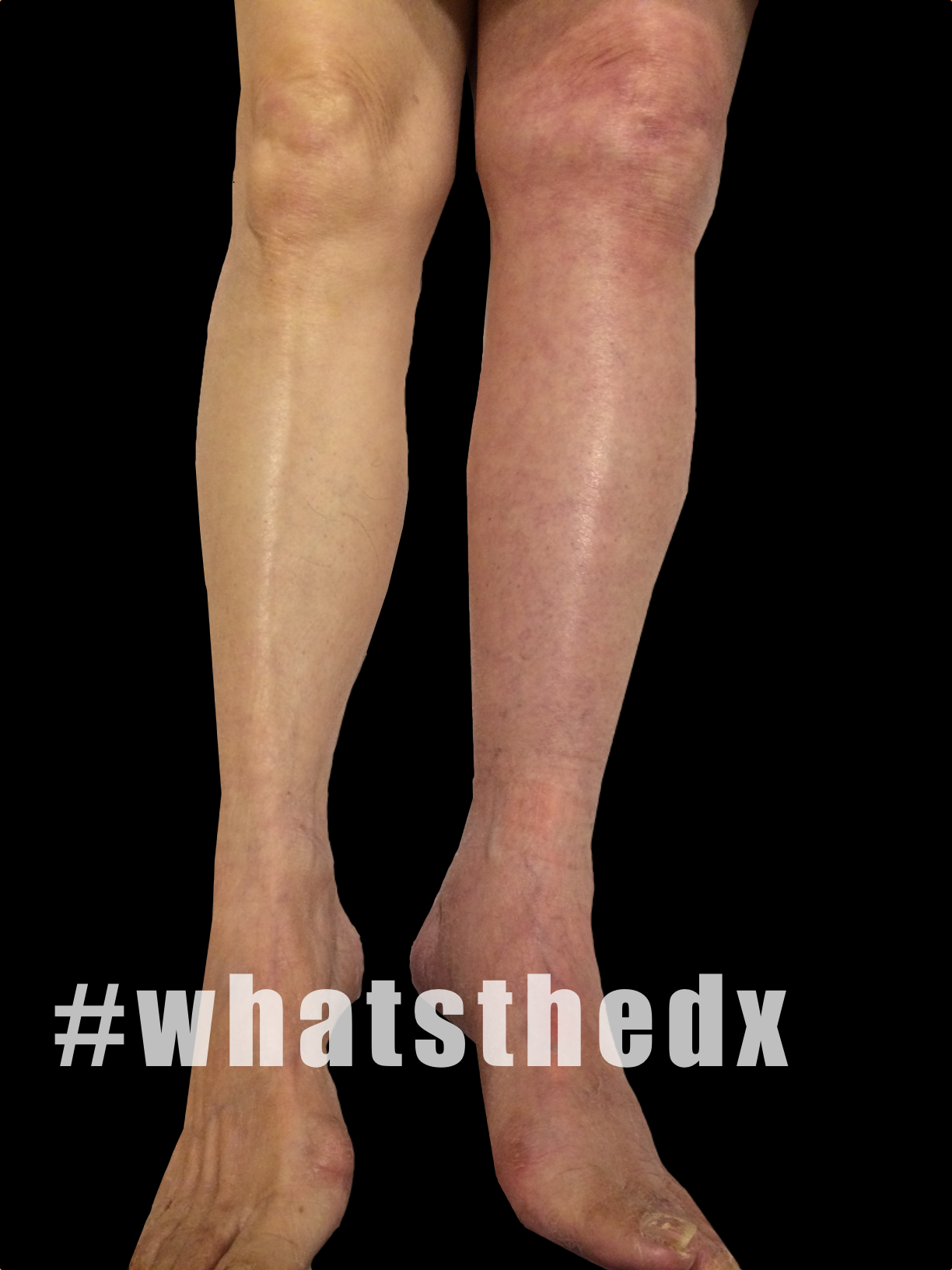
35 year-old female presents with acute leg pain and swelling. What's the diagnosis?
Show Answer

Phlegmasia cerulea dolens; there was extensive clot found in the left external iliac, common femoral, superficial femoral, and popliteal veins.

  • Phlegmasia alba dolens (literally: swollen, white, and painful) is a thrombosis of the major deep veins of the lower extremity. No collateral veins are involved so venous drainage of the leg is reduced but not completely compromised.
  • Phlegmasia cerulea dolens (literally: swollen, blue, and painful) thrombosis of major deep veins with extension to collateral veins. The result is venous congestions, edema, and can progress to gangrene when arterial flow is compromised.
  • Anticoagulation with heparin is recommended, although catheter directed thrombolytics or surgical thrombectomy may be recommended when circulation is threatened.

Show References

Follow me on Twitter (@criticalcarenow) or Google+ (+criticalcarenow)


Title: What's the Diagnosis? Image by Dr. Ahmed Alrasheedi

Category: Visual Diagnosis

Posted: 3/2/2015 by Haney Mallemat, MD

Question

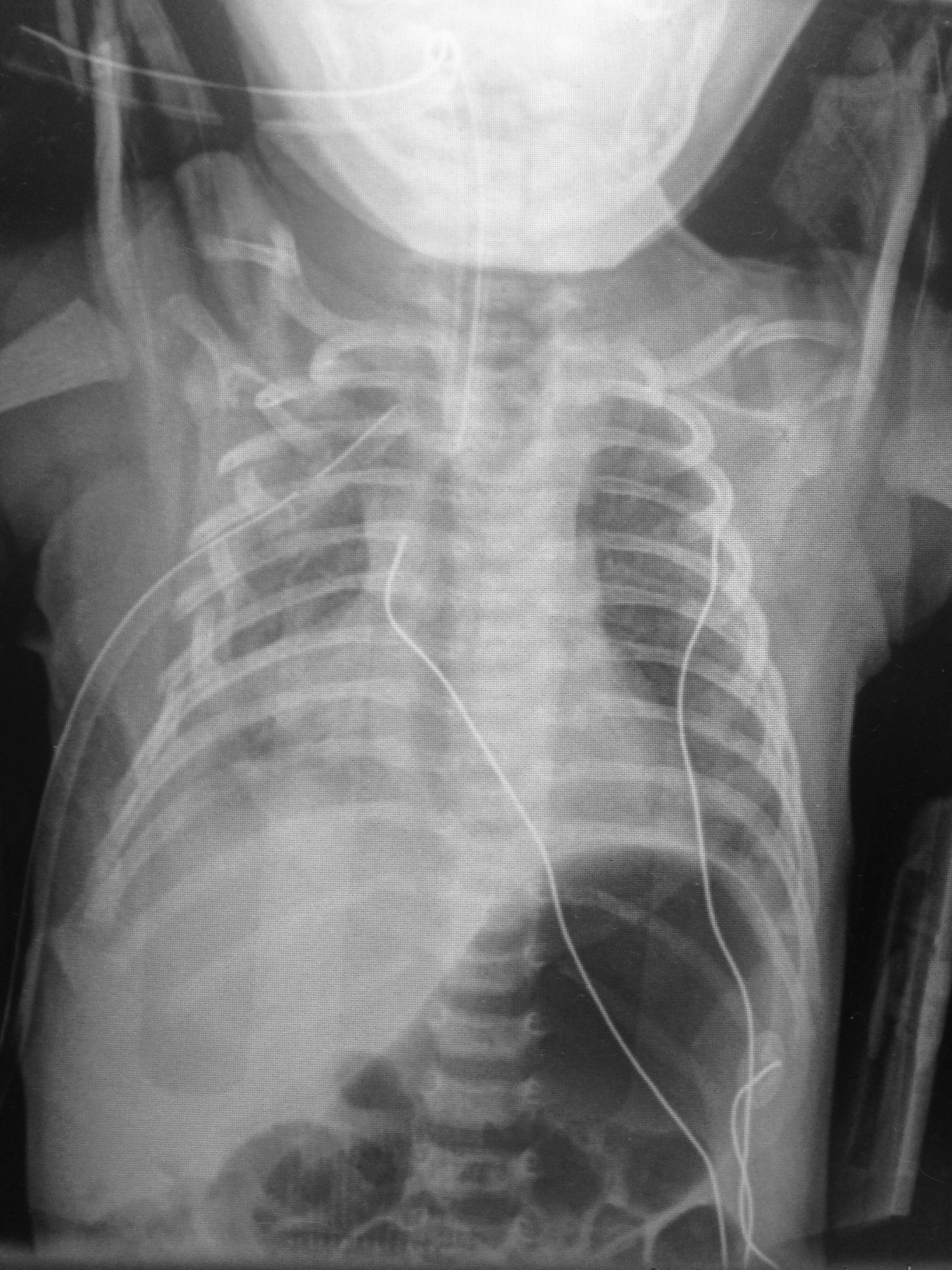
6 day-old child is brought in by parents with 1 day of reduced oral intake and 4 hours of rapid breathing. The child has no fever and no significant birth history. The child is tachycardic, hypotensive, and hypoxic. What’s the diagnosis? 
Show Answer

  • Multiple rib fractures, bilateral clavicle fracture, and hemothorax.
  • This child was ultimately diagnosed with osteogenesis imperfect, but child abuse should be quite high on your differential.

Show References

Follow me on Twitter (@criticalcarenow) or Google+ (+criticalcarenow)


Title: What's the Diagnosis? Image by Dr. Anand Swaminathan

Category: Visual Diagnosis

Posted: 2/23/2015 by Haney Mallemat, MD

Question

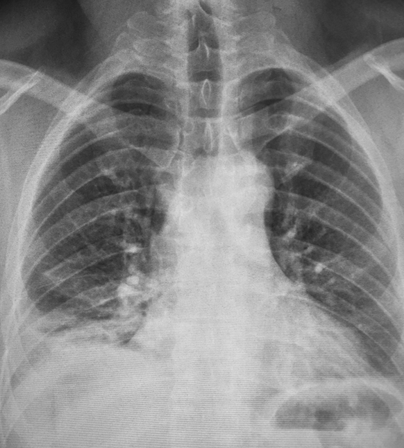
45 year-old male complains of pleuritic chest pain following a "long" flight. What's the diagnosis and what's this sign called?
Show Answer
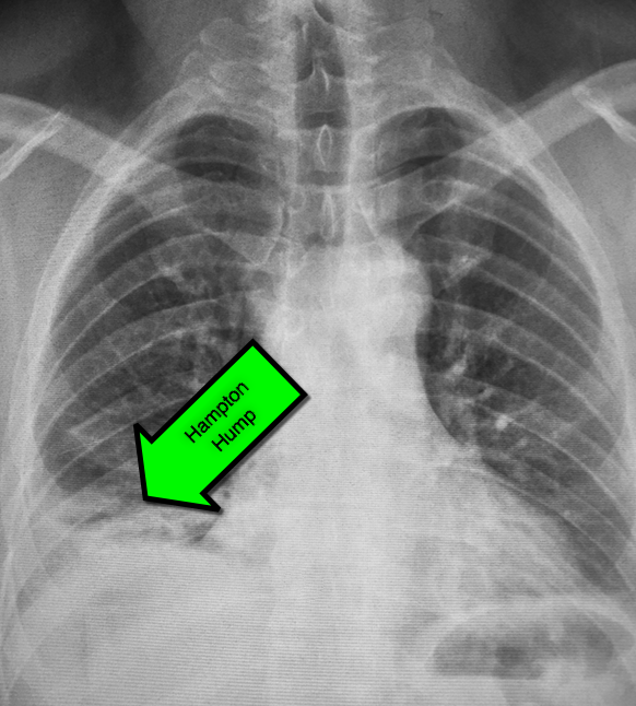
First described by Dr. Aubrey Hampton in 1940, hampton hump is a peripheral (pleural) based opacification found on chest X-ray; it occurs secondary to infarction of the lung

Although commonly associated pulmonary embolism it may occur secondary to other causes of lung infarction

Here are some other infrequent, but not rare signs, of pulmonary embolism on chest X-ray:

  • Westermark sign: area of vascular cutoff distal to a pulmonary embolism
  • Fleishner sign: an enlarged pulmonary artery
  • Pleural effusion

Show References

http://radiopaedia.org

Follow me on Twitter (@criticalcarenow) or Google+ (+criticalcarenow)


Title: Cold on the outside, make 'em warm on the inside

Category: Critical Care

Posted: 2/17/2015 by Haney Mallemat, MD

As the cold and snow rips through the United States, hypothermia is a major concern because each year approximately 1,300 Americans die of hypothermia.

Classification of hypothermia:

  • Mild (32-35 Celsius): shivering, hyperventilation, tachycardia, but patients are usually hemodynamically stable.
  • Moderate (28-32 Celsius): CNS depression, hypoventilation, loss of shivering, risk of arrhythmias, and paradoxical undressing
  • Severe (<28 degrees Celsius): increased risk of ventricular tachycardia/fibrillation, pulmonary edema, and coma

The risk of cardiac arrest increases when the core temperature is less than 32 Celsius and significantly rises when the temperature is less than 28 Celsius. Rapid rewarming is required as part of resuscitation should cardiac arrest occur.

A rescue therapy to consider (when available) is extra corporeal membrane oxygenation (ECMO). ECMO not only provides circulatory support for patients in cardiac arrest, but allows re-warming of patients by 8-12 Celsius per hour.

Some studies quote survival rates of 50% with hypothermic cardiac arrest patients receiving ECMO versus 10% in similar patients who do not receive ECMO.

As winter lingers in the United States, consider speaking to your cardiac surgeons now to plan an Emergency Department protocol for hypothermic patients that may require ECMO.

Show References

Ginty, et. Al. Extracorporeal membrane oxygenation rewarming in the ED: an opportunity for success American Journal of Emergency Medicine 2014 December 3

Follow me on Twitter (@criticalcarenow) or Google+ (+criticalcarenow)


Title: What's the Diagnosis? Case by Dr. Michael Allison (@mgallison15)

Category: Visual Diagnosis

Posted: 2/16/2015 by Haney Mallemat, MD

Question

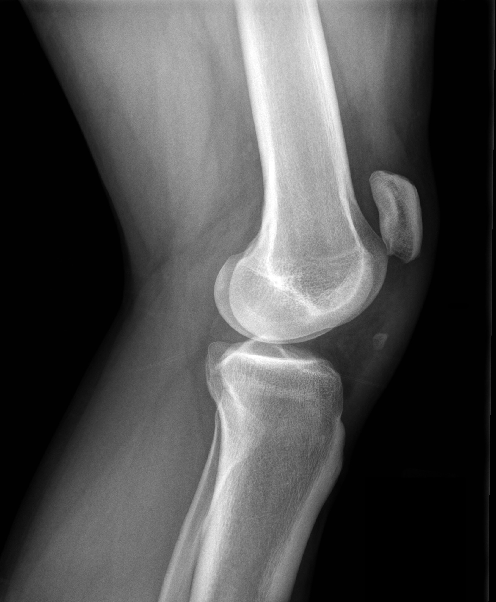
28 year-old male felt his left knee "pop" after landing from a jump. He has limited ability to extend his knee. Xray shown. What's the diagnosis?

Show Answer

Patella Alta secondary to patellar tendon rupture
Patella Alta
  • Mechanism is knee flexion combined with forceful contraction of the quadriceps
  • Swelling, patellar tenderness, and limited extension of knee
  • Radiographs are helpful in diagnosis when using the Insall-Salvatti ratio (see references)

Reference

  • Insall J, Salvati E. Patella position in the normal knee joint. Radiology 1971;101:101
  • Sibley T, Algren D, Ellison S. Bilateral patellar tendon ruptures without predisposing systemic disease or steroid use: a case report and review of the literature. American Journal of Emergency Medicine 2012; 30: 261

Show References

Follow me on Twitter (@criticalcarenow) or Google+ (+criticalcarenow)


Title: What's the Diagnosis? Case by Dr. Jackie Wnek and Dr. Jonathan Hoover

Category: Visual Diagnosis

Posted: 2/2/2015 by Haney Mallemat, MD

Question

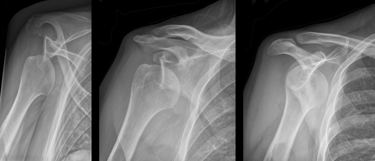
Patient presents with right shoulder pain following minor trauma. What's the diagnosis....and what's the Cunningham technique?
Show Answer

Anterior shoulder dislocation

Cunningham Technique

  • A technique for shoulder reduction that typically does not require sedation.
  • Relies on proper positioning of the patient and gentle massage of the biceps muscle while gentle traction is applied to the antecubital fossa
  • Click here for a step by step tutorial on the technique and click here for videos on the technique

Show References

Follow me on Twitter (@criticalcarenow) or Google+ (+criticalcarenow)


Title: What's the Diagnosis? Image by Dr. Zachary Dezman

Category: Visual Diagnosis

Posted: 1/26/2015 by Haney Mallemat, MD

Question

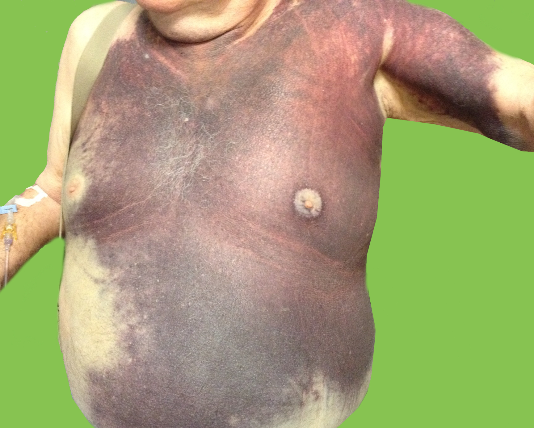
Elderly male presents with the skin findings below. He is also on a medication for atrial fibrillation. What's the diagnosis?
Show Answer

Severe ecchymosis from coumadin overdose

Show References

Follow me on Twitter (@criticalcarenow) or Google+ (+criticalcarenow)


Title: Can you use an intraosseous line for RSI?

Category: Critical Care

Posted: 1/20/2015 by Haney Mallemat, MD

  • Intraosseous (IO) is well-recognized as a venous line for delivering a variety of medications, including vasopressors. However, there is not a wealth of literature to support the use of IOs when administering medications for rapid sequence intubation (RSI).
  • This prospective observational study was conducted to determine whether an IO can be used to reliably and rapidly administers medications during RSI in trauma patients.
  • Thirty-four trauma patients were enrolled in the study and patients had a variety of traumatic mechanisms; blunt, penetrating, burns, and blast. The primate study outcome was the success rate of first-pass intubations using direct laryngoscopy.
  • The authors demonstrated a first pass success rate of 97% with a grade I view on 91% of attempts.
  • Bottom-line: This is yet another study demonstrating that when rapid and reliable access is needed, IO is an excellent option for venous access.

Show References

Follow me on Twitter (@criticalcarenow) or Google+ (+criticalcarenow)

Barnard, et al. Rapid sequence induction of anaesthesia via the intraosseous route: a prospective observational study. Emerg Med J. 2014 Jun 24. pii: emermed-2014-203740.


Previous  |  1 |  2 |  3 |  4 |  5 |  6 |  7 |  8 |  ... |  17 |  Next
University of Maryland School of Medicine logo
  • Departments
  • Programs
  • Research Centers
  • Institutes
  • Graduate Education
  • CIBR Cores
  • Offices of the Dean
    • University of Maryland, Baltimore
    • University of Maryland Medical Center
    • Medical Alumni Association of the University of Maryland
    • Health Sciences and Human Services Library
University of Maryland School of Medicine
655 W. Baltimore Street
Baltimore, MD 21201
Contact Us

© University of Maryland School of Medicine

    • Webmaster
    • Web Accessibility
    • Site Index
    • UMB Hotline
    • Employment
    • Map