
New Imaging Tool Shows Potential for Earlier, More Precise Monitoring of Glaucoma Damage
November 19, 2026 | Ryan J. Fox
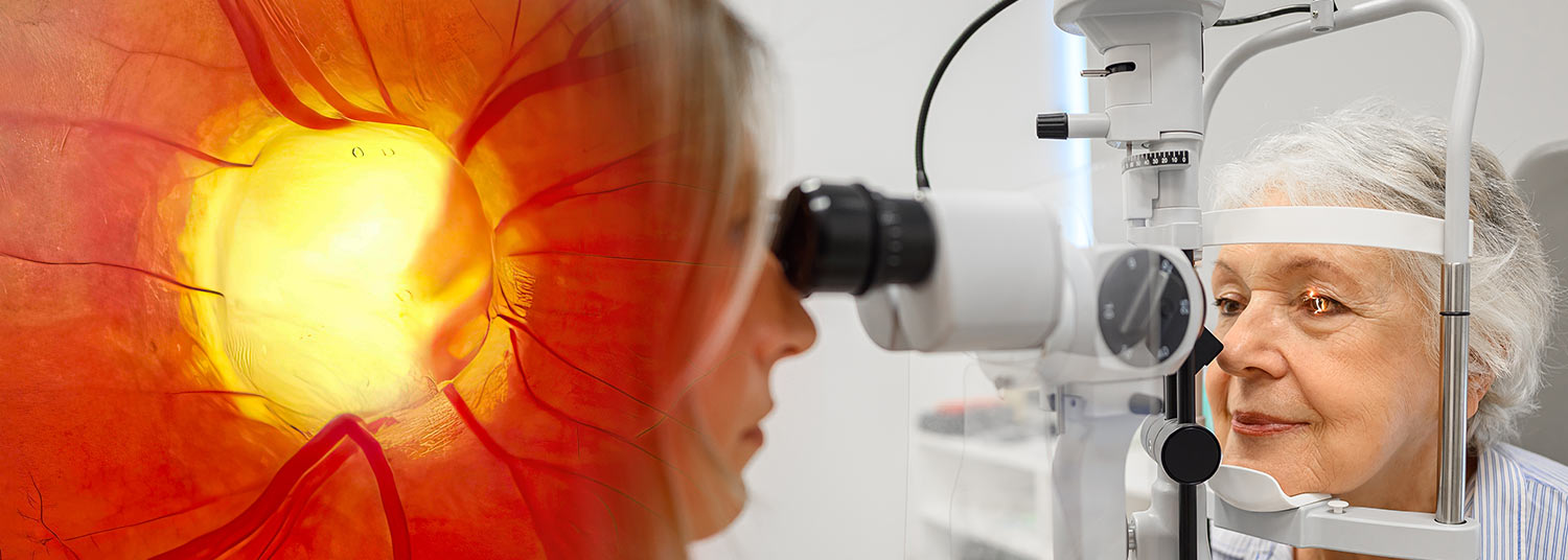
New study by researchers led by the U.S. Food and Drug Administration shows that advanced imaging technology can potentially detect cellular-level damage earlier than conventional clinical tests
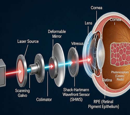
A new study applying advanced adaptive optics imaging technology developed by researchers at the U.S. Food and Drug Administration (FDA) to patients from the University of Maryland is showing promising potential to detect the early signs of glaucoma progression.
In a longitudinal study published in Science Advances, adaptive optics–optical coherence tomography (AO-OCT) – a high-resolution imaging technology engineered by scientists from the FDA’s Center for Devices and Radiological Health (CDRH)– was able to show unprecedented, microscopic changes to cells in adults with glaucoma. The collaboration between the University of Maryland and the FDA indicated that patients with clinical disease determined to be stable by conventional measurements actually had ongoing neuronal degeneration.
Over 4 million people in the United States have glaucoma, and it is a leading cause of irreversible blindness characterized by the progressive loss of retinal ganglion cells (RGCs), neurons that transmit visual information from the eye to the brain. Detecting this neuronal damage early is critical to help prevent vision loss. Most people with glaucoma are unaware they have it until the degenerative loss is permanent. Conventional diagnostic approaches — including traditional optical coherence tomography (OCT) and visual field testing — may not identify progression until substantial damage has occurred. There is currently no treatment for glaucoma, but the disease can be managed in most cases. New treatment strategies are currently under investigation.
 “We are on the verge of a major scientific breakthrough that could lead to early interventions for millions of people with degenerative eye disease,” said Osamah J. Saeedi, MD, Director of Clinical Research, Director of the Glaucoma Division, Vice Chair for Academic Affairs in the Ophthalmology and Visual Science Department at the University of Maryland School of Medicine. “Together with our partners at the FDA, we’re showing the power of collaboration to develop technologies intended to meet the needs of patients, clinicians, and practitioners.”
The study followed five patients with glaucoma and five age-matched healthy volunteers over a three-to-five-year period. One of the challenges the study overcame was distinguishing cellular changes associated with glaucoma from those that occur during normal aging. They did this with a suite of new analysis tools, including ones that employed artificial intelligence to help count thousands of RGCs in the study cohort.
The investigators found that RGC loss was substantially greater in participants with glaucoma than in healthy volunteers, even in those with glaucoma that was measured as stable with conventional methods. Healthy volunteers experienced an average annual cell loss of 0.27 percent, while glaucoma patients demonstrated loss rates that varied across retinal regions, averaging 9.6 percent annually across all patients and locations (ranging widely from 1.2 to 27.9 percent). The more-diseased retinal hemifield experienced an average annual cell loss of 6.9 percent, compared with 4.2 percent in the less-diseased hemifield.
AO-OCT detected statistically significant cellular progression earlier than conventional clinical measurements in this study. Across 24 retinal imaging locations in four glaucoma patients, AO-OCT detected progression approximately 8 months earlier than clinical OCT measurements of ganglion cell layer thickness and about 18 months earlier than visual field pattern deviation testing.
“One of the key findings of this study is the potential for AO-OCT to measure changes in retinal ganglion cell density and morphology much earlier than OCT layer-thickness measurements or subjective functional measures like perimetry,” said Dr. Achyut Raghavendra, Staff Fellow at CDRH and the first author on the study.
The research also identified changes in the morphology of surviving retinal ganglion cells. Glaucoma patients demonstrated enlargement of retinal ganglion cell somas over time, suggesting that surviving neurons may undergo structural remodeling as neighboring cells are lost. Further analysis showed that retinal ganglion cell soma volume declined by 30%, compared with an 11% reduction in overall ganglion cell layer volume, supporting the conclusion that neuronal loss is a major contributor to retinal thinning in glaucoma.
 “When you examine tissue at the cellular level, you can track changes related to disease more precisely and with better sensitivity compared to surrogate measures like thickness,” said Dr. Daniel X. Hammer, senior FDA investigator and the Director of the Division of Biomedical Physics at CDRH. “We expect cellular-level imaging biomarkers will potentially change the way ocular diseases are diagnosed and treated, starting with their use as clinical trial outcome measures.”
AO-OCT currently faces technical limitations, including a relatively small imaging field of view, lengthy imaging sessions, and the need for specialized equipment and image-processing methods. Also, larger scale studies will be needed to validate the findings. The current study used an investigational device at a single site and significant translation and large-scale, multisite clinical studies using FDA-approved devices will eventually need to be conducted. However, this first step shows the incredible promise of adaptive optics-enhanced imaging.
Dr. Saeedi is also an MPower Professor and the Co-Director of the Edward & Jennifer St. John Center for Translational Engineering and Medicine (CTEM), which brings together clinicians from UMSOM and engineers from the A. James Clark School of Engineering at the University of Maryland to tackle health challenges and drive medical innovations, improving treatments for patients and empowering them to live healthier lives.
The Department of Ophthalmology and Visual Sciences has advanced clinical and research grade machines for use in clinical trials and patient care and has recently recruited Andrew Bower, a translational optical engineer to further develop the adaptive optics research program at the University of Maryland, Baltimore.
The research was also supported by the National Institutes of Health through grant 1R01EY031731-01A1.
Contact
Ryan J. Fox
Senior Media and Public Relations Specialist
Ryan.Fox@som.umaryland.edu
Related stories
Wednesday, April 27, 2022
UMSOM Names Noted Faculty-Scientists, Dr. Lisa Schocket and Dr. Osamah Saeedi, To Leadership Appointments in Department of Ophthalmology
UMSOM Dean E. Albert Reece, MD, PhD, MBA, announced today that two senior faculty members in the Department of Ophthalmology and Visual Sciences have been named to leadership positions.