
- The landscape of acute ischemic stroke treatment changed dramatically with endovascular thrombectomy (EVT).
- However, few patients with basilar artery occlusions were included in major EVT trials.
- Basilar artery occlusion accounts for 10% of large vessel occlusions and can result in devastating neurological deficits.
- The recently published BASICS trial evaluated the efficacy of EVT within 6 hours of symptom onset in 300 patients with basilar artery occlusion strokes.
- 44.2% of the EVT group had a good outcome compared to 37.7% of the medical treatment group (p=0.19).
- Good outcome was defined as modified Rankin scale of 0 (no symptoms) to 3 (moderate disability but able to walk without assistance) at 90 days.
- Symptomatic intracranial hemorrhage was higher in the EVT group (4.5% vs. 0.7%, p=0.06).
- History of AFib was more common in the EVT group (28.6% vs. 15.1%).
- It is important to note that this study did not use advanced neuroimaging for patient selection unlike in landmark EVT trials of anterior circulation large vessel occlusion strokes.
Bottom Line: There is no significant difference between endovascular thrombectomy and medical management for basilar artery occlusion strokes within 6 hours of symptom onset.
Show References
A recent pilot study was conducted in two centers (Mayo Clinic & Cleveland Clinic Affiliate) and aimed to evaluate if the administration of oral midodrine in early septic shock could decrease the use of IV vasopressors and decrease ICU and hospital length of stay (LOS). The study was a placebo-controlled, double blinded randomized trial.
This study enrolled:
- 32 adult patients
- within 24 hours of Sepsis 3 definition who continued to have hypotension (MAP < 70mmHg) after antibiotic & 30mL/kg IVF administration
- 3 doses of midodrine 10mg were administered
The study did not find a statistical difference between the two groups in the use of vasopressors or ICU/Hospital LOS. However, there was a trend in the midodrine group which is suggestive of decreased vasopressor use and ICU/Hospital LOS.
It is Important to note the study was not powered to determine clinical significance. Overall the trend noted in the midodrine group should encourage further studies that are clinically powered to determine if there is a statistical difference and therefore a potential benefit to early initiation of oral midodrine in septic shock.
Show References
Vancomycin infusion reactions can manifest as pruritus and an erythematous rash of the neck, face, and torso during or after a vancomycin infusion. This is a histamine reaction caused by degranulation of mast cells and basophils, and can be caused short infusion times <60 min. It is commonly treated with antihistamines and/or a slowing of the infusion rate.
Show References
Norepinephrine is widely considered the first-line vasopressor for patients in septic shock. Vasopressin is often added to norepinephrine in patients requiring escalating doses, but when to add vasopressin, and what exactly the benefit is (as opposed to just further titrating up the norepinephrine) remain unclear. Given the limited evidence for a patient-oriented benefit and the increasing cost of vasopressin, some centers are becoming more judicious in the use of vasopressin. A systematic review in AJEM October 2021 examined the literature on early (< 6 hours of diagnosis) addition of vasopressin to the management of septic shock patients, compared to either no vasopressin or starting it after 6 hours.
Improved with early vasopressin: Need for renal replacement therapy (RRT; secondary outcome)
No difference: mortality, ICU length of stay, hospital length of stay, new onset arrhythmias
Bottom Line: When, and if, to start vasopressin in patients requiring escalating doses of norepinephrine remains controversial. Based on the prior VASST trial, many providers will start vasopressin when norepi doses reach ~ 5-15 mcg/min (approx 0.1-0.2 mcg/kg/min), but there remains limited data to support this practice, and either starting vasopressin or continuing to titrate the norepinephrine as needed are both reasonable approaches in most patients.
Show References
Question
What is the mechanism of action of N-acetylcysteine that is used to treat acetaminophen induced liver injury/toxicity?
Show Answer
Show References
Refractory Anaphylaxis
- Refractory anaphylaxis is defined as anaphylaxis that does not respond to at least 2 doses of 300 mcg of epinephrine given IM into the anterolateral thigh.
- Refractory anaphylaxis can be due to several factors including patient comorbidities, delayed diagnosis, delayed epinephrine administration, or concomitant beta-blocker or ACE-inhibitor medication use.
- Consider the following treatments in patients with refractory anaphylaxis, persistent hypotension, and shock:
- Aggressive IVF resuscitation - patients may need up to 7L of IVFs
- Epinephrine infusion (1-10 mcg/min) to target a MAP of 65 mm Hg
- Glucagon (1-5 mg via slow IV push) for those taking a beta-blocker and who fail to respond to epinephrine
- Norepinephrine, vasopressin, angiotensin II, methylene blue, or dobutamine can be administered with epinephrine for persistent hypotension.
- VA-ECMO - though there is no prospective evidence on the use of ECMO in anaphylaxis.
Show References
A recent article in Pediatrics attempted to estimate the association between fluoroquinolone use and tendon injury in an adolescent population.
Fluoroquinolones are thought to negatively impact tendons and cartilage in the load-bearing joints of the lower limbs through collagen degradation, necrosis, and disruption of the extracellular matrix.
Population: 4.4 million adolescents aged 12–18 years with filled outpatient fluoroquinolone prescription vs. an oral broad-spectrum antibiotic for comparison.
Fluoroquinolones included ciprofloxacin, levofloxacin, moxifloxacin, and gatifloxacin
Comparator antibiotics included amoxicillin-clavulanate, azithromycin, cefalexin, cefixime, cefdinir, nitrofurantoin, and bactrim.
Outcomes: Primary outcome was 90-day tendon rupture (Achilles, patellar, quadricep, patellar, tibial) identified by diagnosis and procedure codes. Secondary outcome was tendinitis.
Results: The weighted 90-day tendon rupture risk was 13.6 per 100 000 fluoroquinolone-treated adolescents and 11.6 per 100 000 comparator-treated adolescents.
Fluoroquinolone-associated excess risk: 1.9 per 100 000 adolescents; the corresponding number needed to treat to harm was 52 632.
The weighted 90-day tendinitis risk was 200.8 per 100 000 fluoroquinolone-treated adolescents and 178.1 per 100 000 comparator-treated adolescents
Fluoroquinolone-associated excess risk excess risk: 22.7 per 100 000 adolescents; the corresponding number needed to treat to harm was 4405.
Conclusion:
The excess risk of tendon rupture associated with fluoroquinolone treatment was extremely small, and these events were rare. On average, 50,000 adolescents would need to be treated with a fluoroquinolone for 1 additional tendon rupture to occur
The excess risk of tendinitis associated with fluoroquinolone treatment though larger was also small.
Besides tendon rupture, other more common potential adverse drug effects may be more important to consider for treatment decision-making, in adolescents without other risk factors for tendon injury.
Show References
During cardiac arrest, metabolic acidosis develops because of hypoxia-induced anaerobic metabolism and decreased acid excretion caused by inadequate renal perfusion. Sodium bicarbonate (SB) administration was considered as a buffer therapy to correct metabolic acidosis. However, SB has several side effects such as hypernatremia, metabolic alkalosis, hypocalcemia, hypercapnia, impairment of tissue oxygenation, intracellular acidosis, hyperosmolarity, and increased lactate production. The 2010 Pediatric Advanced Life Support (PALS) guideline stated that routine administration of SB was not recommended for cardiac arrest except in special resuscitation situations, such as hyperkalemia or certain toxidromes. An evidence update was conducted in the 2020 Pediatric Life Support (PLS) guideline and the recommendations of 2010 remain valid. This article was a systematic review and meta-analysis of observational studies of pediatric in hospital cardiac arrests. The primary outcome was the rate of survival to hospital discharge after in hospital cardiac arrests. The secondary outcomes were the 24-hour survival rate and neurological outcomes.
Show References
- Post-lumbar puncture (LP) headache, reported in up to 33% of patients, is due to a persistent CSF leak causing intracranial hypotension.
- A recent review by Cognat et al. looked to answer several frequently asked clinical questions:
- Who is at decreased risk of post-LP headache?
- Infants and children have a similar prevalence compared to adults.
- Older patients have a lower risk, with an incidence of <5% in those over 60 years old.
- Does needle choice minimize the risk of post-LP headache?
- Atraumatic non-cutting ("Whiteacre" or "Sprotte") needles have lower rates (RR 0.4, 0.34-0.47).
- The use of atraumatic needles does not affect the rate of success, success on first attempt, or duration of the LP.
- Does performing the LP in a specific way prevent post-LP headache?
- LPs performed in the lateral decubitus position and at a higher intervertebral space have a lower incidence.
- Difficult LPs (e.g. multiple attempts, traumatic tap) do not appear to affect the rate.
- The volume of CSF removed does not affect the rate.
- Do any treatments after the LP reduce post-LP headache occurrence?
- Bed rest after LP does not reduce and may in fact worsen the likelihood.
- Fluids and caffeine do not prevent post-LP headaches.
- Who is at decreased risk of post-LP headache?
Bottom Line: The use of atraumatic needles is most effective in reducing the risk of post-LP headaches. These needles are easy to use and have similar rate of success as cutting needles.
Show References
RECAP: RECOVERY trial is a large, randomized, open label, adaptive trial studying different treatments on COVID-19. Most well known is the use of dexamethasone which reduced mortality by 1/3 in COVID patients requiring mechanical ventilation and by 1/5 in those requiring oxygen, with no benefit on those patients not requiring oxygen.
They recently published results in the Lancet on the use of tocilizumab.
Population:
- Up to 21 days after main randomization, regardless of treatment, RECOVERY trial patients with progressive COVID-19 were eligible for tocilizumab.
Inclusion:
- April 23rd 2020 to Jan 24th 2021-- 21,550 patients with hypoxia (<92% on RA or requiring O2), systemic inflammation (CRP > 75 mg/L) eligible for standard care or standard care plus toci 400-800 mg (dosing based on weight), second dose 12-24 hours later if no improvement
Outcomes:
- Primary outcome 28 day mortality followed by:
- Hospital discharge within 28 days
- Rate of mechanical ventilation
Results:
- 621 (31%) tocilizumab patients and 729 (35%) of usual care patients died within 21 days (RR 0.85, p=0.0028). Consistent even in those receiving steroids (83%).
- Tocilizumab group more likely to be discharged from the hospital, less likely to receive invasive mechanical ventilation (35% vs 42%).
Conclusion:
- Tocilizumab improved survival and other clinical outcomes- by 1/3 for those on simple oxygen, and by ½ for those receiving invasive mechanical ventilation.
- Added to the additional benefit of steroids.
- Findings support the earlier REMAP-CAP trial on the effectiveness of tocilizumab for ICU COVID patients
Show Additional Information
Show References
Both erythrocyte sedimentation rate (ESR) and C-reactive protein (CRP) are highly sensitive (84-100%) for spinal infections and are observed in >80% with vertebral osteomyelitis and epidural abscesses.
ESR
Most sensitive and specific serum marker, usually elevated in both spinal epidural abscess (SEA) and vertebral osteomyelitis.
ESR was elevated in 94-100% of patients with SEA vs. only 33% of non-SEA patients
Mean ESR in patients with SEA was significantly elevated (51-77mm/hour)
CRP
Not highly specific
Less useful for acute diagnosis since CRP levels rise faster and return to baseline faster than ESR (elevated CRP seen in 87% of patients with SEA as well as in 50% of patients with spine pain not due to a SEA)
Better used as a marker of response to treatment.
Show References
Background:
Cardiac arrest from massive pulmonary embolism (PE) can be up to 90% (1). A recent systemic review evaluated the efficacy of Venoarterial-Extracorporeal Membrane Oxygenation (VA-ECMO) for PE-related cardiac arrest.
Results:
The authors screened 1115 articles and included 77 articles, including gray literature. The authors performed a quantitative analysis of a total of 301 patients.
Overall, 183/301 (61%) patients survived to hospital discharge, a significant improvement from 90%.
Patients who were cannulated during chest compression were associated with 7x higher odds of death (OR, 6.84; 95% CI, 1.53–30.58; p = 0.01), compared to those who were cannulated after ROSC. However, cannulation in the ED was not associated with improved outcomes, compared with other cannulation site.
No increased risk of death among patients who received tPA prior to VA-ECMO vs. those who did not (OR, 0.78; 95% CI, 0.39–1.54; p = 0.48).
Patients whose age > 65 years of age were associated with 3X risk of death, compared to those with age < 65 years (OR, 3.56; 95% CI, 1.29–9.87; p = 0.02).
Take-home points
Please consider “early” VA-ECMO for eligible patients who have cardiac arrest from massive PE. However, it will take great convincing to push the PERT team to cannulate for VA-ECMO while the patient is still receiving chest compression.
Show References
Background:
Cardiac arrest from massive pulmonary embolism (PE) can be up to 90% (1). A recent systemic review evaluated the efficacy of Venoarterial-Extracorporeal Membrane Oxygenation (VA-ECMO) for PE-related cardiac arrest.
Results:
The authors screened 1115 articles and included 77 articles, including gray literature. The authors performed a quantitative analysis of a total of 301 patients.
Overall, 183/301 (61%) patients survived to hospital discharge, a significant improvement from 90%.
Patients who were cannulated during chest compression were associated with 7x higher odds of death (OR, 6.84; 95% CI, 1.53–30.58; p = 0.01), compared to those who were cannulated after ROSC. However, cannulation in the ED was not associated with improved outcomes, compared with other cannulation sites.
No increased risk of death among patients who received tPA prior to VA-ECMO vs. those who did not (OR, 0.78; 95% CI, 0.39–1.54; p = 0.48).
Patients whose age > 65 years of age were associated with 3X risk of death, compared to those with age < 65 years (OR, 3.56; 95% CI, 1.29–9.87; p = 0.02).
Take-home points
Please consider “early” VA-ECMO for eligible patients who have cardiac arrest from massive PE. However, it will take great convincing to push the PERT team to cannulate for VA-ECMO while the patient is still receiving chest compression.
Show References
- Electronic cigarette (E-cigs) smoking (vaping) continues to be a major concern among adolescents and teens, who mistakenly think it is safer than smoking traditional cigarettes or don't consider it as smoking at all.
- Typically, they contain nicotine which is highly addictive and can cause harm in the developing brain, but can also contain other dangerous chemicals, flavorings and drugs.
- They often contain higher amounts and concentrations of nicotine. 1 JUUL pod can contain the equivalent of 20 packs of nicotine cigarettes.
- Inhaled aerosols of the various chemicals, flavorings, and heavy metals have resulted in lung disease and acute respiratory failure. Bilateral infiltrates on chest imaging is a common finding.
- Nicotine toxicity can also occur. Symptoms include vomiting, diarrhea, abdominal pain, salivation, headache, dizziness, confusion, and seizures. Hypertension and tachycardia acutely, followed by hypotension and bradycardia can be expected.
- Bottom Line: Ask specifically about electronic cigarette use in adolescents and teens who present with acute complaints. One study found that of those who regularly used and presented for evaluation of symptoms, 98% were respiratory, 81% were gastrointestinal, and 100% were constitutional in nature.
Show References
- Functional neurological disorders (FND) are unintentional and involuntary.
- Imaging and electrophysiological studies have shown cerebral dysfunctions in attention and perception, which may explain why symptoms often improve with distraction.
- Diagnosis requires demonstration of inconsistency and/or incongruency with recognized neurological or medical conditions.
- No clinical sign alone is diagnostic.
- Patients may have comorbidities such as multiple sclerosis, stroke, or epilepsy.
- Hoover’s sign and drift without pronation have been described as positive signs for FND.
- These can also be seen in patients with pain, neglect, or apraxia.
- Avoid maneuvers that may harm the patient, such as dropping their arm onto their face.
- A high-pitched tuning fork applied to the nostrils is an effective stimulus to assess responsiveness.
- Avoid using terms like non-organic, psychogenic, or pseudoseizure.
- When counseling a patient, avoid only explaining what conditions they do not have or attributing symptoms to psychological problems or stress.
- Instead, name the diagnosis, explain that their symptoms are real and common, and emphasize that symptoms are potentially reversible.
- Early diagnosis of FND is associated with improved physical and psychological outcomes.
Bottom Line: Functional neurological disorders (FND) are commonly encountered in the ED. A thorough neurological exam may reveal positive signs suggestive of FND. Early diagnosis and referral to specialists may improve outcomes.
Show References
Erythrocyte sedimentation rate (ESR) for spinal infection
Sensitive for spinal infection but not specific
Elevated ESR is observed in greater than 80% of patients with vertebral osteomyelitis and epidural abscess
ESR is the most sensitive and specific serum marker for spinal infection
Usually elevated in acute presentations of SEA and vertebral osteomyelitis
ESR was elevated in 94-100% of patients with SEA vs. only 33% of non-SEA patients
Mean ESR in patients with SEA was significantly elevated (51-77mm/hour)
Infection is unlikely in patients with an ESR less than 20 mm/h.
Incorporating ESR into an ED decision guideline may improve diagnostic delays and help distinguish patients in whom MRI may be performed on a non-emergent basis
Show References
Supplemental oxygen therapy is frequently required for patients presenting with acute respiratory distress and COPD exacerbation. Over-oxygenation can derail compensatory physiologic responses to hypoxia,1 resulting in worsening VQ mismatch and, to a lesser degree, decreases in minute ventilation, that cause worsened respiratory failure.
The 2012 DECAF (Dyspnea, Eosinopenia, Consolidation, Acidaemia, and Atrial Fibrillation) score was found to predict risk of in-hospital mortality in patients admitted with acute COPD exacerbation.2,3 Data from the DECAF study’s derivation and external validation cohorts were examined specifically to look at outcome associated with varying levels of oxygen saturation.
- 1027 patients from 6 UK hospitals receiving supplemental oxygen at admission
- Lowest in-hospital mortality seen in the 88-92% cohort
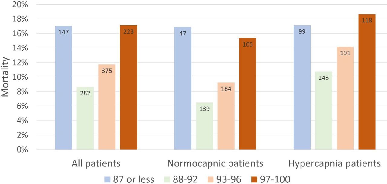
- Adj OR for in-hospital mortality in ≥97% vs 88-92% group: 2.97 (95% CI 1.58-5.58, p=0.001)
- Adj OR for in-hospital mortality in 93-96% vs 88-92% group: 1.98 (95% CI 1.09-3.60, p=0.025)
- Surprisingly, mortality risk seen more in normocapnic than hypercapnic patients
- Association between admission SpO2 and mortality persisted after adjusting for baseline risk and disease severity using the DECAF and NEWS 2 score
Bottom Line
In patients presenting to the ED with acute COPD exacerbation requiring oxygen supplementation, a target oxygen saturation of 88-92% is associated with the lowest in-hospital mortality, and higher oxygen saturations should be avoided independent of patients' PCO2 levels.
Show References
Show References
- Alteplase administration in acute ischemic stroke (AIS) has been associated with errors.
- A recent study looked at the types of errors that occurred at regional hospitals and Comprehensive Stroke Centers.
- 19.8% (133/676) of patients had an error associated with alteplase administration with the majority occurring at regional hospitals without stroke certification.
- The most common error was over-dosage of alteplase, occurring in 5% (36) of patients.
- Other common errors included under-dosage, infusion errors, and apparent contraindications.
- The most common contributing factor leading to the error was incorrect calculation.
- Administration error was associated with higher rate of hemorrhagic conversion (12.7% vs. 7.1%, p=0.04).
Bottom Line: Alteplase administration in acute ischemic stroke is associated with errors, most commonly with over-dosage of the medication.
Show References
Study Question: A recent study investigated whether adult patients presenting to the ED with a diagnosis of mTBI prescribed light exercise were less likely to develop persistent postconcussion symptoms.
Setting: Randomized controlled trial conducted in three Canadian EDs. Consecutive, adults (18–64 years) seen in ED with a mTBI sustained within the preceding 48 hours.
The intervention group received discharge instructions prescribing 30 minutes of daily light exercise.
The control group was given standard mTBI instructions advising gradual return to exercise following symptom resolution.
Outcome: The primary outcome was the proportion of patients with postconcussion symptoms at 30 days,
A total of 367 patients were enrolled. Median age was 32 years Male 43%/Female 57%.
Result: There was no difference in the proportion of patients with postconcussion symptoms at 30 days. There were no differences in median change of concussion testing scores, median number of return PCP visits, median number of missed school or work days, or unplanned return ED visits within 30 days. Participants in the control group reported fewer minutes of light exercise at 7 days (30 vs 35).
Conclusion
Prescribing light exercise for acute mTBI, demonstrated no differences in recovery or health care utilization outcomes.
Extrapolating from studies in the athletic population, there may be a patient benefit for light exercise prescription.
Make sure that the patient is only exercising to their symptomatic threshold as we recommend with concussed athletes. Previous studies have shown that athletes with the highest post injury activity levels had poorer visual memory and reaction time scores than those with moderate activity levels.