
- Many infants w/cyanotic heart disease only survive w/early surgical intervention
- The most rapid & effective first-line therapy for stabilization of the crashing neonate is IV prostaglandin E1 (PGE1)
- PGE1 serves to reopen the ductus arteriosus allowing partially desaturated systemic arterial blood to enter the pulmonary artery and be oxygenated
- The widespread use of this agent has profoundly decreased morbidity & mortality
- The initial dose of PGE1 is 0.1 mg/kg/min
- ADR for PGE1 include: apnea, hypotension, edema, and low grade fever
Show References
Hematoma Block
Provides good aesthesia for reduction of fractures. Onset in approximately 5 minutes
Benefits: No need for NPO, simple and easy to perform & can be done without additional personnel (unlike w/ procedural sedation)
Contraindications: Open fractures, dirty or infected overlying skin
1) Identify fracture site with x-ray and palpation
2) Clean skin w/ Betadine
3) Insert needle into the hematoma. * Confirm placement by aspirating blood *
4) Inject anesthetic (lidocaine 1 or 2%) into the fracture cavity and adjacent periosteum
http://www.youtube.com/watch?v=tjnsdjfwMmY
Cyclophosphamide-induced hemorrhagic cystitis is a well known to oncologists. This unique complication of this chemotherapeutic drug has a defined mechanism and could be seen in your Emergency Department.
- Hemorrhagic cystitis occurs in 46% of patients that receive cyclophosphamide
- Can occur even months after administration
- 5% can actually die from the hemorrhage
- Treatment: Bladder irrigation, hydration, supportive. Oral adminsitration of MESNA (2mercaptoethan sulfonate) and bladder irrigation with prostaglandins and even methylene blue have been attempted.
Question
A 38 year old man is brought in by ambulance for a seizure. His medical history is not known. On exam he is post-ictal and otherwise has a non-focal neurologic exam. He has an abrasion above the right eye, a small tongue laceration, and was incontinent of urine. A head CT was done and is shown below. What was the cause of this man's seizure?

Show Answer
Show References
Attachments
Postintubation Hypotension
- It is clear that preintubation hypotension is associated with increased mortality in critically ill patients who require mechanical ventilation.
- Unfortunatley, the literature is less clear on the frequency and impact of hypotension that develops after intubation.
- Two recent publications in the Journal of Intensive Care provide valuable information on postintubation hypotension. Some highlights of the studies include:
- Retrospective cohorts of over 300 patients who developed postintubation hypotension, defined as a SBP < 90 mm Hg within 60 min of intubation.
- Postintubation hypotension occurred in almost 25% of patients.
- Median time to hypotension was 11 minutes.
- Patients with postintubation hypotension had a higher inhospital mortality (33% vs. 23%).
- A preintubation Shock Index > 0.8 was the strongest predictor of cardiovascular collapse after intubation.
- Take Home Point: Postintubation hypotension occurs frequently and may be associated with worse outcomes.
Show References
Question
45 year-old male complains of chest pain and cough. He also tells you, "...oh, and by the way doc, I just smoked something." What's the diagnosis?

Show Answer
· Cyanosis in the newborn is defined as an arterial saturation <90% and a PO2 <60 torr
· To help differentiate between cardiogenic and non-cardiogenic causes initially obtain an arterial saturation on room air and obtain a subsequent measurements on 100% oxygen
· Infants w/neurogenic or pulmonary causes of cyanosis will demonstrate increases in arterial blood saturation on 100% oxygen while infants with congenital heart disease show minimal elevation
· There are 3 general sources of arterial desaturation in neonates with structural heart disease:
1.) Lesions with decreased pulmonary blood flow (tetralogy of Fallot, severe pulmonary stenosis/atresia, and tricuspid atresia)
2) Admixture lesions, in which desaturated systemic venous blood mixes with intracardiac blood, and then enters the aorta (transposition of great vessels, partial anomalous pulmonary venous drainage)
3) Lesions with increased pulmonary blood flow and pulmonary edema, in which diffusion barriers and intrapulmonary shunting prevent proper oxygenation (truncus arteriosus)
Show References
Question
Patient: A 10 year old female is brought to the ED after swallowing 2 beads (see image). Based on the findings, what are your concerns and what is the disposition?

Show Answer
SSRIs and SNRIs like venlafaxine and sertraline are well known to cause hyponatremia. Usually considered safe, this adverse drug event can lead to weakness, confusion, seizure and even cerebral edema. Elderly are more susceptible to this adverse effect.
ADH is regulated by serotonin and thus the mechanism for the Hyponatremia is SIADH.
Tolvaptan, a vasopressin receptor antagonist, has been a new treatment that has been used anecdotally in Europe. Waiting for the first US case report.
Show References
More than 1.2 billion people are infected with at least one species.
Most helminth infections are contracted by ingesting the eggs, except strongyloides and hookworm whose larvae penetrate bare skin when it is contact with the soil.
The roundworm (Ascaris lumbricoides) life cycle involves migration through the lung tissue which can cause pneumonitis. Patients can present with interstitial infiltrates, wheeze, and blood tinged sputum. Ascaris than migrates to the intestines where it can cause partial small bowel obstruction. In pediatric patients, the appendix may be invaded causing gangrene with symptoms indistinguishable from appendicitis. In adults, the worms can invade the biliary tract and cause biliary disease or pancreatitis. Fever causes this helminth to migrate and it can emerge from the nasopharynx or the anus.
Whipworms (Trichuris trichiura) present as colitis or symptoms similar to inflammatory bowel disease. Chronic illness can involve anemia and clubbing. In severe cases, trichuris can cause dysentery and rectal prolapse.
Hookworms (Necator americanus or Ancylostoma duodenale) also have a pulmonary phase, but with milder symptoms than Ascaris. Eventually hookworms cause iron deficiency anemia and malnutrition. They can be a primary cause of anemia in pregnancy in endemic areas.
Threadworm (Strongyloides stercoralis) can cause a wide spectrum of disease presentations. The infection can start with a rash, larva currens. The infection may be subclinical or may invade the lung, intestinal wall, or the nervous system. Eventually hyperinfection may develop which is a very large increase in worm burden and then the infection becomes disseminated.
Toxocara canis or toxocara cati have affected approximately 14% of the US population. These helminthes reproduce in dogs or cats, and human infection is not part of the normal life cycle. Most infections are subclinical but it can produce a mild pneumonitis that is very similar to asthma. There can be pain and inflammation as the helminthes travel through organs such as the liver or lung and is called visceral larva migrans. The helminth may also move through the eye and optic never causing an ocular form of the disease, ocular larva migrans.
Pinworms (Enterobius vermicularis) are the cause of most common helminth infection in US and can present with anal pruritus leading to trouble sleeping. When an infection is identified, everyone in the household should be treated, regardless of symptoms.
University of Maryland Section of Global Emergency Health
Author: Jenny Reifel Saltzberg, MD, MPH
Show References
Intra-aortic balloon pumps (IABP) are devices that provide hemodynamic support during cardiogenic shock; the balloon inflates during diastole (improving coronary artery perfusion) and deflates during systole (reducing afterload and improving systemic perfusion). Click here to see a 41 second video illustrating how it works.
Several guidelines recommend placement of an IABP for patients in cardiogenic shock secondary to acute myocardial infarction (AMI), if early revascularization (e.g., CABG) is planned (Class I recommendation). Data behind this recommendation, however, is limited.
The IABP-SHOCK II trial was a randomized, multi-center, open-label study that enrolled 600 patients (598 in the analysis) with cardiogenic shock secondary to AMI (STEMI or NSTEMI). Patients were randomized to the control group (receiving standard therapy; N=298) or the experimental group (receiving IABP; N=300).
No significant difference was found between groups with respect to 30-day mortality (primary end-point), secondary end-points (e.g., time to hemodynamic stabilization, renal function, lactate levels, etc.), or complications (e.g., major bleeding, peripheral ischemic complications, etc.).
Bottom line: Perhaps it is time to reassess the approach to cardiogenic shock secondary to AMI when early revascularization is planned. At this time consultation with local expertise is recommended.
Show References

- Ventricular assist devices (VAD) have become an option as bridge to transplant or destination therapy in many patients (prevalence heart failure in US 5.7 million)
- VADs have significantly improved quality of life by NYHA class & 6 min walk distance
- 2 main types of VAD exist, pulsatile (PF) and continuous flow (CF), with 98% being CF
- Both bleeding and thrombosis are frequently encountered complications
- Although required systemic anticoagulation increases the risk of bleeding, there is a inherent association between CF VADs and GI AVMs
- Hypotension a common complication, which should be assessed by ruling out: bleeding, thrombosis, mechanical obstruction, sepsis, and RV failure
Show References
"When can my child get back out on the field doc?"
Return to play
▸ Concussion symptoms should be resolved before returning to exercise.
▸ A RTP progression involves a gradual, step-wise increase in physical
demands, sports-specific activities and the risk for contact.
▸ If symptoms occur with activity, the progression should be halted and
restarted at the preceding symptom-free step.
▸ RTP after concussion should occur only with medical clearance from a
licenced healthcare provider trained in the evaluation and management
of concussions.
Short-term risks of premature RTP
▸ The primary concern with early RTP is decreased reaction time leading
to an increased risk of a repeat concussion or other injury and
prolongation of symptoms.
Long-term effects
▸ There is an increasing concern that head impact exposure and
recurrent concussions contribute to long-term neurological sequelae.
▸ Some studies have suggested an association between prior concussions
and chronic cognitive dysfunction. Large-scale epidemiological studies are
needed to more clearly define risk factors and causation of any long-term
neurological impairment.
Show References
- CDC recommends pediatric influenza antiviral treatment for those at higher risk for influenza complications, and include the following:
- less than 2 years of age;
- chronic diseases including: pulmonary (ie asthma), cardiovascular (except hypertension alone), renal, hepatic, hematologic (ie sickle cell disease), metabolic (ie diabetes), neurologic/neurodevelopmental (ie cerebral palsy, epilepsy), and intellectual disability (ie mental retardation)
- immunosuppression (ie HIV)
- less than 19 years of age and on chronic aspirin treatment;
- morbid obesity (BMI>40)
- adamantanes (amantadine and rimantadine) should not be used due to high levels of resistance to influenza A
- neuraminadase inhibitors (oseltamivir and zanamivir) should be started within 48 hours of illness onset to reduce the duration and severity of disease
- oseltamivir can be used in children as young as 2 weeks of age at a dose of 3mg/kg twice daily for 5 days.
Show References
Several medications can produce a false-positive result for methadone on the urine drug screen: diphenhydramine, doxylamine, clomipramine, chlorpromazine, quetiapine, thioridazine, and verapamil.
Add a new one to the list. Tapentadol, a relatively new opioid analgesic similar to tramadol, can also produce a false-positive result for methadone on certain immunoassays.
A separate study concluded that tapentadol does not affect the amphetamine screen.
Show References
Question
These two Ethiopian boys present with “back problems”. What are the diagnoses and what do you need to worry about with each of them?
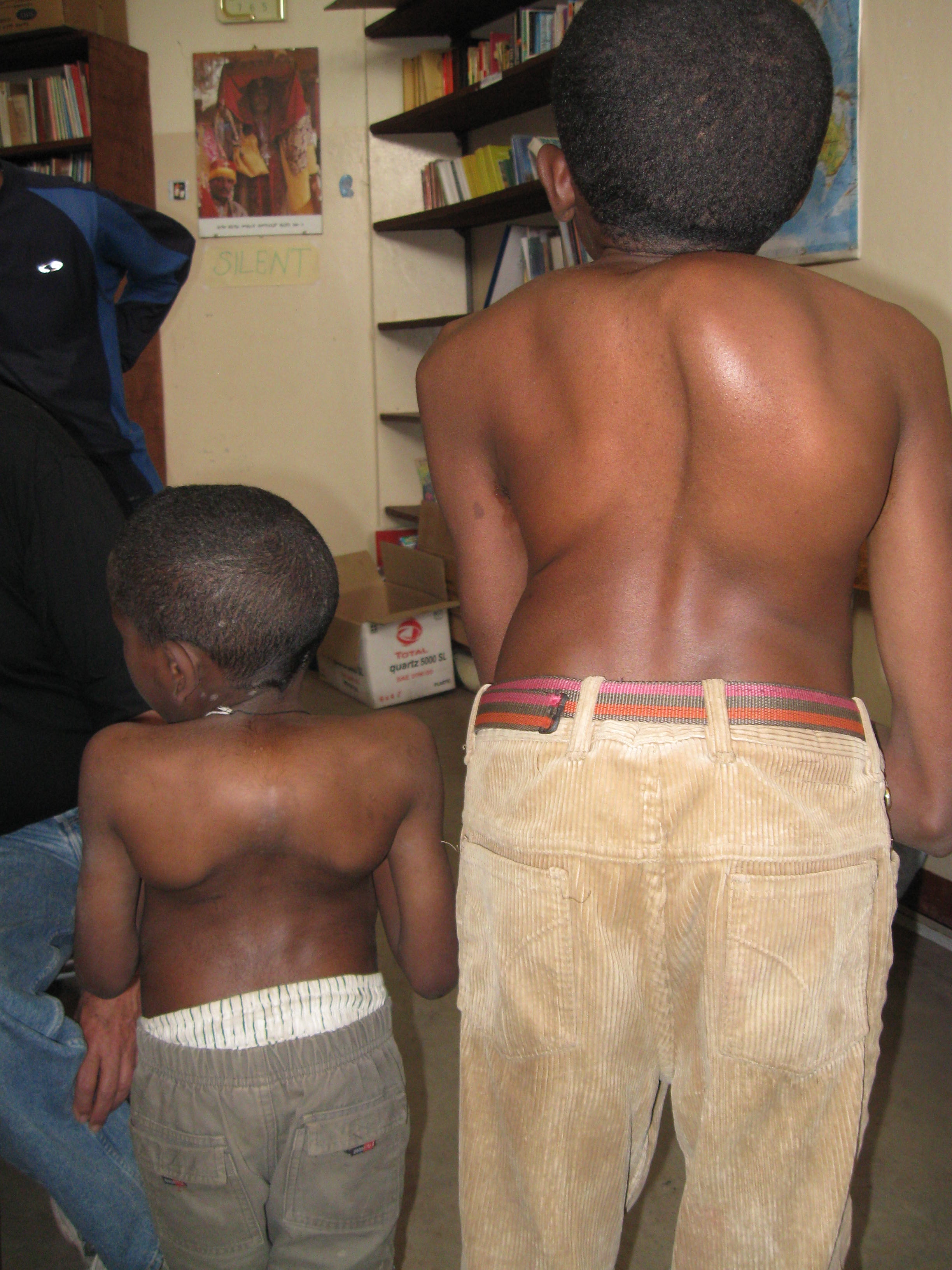
Show Answer
Show References
Attachments
The Crashing Cardiac Transplant Patient
- Approximately 2000 patients receive a cardiac transplant each year in the United States.
- With improvements in surgical techniques, immunosuppression, and management of complications, graft half-life is now approximately 13 years; thereby increasing the likelihood that a cardiac transplant patient will show up in your ED.
- In the crashing cardiac transplant patient, think of the following causes for acute decompensation:
- Acute rejection
- Primary graft failure
- RV failure
- Sepsis
- For patients with primary graft failure initiate inotropic support with dobutamine, epinephrine, milrinone, or isoproteronol. Those failing standard inotropes will likely require mechanical circulatory support (VAD) or ECMO.
- Patients with acute RV failure will often require the combination of a pulmonary vasodilator (inhaled NO, prostaglandins) and inotropic agent. In addition, it is critical to avoid hypercapnia and hypoxia.
Show References
Question
4 year-old female with the post-procedural CXR shown below. What's the diagnosis? (Hint: use the zoom...this one is tricky)

Show Answer

Show References
- BCI results in a spectrum of outcomes from asymptomatic to sudden cardiac death
- Normal screening ECG is associated with a 98% negative predictive value
- Sinus tachycardia is the most common ECG abnormality among trauma victims
- Myocardial contusion (MC) is the most common & ambiguous diagnosis following BCI
- MC has no consensus definition or uniform diagnostic criteria and can be loosely defined as BCI w/mild increase in cardiac biomarkers or frank cardiac dysfunction (e.g. wall motion abnormalities, arrhythmias, conduction disturbances, or SCD)
- BCI w/ a normal ECG & stable hemodynamics have a benign clinical course and rarely require further diagnostic testing or long periods of close observation
- Individuals w/ECG abnormalities, hemodynamic instability, or rapid deceleration injury concerning for blunt aortic injury (BAI) warrant imaging of heart and great vessels by echocardiogram and CT scan