
Clinical Presentation:
A 35-year-old female presents to your emergency department complaining of fever, malaise, myalgias, headache and an urticarial rash. Her physical exam reveals a papular rash and hepatosplenomegaly. You also find out that she traveled to Sudan 6 weeks earlier. She stayed mostly in Kharotum, but while there, she swam in the Nile. You send a smear for malaria, which is negative. What other major parasite should you consider?
Diagnosis:
- Schistosomiasis, also know as bilharzia, is a disease caused by parasitic worms.
- While not found in the US, it impacts more than 200 million people globally, and is second only to malaria as a major parasitic infection.
Discussion:
While the acute presentation is generally non-specific, chronic complications may be more serious. Many organ systems can be impacted and symptoms of chronic infection can include liver dysfunction, including portal hypertension and esophageal varacies or hematuria and renal failure.
Treatment:
- Treatment is a one-day course of Praziquantel but must be initiated 6-8 weeks after infection. (It's most effective against the adult worm so timing is key!)
Bottom Line:
Consider a broader differential in travelers. There are many infectious killers that can be easily treated.
University of Maryland Section of Global Emergency Health
Author: Jon Mark Hirshon, MD, MPH, PhD
Show References
Thrombelastography for Management of Non-Traumatic Hemorrhagic Shock
The use of thrombelastography (TEG, ROTEM) has traditionally been utilized and studied in the management of acute coagulopathy of trauma (ACoT) developed by patients in hemorrhagic shock secondary to trauma.
Functional coagulation tests such as the TEG may provide valuable information when resuscitating the hemorrhaging patient, especially if there is any concern for an underlying coagulopathy.
The following is a TEG recently returned during the resuscitation of a 60 y/o male with a history of HCV cirrhosis presenting with hemorrhagic shock secondary to a massive upper GIB. The University's Massive Transfusion Protocol was promptly activated and at this point, the patient had received approximately 4 units of PRBCs & FFP along with 1 liter of crystalloid. His Hgb was 5, PT/PTT/INR were undetectable, and his fibrinogen was 80.

Below is a table that simplifies the treatment, based on the test's abnormalities:
- Prolonged R: Fresh frozen plasma
- Prolonged K or reduced α angle: Cryoprecipitate
- Low MA: Platelets, desmopressin (DDAVP)
- Elevated LY 30%: Consider antifibrinolytics (aminocaproic acid, TXA)
After reviewing the initial TEG, all perameters were abnormal in addition to the presence of significant fibrinolysis. The patient was given an additional 4 units of FFP, DDAVP, cryoprecipitate, a unit of platelets, and aminocaproic acid. The patient still required significant resuscitation, however bleeding had significantly decreased as well has his pressor requirement. Below is the patient's follow-up TEG 2 hours later.

There is growing enthusiasm for the use of functional coagulopathy testing in the patient with hemorrhagic shock. Early resuscitation with blood products as your fluid of choice with limited fluid administration while arranging for definitive source control are critical, but also consider early thrombelastography to detect additional causes for uncontrolled hemorrhage.
References
- Walsh M, Thomas SG, Howard JC, et al. Blood component therapy in trauma guided with the utilization of the perfusionist and thromboelastography. Journal of Extra-Corporeal Technology. 2011 Sep; 43(3):162-7.
- The Use of TEG & Goal Directed Blood Component Therapy. MarylandCCProject.org
Follow Me On Twitter: @JohnGreenwoodMD
email: johncgreenwood@gmail.com
Show References
Question
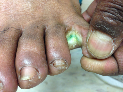
41year-old male without past medical history presents with the image below. What's the diagnosis and what's the most likely causative organism?
Show Answer
Show References
Methadone prolongs the QTc interval. Is the degree of QTC widening correlated to worse outcomes after overdose?
The authors of a new study concluded the triage QTc can predict death, intubation, and respiratory arrest. QTc thresholds of 470, 447.5, and 450 msec had sensitivity (95 % CI) and specificity (95 % CI) of 87.5 (47.3-99.7), 86.8 (74.7-94.5), and 77.3 (62.2-88.5), respectively.
My Thoughts
Respiratory depression is the predominant cause of death in methadone overdoses. QTc interval prolongation may have the potential to help predict outcomes, but the QTc thresholds in this study were really not that prolonged. Patients on chronic methadone without overdose have baseline QTc intervals longer than those in this study after overdose.
Application to Clinical Practice
Many factors contribute to the ultimate disposition of methadone overdose cases. Even if QTc widening is correlated to outcomes, it really won't change our management.
Show References
Case Presentation:
A 64 yo male with a history of IDDM presents with generalized fatigue. He felt so weak last night that he missed his pm dose of Lantus and vomited this morning. He arrived with a critically elevated BG of 590. He flew to the US from Sierra Leone 3 days ago.
Labs include:
Wbc 3.5 Plt 34 Hb 12 Hct 36
Na 125 CL 93 Co2 14 K 4.5 BUN 25 Cr 1.9 Glu 590 AG 18
VBG pH 7.23
Clinical Question:
Other than treating his diabetic ketoacidosis and renal failure, would you send any further tests?
Answer:
Thick smear for Malaria.
Bottom Line:
- DKA is often a symptom of an underlying metabolic or infectious insult
- Have a high suspicion for malaria in those travelers coming from endemic regions and don't forget malaria symptoms are often insidious presenting with myalgia and fatigue, as well as the traditional undulating fevers and chills
- In addition to anemia, thrombocytopenia has been seen in P. vivax and P. falciparum. (In the at-risk traveler, this finding should raise your suspicion for malaria.)
University of Maryland Section of Global Emergency Health
Author: Bradford Schwartz, MD
Show References
- A feared complication of patients taking vitamin K antagonists (VKA) is life-threatening bleeding (LTB), including intracranial hemorrhage (ICH).
- Prothrombin complex concentrate (PCC; containing factors 2,7,9,and 10) rapidly reverses VKA-associated bleeding. Despite a rapid reversal of the INR, there is little literature demonstrating a mortality benefit.
- The EPAHK study was observational-cohort that examined the 7-day mortality of guideline-concordant administration of PCC and vitamin K (GC-PCC-K) for multiple-types of patients with warfarin-associated bleeding.
- The study demonstrated patients who received GC-PCC-K within 8 hours of presentation had a two-fold decrease in 7-day morality; there was a three-fold reduction when only ICH was considered.
Show References
Question
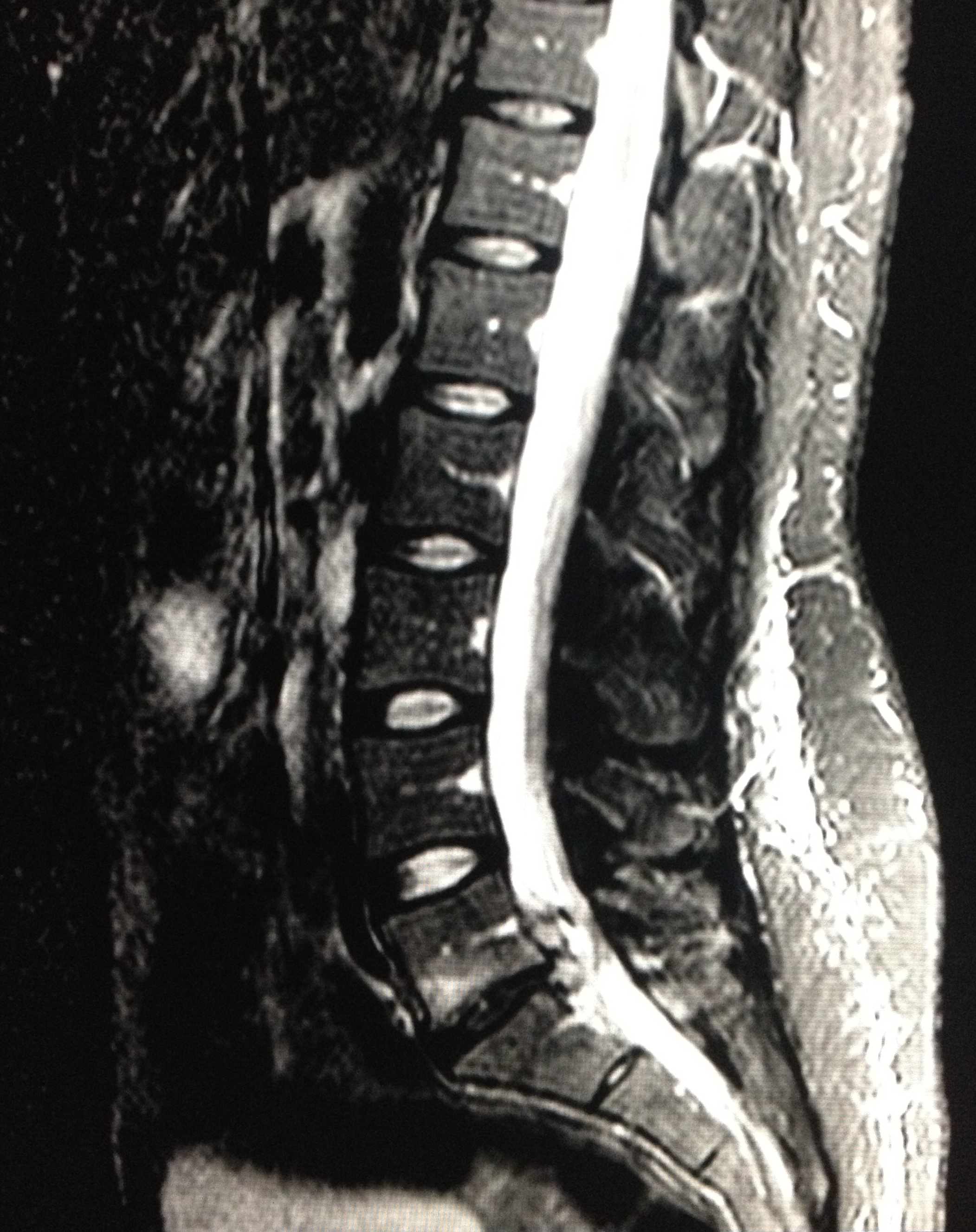
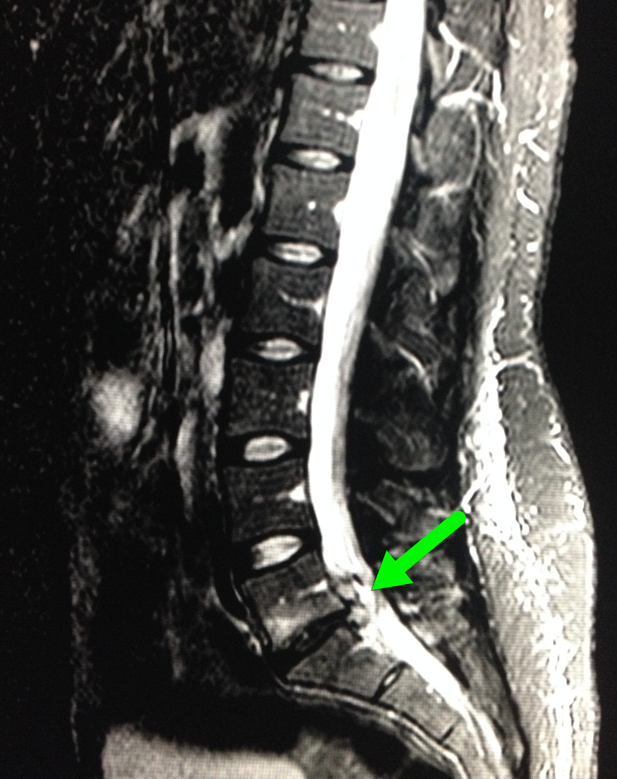
28 year-old female with history of chronic back pain presents with right-foot numbness and inability to move her foot at the ankle joint. What’s the diagnosis and what neurologic finding would you expect to find?
Show Answer
Show References
In a prospective cohort of 598 ED patients, 5 risk factors were independently associated with uncomplicated cellulitis patients who fail initial antibiotic therapy as outpatients and require a change of antibiotics or admission to hospital:
- Fever (temperature > 38°C) at triage (OR = 4.3, 95% CI = 1.6 to 11.7)
- Chronic leg ulcers (OR = 2.5, 95% CI = 1.1 to 5.2)
- Chronic edema or lymphedema (OR = 2.5, 95% CI = 1.5 to 4.2)
- Prior cellulitis in the same area (OR = 2.1, 95% CI = 1.3 to 3.5)
- Cellulitis at a wound site (OR = 1.9, 95% CI = 1.2 to 3.0)
Patients presenting with uncomplicated cellulitis and any of these risk factors may need to be considered for observation +/- IV antibiotics.
Show References
This is a must memorize simple table of the toxic doses of local anesthetics. Toxicity of local anesthetics starts with slurred speech, lethargy to seizures and lethal cardiac dysrhythmias. There should be zero tolerance to actual cause toxicity when repairing a laceration or performing a fascia iliaca block.
Remember that a Bupivcaine solution 0.5% = 0.5 g/dL (%=g/dL) so a 70kg person, you can use a maximum of 2mg/kg x 70kg person. You can inject 140 mg in a 70kg person. This is a maximum volume injection of 28 mL if you were doing a fascia iliaca block. You can double the volume if you use a more dilute solution of 0.25%.
| Local Anesthetic | mg/kg |
| Bupivacaine | 2 |
| Ropivacaine | 3 |
| Lidocaine | 4 |
| Lidocaine with Epinephrine | 6 |
| Prilocaine | 6 |
Treatment for cardiac dysrhythmias due to local anesthetics is 20% lipid emulsion therapy - don't follow ACLS protocol as epinephrine or other antidysrhythmics (especially lidocaine) will be lethal.
General Information:
This year there have been over 280 cases of measles in the US, spanning 18 states; early recognition is key to preventing transmission.
Remember the 3 c's for recognition:
Cough, Coryza (runny nose), Conjunctivitis + febrile rash
-Incubation period is 10-12 days
-Symptoms usually start with fever, followed by rash 2-3 days later starting from the hairline and spreading to the trunk and extremities
-Completion of the first series of vaccines provides 90-95% immunity from measles
Relevance to the EM Physician:
-Immediately place any patient suspected of having measles on airborne precautions
-Look for koplik spots on the oral mucosa (commonly described as appearing like small grains of salt)
-Complications include diarrhea, otitis media, “measles croup,” pneumonia, encephalitis (1/1000 cases), and death (2-3/1000 cases)
-Post exposure prophylaxis (PEP) is recommended for unvaccinated exposed individuals and is effective up to 72 hours after exposure; however, vaccination is contraindicated in pregnant women
Bottom Line:
-The incidence of measles is rising sharply in the US. Vaccination, early detection, and post exposure prophylaxis for exposed individuals is key to reversing this trend.
University of Maryland Section of Global Emergency Health
Author: Bradford Schwartz, MD
Show References
Emergent reversal of Dabigatran
What is it:
Direct thrombin inhibitor used for stroke prevention in non-valvular atrial fibrillation
When do I worry about reversal:
Patients can have clinically important bleeding (GI hemorrhage, or Intracranial bleeding) or need reversal for emergent surgery
Patients with renal failure can have a prolonged medication effect
What can I do:
1. Activated charcoal: good for recent overdose or recent ingestion (within 2 hours)
2. Hemodialysis: around 60-65% can be removed within 2-4 hrs; putting in a dialysis line can be…bloody
3. FFP: in rat studies, has been shown to reduce the volume of intracranial hemorrhage. Unknown in humans. No good evidence of use based on coagulation mechanisms. Still worth a try though.
4. Recombinant activated factor VII: Has been shown to correct the bleeding time in animal studies. Probably the best bet in severe bleeding
5. Pro-thrombin complex concentrate: has been shown to decrease the bleeding time in animal studies
How do I monitor effect?
No great way here. Check aPTT and thrombin time (TT). At supra-therapeutic doses there is no good test.
Coming attractions: Dabigatran-fab for emergent reversal (see previous pearl: https://umem.org/educational_pearls/2415/)
Show References
Question
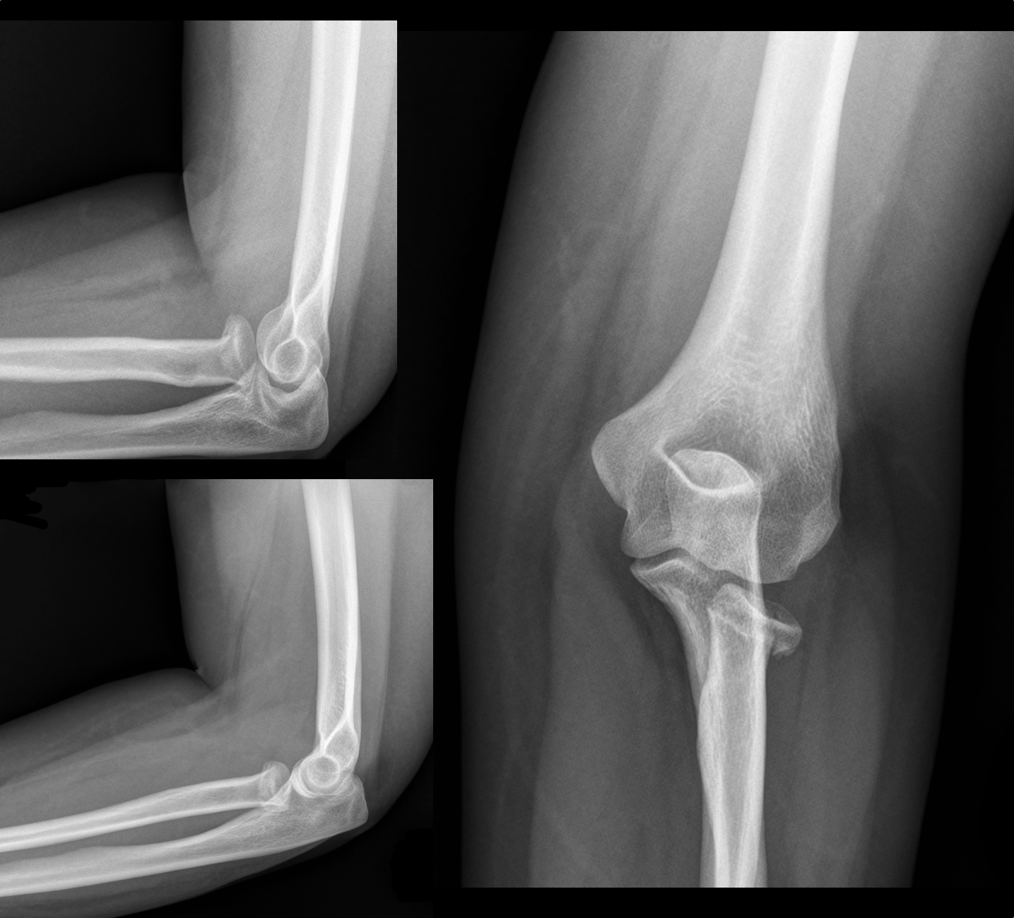
30 year-old female with elbow pain following a fall. What's the diagnosis? (bonus points if you name the fracture with an associated radio-ulnar joint dislocation)
Show Answer
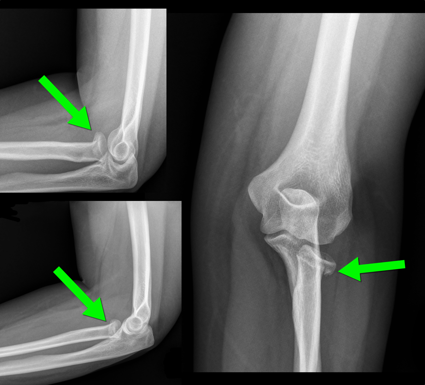
 d
dShow References
When examining a knee for a meniscal injury the commonly described tests are the McMurray Test and Apley Test. However, these tests have sensitivities of 48-68% and 41% respectfully, and specificities of 86-94% and 86-93% respectfully. Depending on whether you are looking at the medical or lateral meniscus.
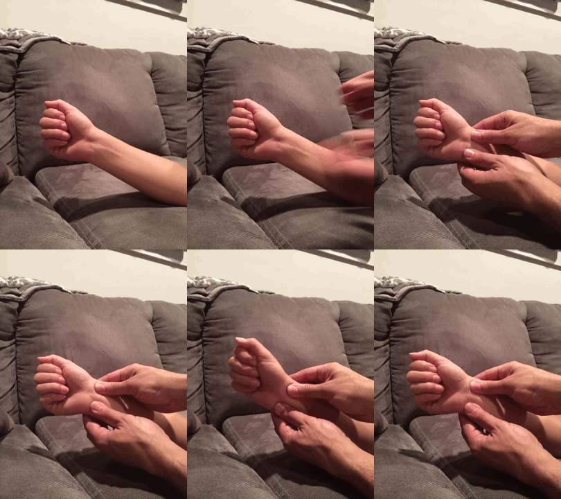
The Thessaly Test that was first described in 2005 can be performed with knee in either 5 or 20 degrees of flexion and has a senstivity of 89-92% and specificity of 96-97% when performed in 20 degrees flexion. The test also tends to be easier to perform.
To perform the test:
- Stand on affected leg only with the other leg held up in the air. The examiner holds hands for balance.
- Flex knee to be test to 20 degrees, while the other leg is held in the air
- Internally and Externally Rotate Knee
- Positive test is pain at medial or lateral joint line with possible locking/catching sensation
Essentially you and your patient will look like you are doing the twist as they rotate their knee with you holding their hands.
A video of the technique can be found at http://youtu.be/R3oXDvagnic
Show References
Seizure is a very common effect seen in many overdoses. Think about the following drugs which have a higher propensity for seizure as noted in a Swiss study of over 15000 patients and isolating to single drug overdoses:
The most prevalent pharmaceuticals were mefenamic acid (51 of the 313 cases), citalopram (34), trimipramine (27), venlafaxine (23), tramadol (15), diphenhydramine (14), amitriptyline (12), carbamazepine (11), maprotiline (10), and quetiapine (10).
The drug mefenanamic acid is not used much in the USA but citalopram, venlafaxine and tramadol as well as the most prelavent bupropion which was number one in the study are all commonly prescribed in the USA. Keep a watchful eye if you see any of these drugs on a drug list or as an overdose.
Show References
General Information:
- Acute diarrheal illness is a common cause of morbidity and mortality disproportionately affecting low and middle income countries
- Acute diarrhea poses the greatest threat to the immunocompromised, children, and the elderly
- Stool samples are costly and frequently don’t provide information altering the course of treatment in acute, non-severe diarrhea
- However, for acute diarrhea, a single stool sample should be obtained when diarrhea is associated with:
- fever (≥38.5°C)
- a severe coexisting condition in a hospitalized patient on antibiotics
- persistent diarrhea (≥14 days)
- profuse cholera-like watery diarrhea
- dehydration
- dysentery
- an elderly or immunocompromised patient
- food handlers, nursing home residents, and daycare workers
- The stool sample must be processed by the lab within 4 hours to directly visualize parasites and within 12 hours for routine microbiologic staining.
Bottom Line for the EM Physician: Use these guidelines to test stool only when helpful to patient care and avoid flushing resources down the toilet.
University of Maryland Section of Global Emergency Health
Author: Alex Skog
Show References
Are Intermediate Lactate Levels Concerning in Patients with Suspected Infection?
- It is well known that lactate levels > 4 mmol/L are associated with increased mortality in patients with suspected infection.
- What is unclear, however, is the prognostic value of intermediate lactate levels (2.0-3.9 mmol/L) in patients with suspected infection.
- Puskarich, et al. performed a systematic review to determine the risk associated with intermediate lactate levels.
- 8 studies (> 11,000 patients) were included in the analysis
- Mortality for patients with intermediate lactate levels but without hypotension was 15%
- Mortality was > 30% for hypotensive patients with intermediate levels of lactate.
- Take Home Point: Patients with intermediate lactate levels have an increased risk of mortality.
- Though no current guidelines exist for the optimal care of these patients, aggressive care should continue until repeat levels demonstrate normalization.
Show References
Question
Diabetic patient with active intravenous drug use presents with hypotension, fever, and tenderness of right arm. What's the diagnosis and what antibiotic(s) would you start?

Show Answer
Show References
Happy Memorial Day! With all the recent attention in the news about swimming and drowning I thought I would share this article
The Instinctive Drowning Response—so named by Francesco A. Pia, Ph.D., is what people do to avoid actual or perceived suffocation in the water. And it does not look like most people expect. There is very little splashing, no waving, and no yelling or calls for help of any kind. To get an idea of just how quiet and undramatic from the surface drowning can be, consider this: It is the No. 2 cause of accidental death in children, ages 15 and under (just behind vehicle accidents)—of the approximately 750 children who will drown next year, about 375 of them will do so within 25 yards of a parent or other adult. In some of those drownings, the adult will actually watch the child do it, having no idea it is happening.* Drowning does not look like drowning—Dr. Pia, in an article in the Coast Guard’s On Scenemagazine, described the Instinctive Drowning Response like this:
- “Except in rare circumstances, drowning people are physiologically unable to call out for help. The respiratory system was designed for breathing. Speech is the secondary or overlaid function. Breathing must be fulfilled before speech occurs.
- Drowning people’s mouths alternately sink below and reappear above the surface of the water. The mouths of drowning people are not above the surface of the water long enough for them to exhale, inhale, and call out for help. When the drowning people’s mouths are above the surface, they exhale and inhale quickly as their mouths start to sink below the surface of the water.
- Drowning people cannot wave for help. Nature instinctively forces them to extend their arms laterally and press down on the water’s surface. Pressing down on the surface of the water permits drowning people to leverage their bodies so they can lift their mouths out of the water to breathe.
- Throughout the Instinctive Drowning Response, drowning people cannot voluntarily control their arm movements. Physiologically, drowning people who are struggling on the surface of the water cannot stop drowning and perform voluntary movements such as waving for help, moving toward a rescuer, or reaching out for a piece of rescue equipment.
- From beginning to end of the Instinctive Drowning Response people’s bodies remain upright in the water, with no evidence of a supporting kick. Unless rescued by a trained lifeguard, these drowning people can only struggle on the surface of the water from 20 to 60 seconds before submersion occurs.”
This doesn’t mean that a person that is yelling for help and thrashing isn’t in real trouble—they are experiencing aquatic distress. Not always present before the Instinctive Drowning Response, aquatic distress doesn’t last long—but unlike true drowning, these victims can still assist in their own rescue. They can grab lifelines, throw rings, etc.
Look for these other signs of drowning when persons are in the water:
- Head low in the water, mouth at water level
- Head tilted back with mouth open
- Eyes glassy and empty, unable to focus
- Eyes closed
- Hair over forehead or eyes
- Not using legs—vertical
- Hyperventilating or gasping
- Trying to swim in a particular direction but not making headway
- Trying to roll over on the back
- Appear to be climbing an invisible ladder
So if a crew member falls overboard and everything looks OK—don’t be too sure. Sometimes the most common indication that someone is drowning is that they don’t look like they’re drowning. They may just look like they are treading water and looking up at the deck. One way to be sure? Ask them, “Are you all right?” If they can answer at all—they probably are. If they return a blank stare, you may have less than 30 seconds to get to them. And parents—children playing in the water make noise. When they get quiet, you get to them and find out why.
Show References
Methylene blue is an extremely effective antidote for acquired methemoglobinemia but has important adverse effects if given in excess of recommended dose.
Below is the usual dose of methylene blue for treatment of methemoglobinemia
1-2 mg/kg of 1% solution IV with a repeat dose given if there is inadequate response to the first one
Adverse effects include:
- >4 m/kg -- Reversible skin, feces, and urine discoloration
- 5-7 mg/kg -- EKG abnormalities (T-wave inversions, diminished R-waves), shortness of breath, chest discomfort, diaphoresis, nausea, diarrhea, abdominal discomfort
- Paradoxically, between 4 and 15 mg/kg, it may cause methemoglobinemia
Show References
General Information:
- There is a growing incidence of chronic kidney disease (CKD) in Central America referred to as Mesoamerican Nephropathy
- Patients tend to be young (30-50 years old), male, agricultural workers, and do not have a history of diabetes, hypertension, or obesity
- Etiology remains unconfirmed but is likely multifactorial with contributors including: repeated dehydration, excessive NSAID use, toxins from sugarcane derived alcohol, and mild Leptospirosis infection.
Area of the world affected:
- Highest prevalence in El Salvador and Nicaragua
- Lower prevalence in Costa Rica and Guatemala
Relevance to the US physician:
- Immigrants with Mesoamerican Nephropathy may present to the ED with acute on chronic kidney disease
- Treatment guidelines are the same as for other CKD etiologies
- Council patients on proper hydration during exertion, limiting NSAID use, and avoiding homemade alcohol consumption
Bottom Line:
- Mesoamerican Nephropathy should be considered in Central American immigrants presenting to the ED with clinical and laboratory signs of CKD but without traditional risk factors.
University of Maryland Section of Global Emergency Health
Author: Emilie J.B. Calvello, MD, MPH & Alex Skog