University of Maryland School of Medicine logo
Emergency Medicine
All Departments
UMSOM Assistant
UMEM Educational Pearls
  • Home
  • Departments
  • Emergency Medicine
  • CME
  • UMEM Educational Pearls
  • CME
  • Courses and Conferences
  • Free Open Access Medical Education
  • UMEM Educational Pearls

Search

141-160 of 245 results with category "Visual Diagnosis"

Previous  |  1 |  ... |  3 |  4 |  5 |  6 |  7 |  8 |  9 |  10 |  11 |  12 |  13 |  Next

Title: What's the Diagnosis? Case by Dr. Zachary Dezman

Category: Visual Diagnosis

Posted: 9/16/2013 by Haney Mallemat, MD

Question

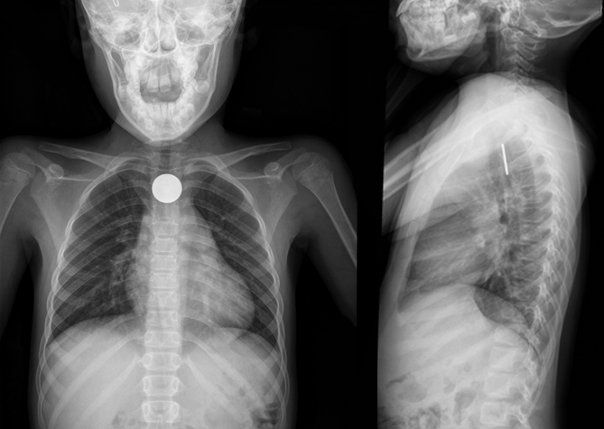
8 year-old girl presents with dysphagia and drooling, Xray is shown. What’s the diagnosis (and where is it located)?
 

Show Answer
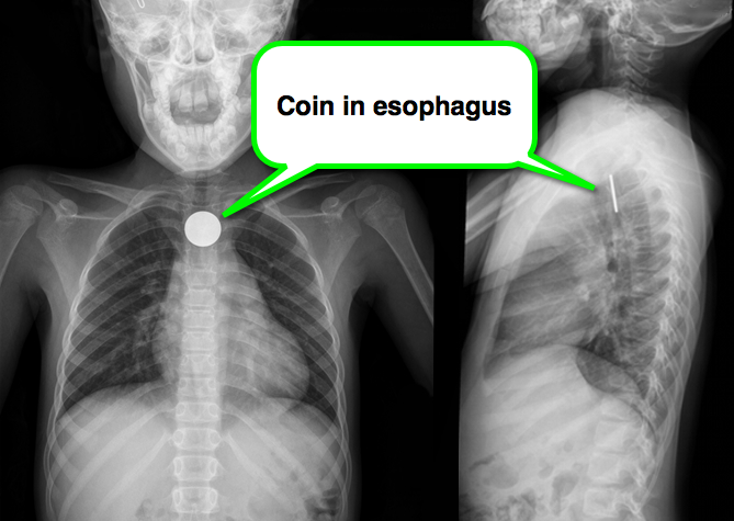
A coin located in the esophagus at the level of the cricopharyneus muscle

Foreign body (FB) pearls

  • The cricopharyngeus muscle is the most common site of esophageal obstruction in children
  • An "end-on" or a “slit-like" appearance on PA CXR sugggests a foreign body in the trachea
  • A “double-ring” sign would identify a button battery rather than a coin
  • FBs should generally be removed, or seen to pass into the stomach, before ED discharge. In this case, however, there were signs of obstruction (drooling) so the risk of perforation is high.
  • Items in the proximal third of the esophagus should be removed emergently via endoscopy.

Show References

Kenton, Foreign Bodies in the Gastrointestinal Tract and Anorectal Emergencies, Emerg Med Clin N Am 29 (2011) 369–400

Follow me on Twitter (@criticalcarenow) or Google+ (+criticalcarenow)


Title: What's the Diagnosis? Case submitted by Dr. Mak Moayedi

Category: Visual Diagnosis

Posted: 9/9/2013 by Haney Mallemat, MD (Updated: 9/9/2013)

Question

This week's case is challenging, but very interesting...

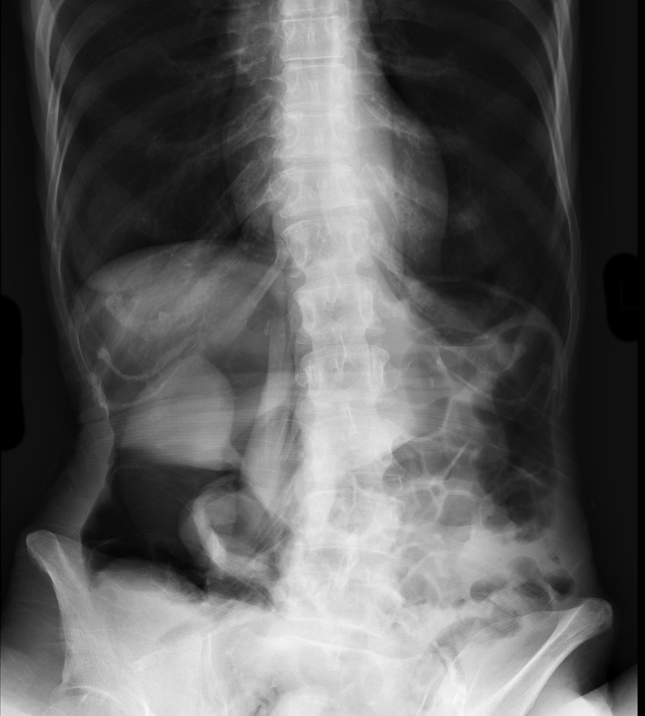
An elderly patient presents with a history of significant weight loss and chronic constipation; abdominal Xray is below. What's the diagnosis? (Hint: why is the right kidney and psoas muscle so well defined?)
 

Show Answer
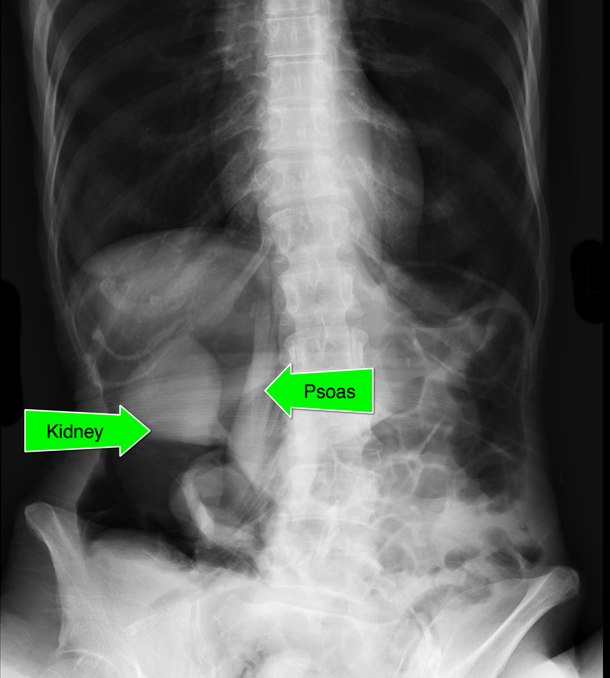
  • Massive retroperitoneal and peritoneal free-air from ascending colon rupture; colon cancer was eventually diagnosed in the operating room.
  • Notice that the kidney and psoas muscle outlined by the free-air below.

Show References

Follow me on Twitter (@criticalcarenow) or Google+ (+criticalcarenow)


Title: What's the Diagnosis? Case by Dr. Kathleen Stephanos and Dr. Michael Bond

Category: Visual Diagnosis

Posted: 9/2/2013 by Haney Mallemat, MD

Question

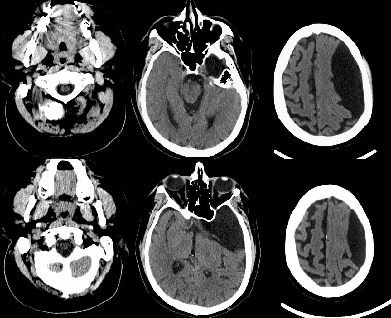
Elderly male presents with headache, confusion, and trouble with gait. What's in your differential diagnosis?
Show Answer
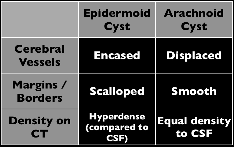
Based on the CT scan shown, the differential here includes epidermoid and arachnoid cyst

Arachnoid cysts (AC) occur within the cerebrospinal axis and do not communicate with the ventricular system. Most occur in the middle cranial fossa and are typically benign; continuing cerebrospinal fluid

The majority of AC occurs from abnormalities in development, but a small portion occurs secondary to post-surgical adhesions or in association with cancer.

MRI is the test of choice to help define the extent of the cyst as well as determine alternative diagnoses.

Treatment is variable with some experts stating that only symptomatic ACs should be treated with others recommending removal to avoid future complications. 

The patient in the stem presented with symptoms secondary to complications from the AC.

Show References

Follow me on Twitter (@criticalcarenow) or Google+ (+criticalcarenow)

Table adapted from Radiology Review Manual, 7th edition, by Wolfgang Dähnert


Title: What's the Diagnosis? Case by Dr. Yemi Adebayo

Category: Visual Diagnosis

Posted: 8/26/2013 by Haney Mallemat, MD (Updated: 8/26/2013)

Question

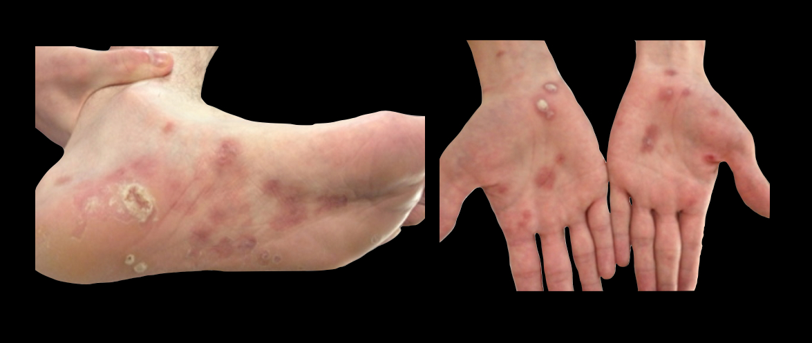
23 year-old patient presents with a rash on his palms and soles. He also states that he had a something strange on his genitals several weeks before. What's the diagnosis and what’s the treatment (including dosing) for this disease?
Show Answer

  • This is secondary syphilis
  • The preferred treatment is Benzathine Penicillin G
    • 2.4 million units intramuscularly
    • Note: this dose is also the same primary syphilis
  • Patients with a true penicillin allergy should receive either:
    • Doxycycline 100mg BID x 14 days
    • Tetracycline 500mg QID x 14 days
  • A one-time dose of Azithromycin was previously recommended because of increased patient compliance. Many studies, however, suggest resistant species emerging and subsequent risk of treatment failure.

Show References

Centers for Disease Control and Prevention, National Center for HIV/AIDS, Viral Hepatitis, STD, and TB Prevention, Division of STD Prevention. 2010 Treatment Update.

http://www.cdc.gov/std/syphilis/treatment.htm

Follow me on Twitter @criticalcarenow or Google+ (+criticalcarenow)


Title: What's the Diagnosis? The ECHO Edition

Category: Visual Diagnosis

Posted: 8/19/2013 by Haney Mallemat, MD

Question

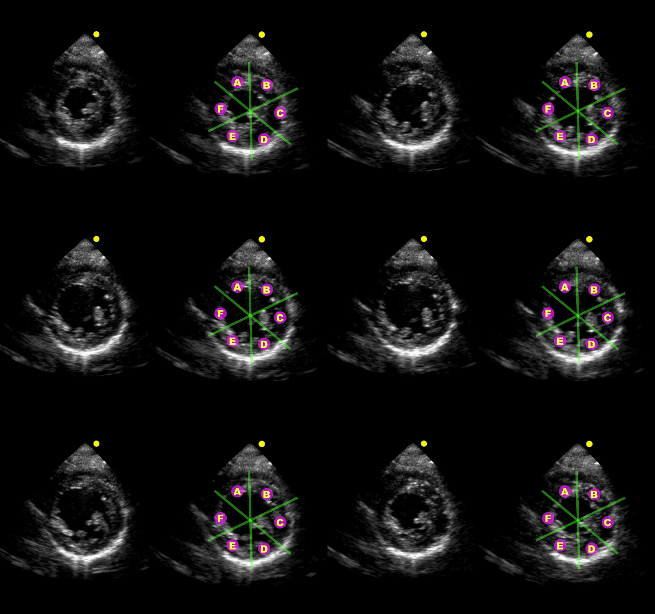
Which echocardiographic view of the heart is this and can you name all 6 segments of the left ventricle? (Hint: A = Anteroseptal wall)
Show Answer

Parasternal short-axis view at the level of the papillary muscles

  • A = Anteroseptal wall
  • B = Anterior wall
  • C = Anterolateral wall
  • D = Inferolateral wall
  • E = Inferior wall
  • F = Inferoseptal wall


Title: What's the diagnosis?

Category: Visual Diagnosis

Posted: 8/12/2013 by Haney Mallemat, MD (Updated: 8/12/2013)

Question

Patient with liver disease presents with dyspnea, fever, and the following ultrasound? What's the diagnosis? (Hint: there are two)?
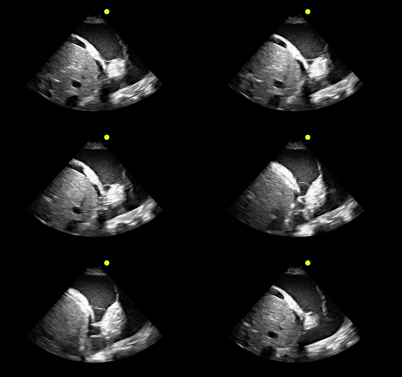
 

Show Answer
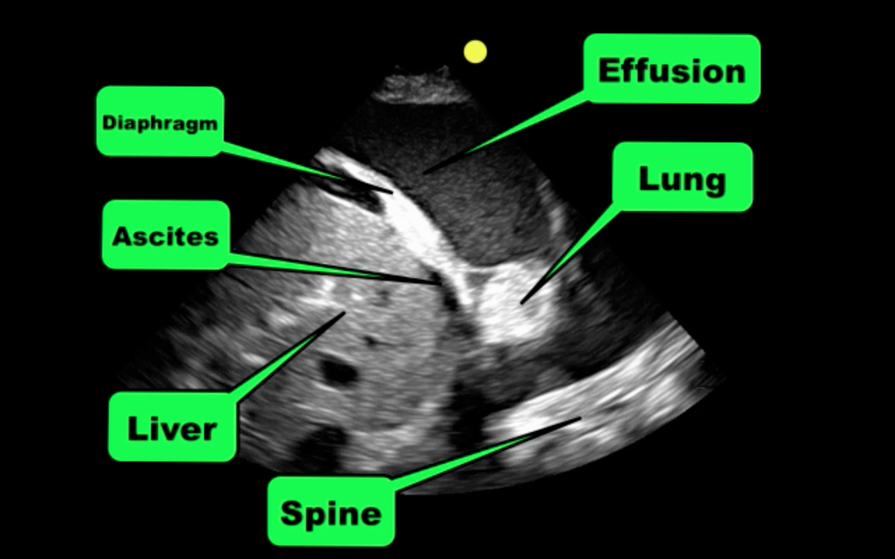
Answer: Complex pleural effusion and ascites

The take away point from this case is to always place the diaphragm in the center of the screen in order to distinguish peritoneal from thoracic fluid. Fluid in both compartments will sometimes be present (as in this case).

Complex pleural effusions

  • Ultrasound can help differentiate simple versus complex effusions
  • Simple effusions tend to be consistent with:
    • Acute volume overload from resuscitation
    • Heart failure
    • Conditions with low oncotic pressure (e.g., nephrotic syndrome)
    • Hepatic hydrothorax
  • Complex effusions tend to be consistent with:
    • Hemothorax
    • Empyema
    • Malignancy 

Show References

Follow me on Twitter (@criticalcarenow) or Google+(+criticalcarenow)


Title: What's the Diagnosis? Case by Dr. Zachary Dezman

Category: Visual Diagnosis

Posted: 8/5/2013 by Haney Mallemat, MD

Question

45 year-old man presents after he cannot close his left eye. In the photo below, he is trying to simultaneously raise his forehead and smile. Of note, he was also started on doxycycline recently for Lyme disease. What two medications should he receive?
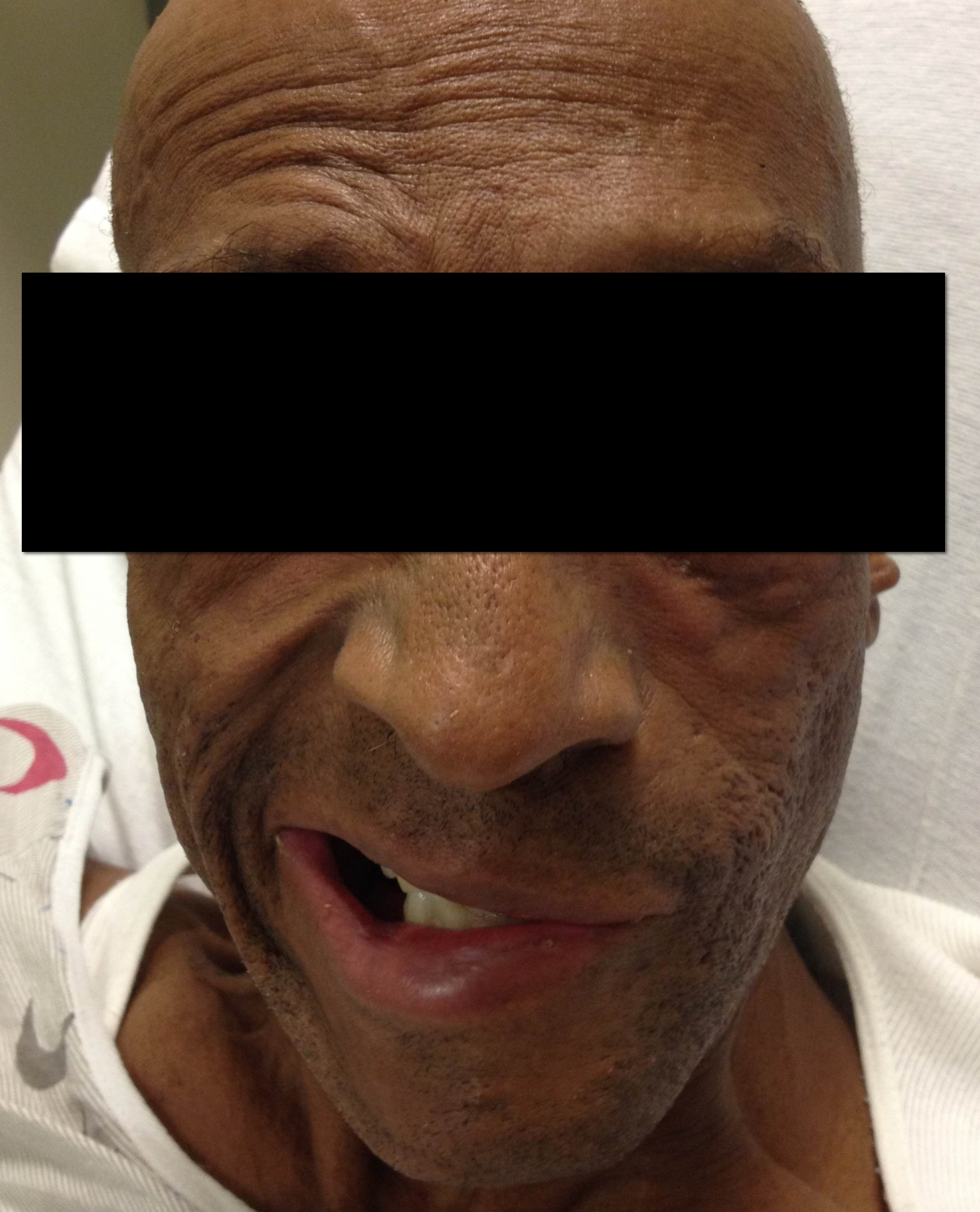
Show Answer
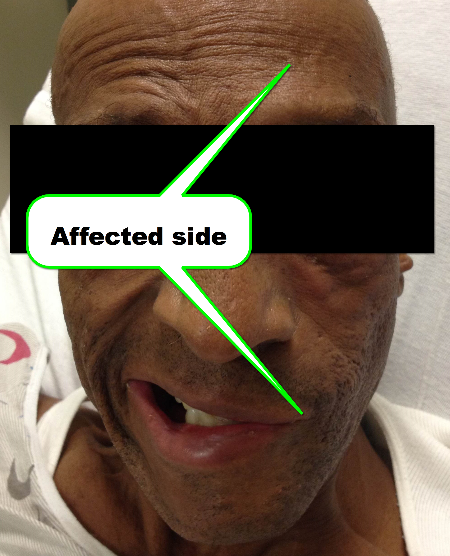
  1. Prednisone
  2. Eye drops for lubrication of the affected eye

Bell Palsy

  • Etiologies include Lyme disease, Herpes Simplex Virus, Guillain-Barre, Epstein-Barr Virus, etc.
  • Critical action is to distinguish Bell palsy from a stroke
    • Bell palsy affects peripheral portion of 7th cranial nerve (motor and sensory). Inability to wrinkle forehead (shown here), close eyelid, and an asymmetric smile is classic and taste to anterior 2/3 of tongue (sensory) is also affected.
    • Stoke affects the central 7th cranial nerve causing an asymmetric smile, but the ability to wrinkle the forehead and close eyelid is preserved (due to bilateral cross-over of the peripheral 7th nerve fibers)
  • The two medications that should be given are:
    • Prednisone for 7-14 days
    • Eye drops / ointment to maintain eye moisture and lubrication
  • Don’t forget to discharge with instruction to patch their eye during sleep to protect their eye
  • About 85% of patients recover within three weeks.

Show References

Follow me on Twitter (@criticalcarenow) or Google+ (+criticalcarenow)


Title: What's the Diagnosis?

Category: Visual Diagnosis

Posted: 7/29/2013 by Haney Mallemat, MD

Question

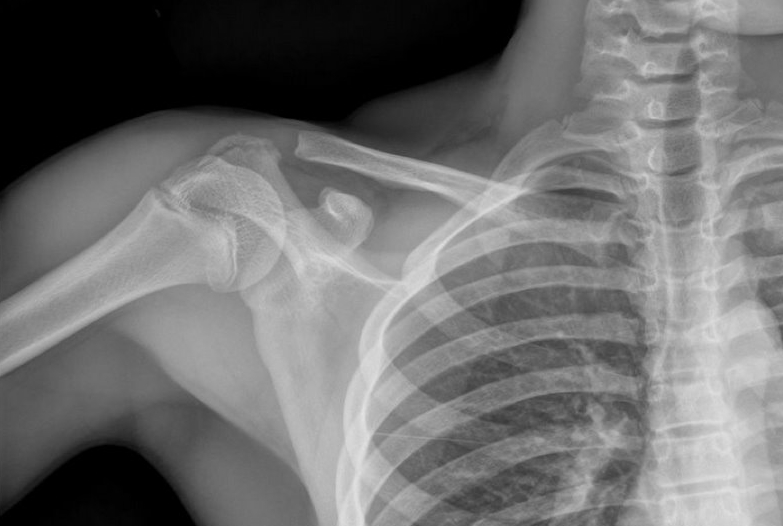
13 year-old female fell on right shoulder while catching a rebound during a basketball game. The patient is holding her arm in adduction and has exquisite scapular tenderness on exam. What’s the next step in management? …oh, and what’s the diagnosis?
Show Answer
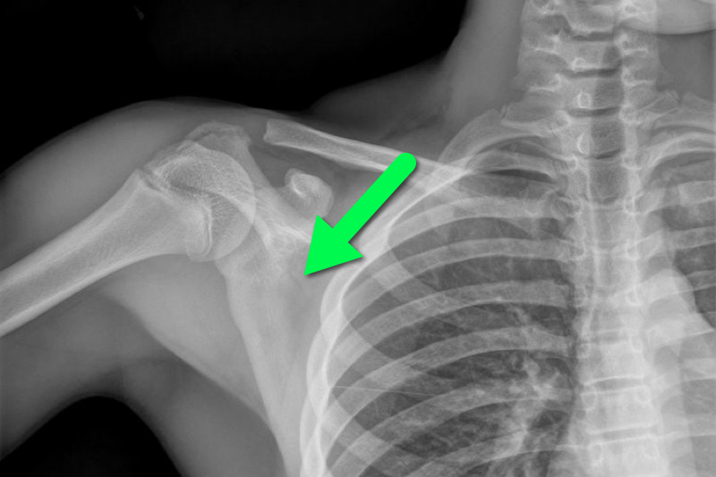
Answer: Non-displaced scapular fracture

Treatment:

  • Analgesics (NSAIDs)
  • Non-displaced fractures require sling and swath immobilization
  • Orthopedic reduction for displaced fractures.
  • Early range-of-motion exercises
  • Beware post-traumatic arthritis or bursitis as complications

Bonus Pearls: #foam4yrdome

This installment of #foam4yrdome will focus on freeemergencytalks.net which is quite possibly the best Critical Care and Emergency Medicine FREE lecture website.

The website was founded and is maintained by Professor Joe Lex (@joelex5); the website hosts hundreds of free talks.

Check out talks from all the major conferences featuring the best speakers in Emergency and Critical care medicine today.

Show References

Follow me on Twitter (@criticalcarenow) or Google+ (+criticalcarenow)


Title: What's the Diagnosis? Case by Dr. Zachary Dezman

Category: Visual Diagnosis

Posted: 7/22/2013 by Haney Mallemat, MD

Question

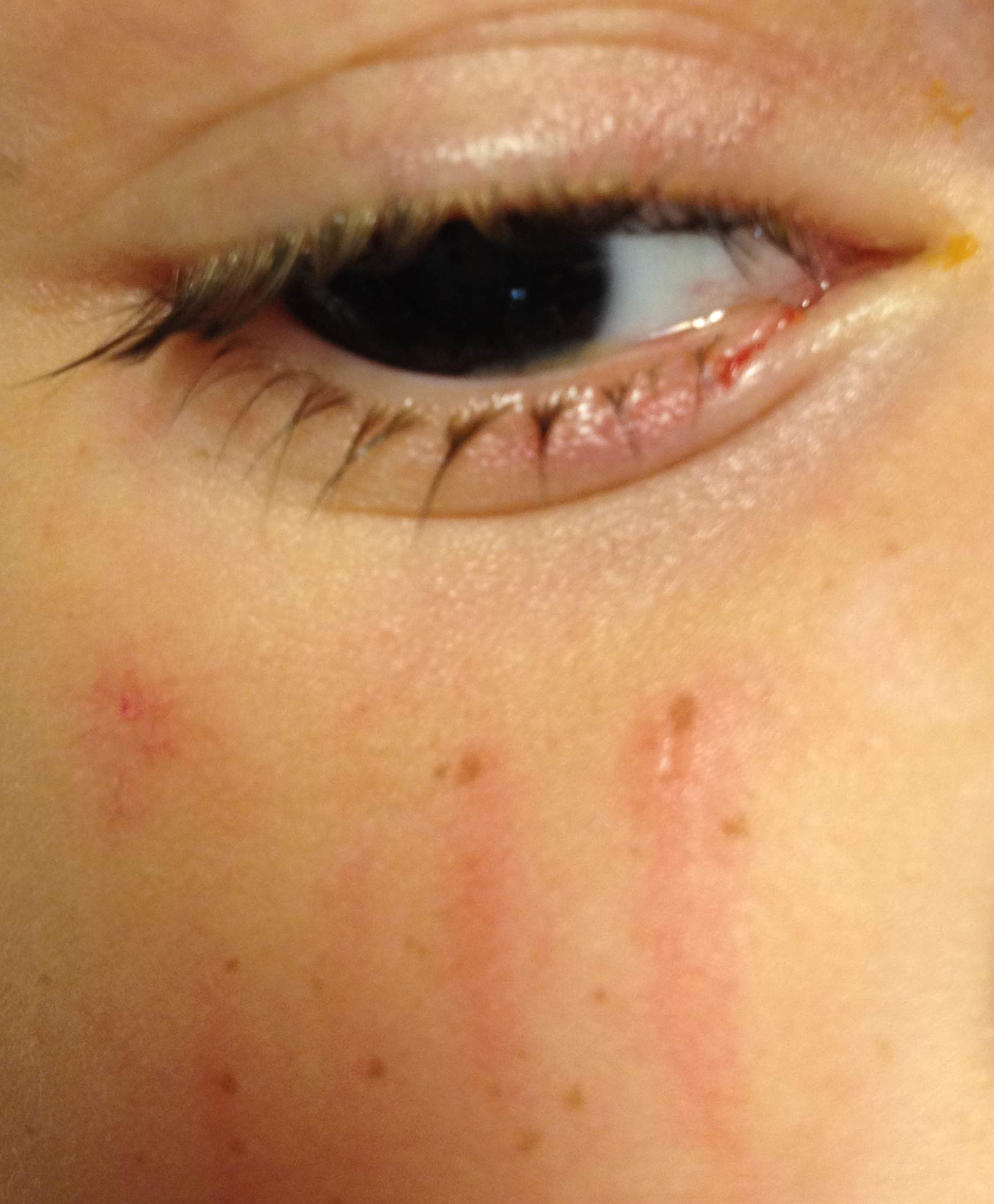
A 3 year-old boy was attacked by a dog and sustained the injury below. Name one injury that should be strongly considered (Hint: there are several)
Show Answer
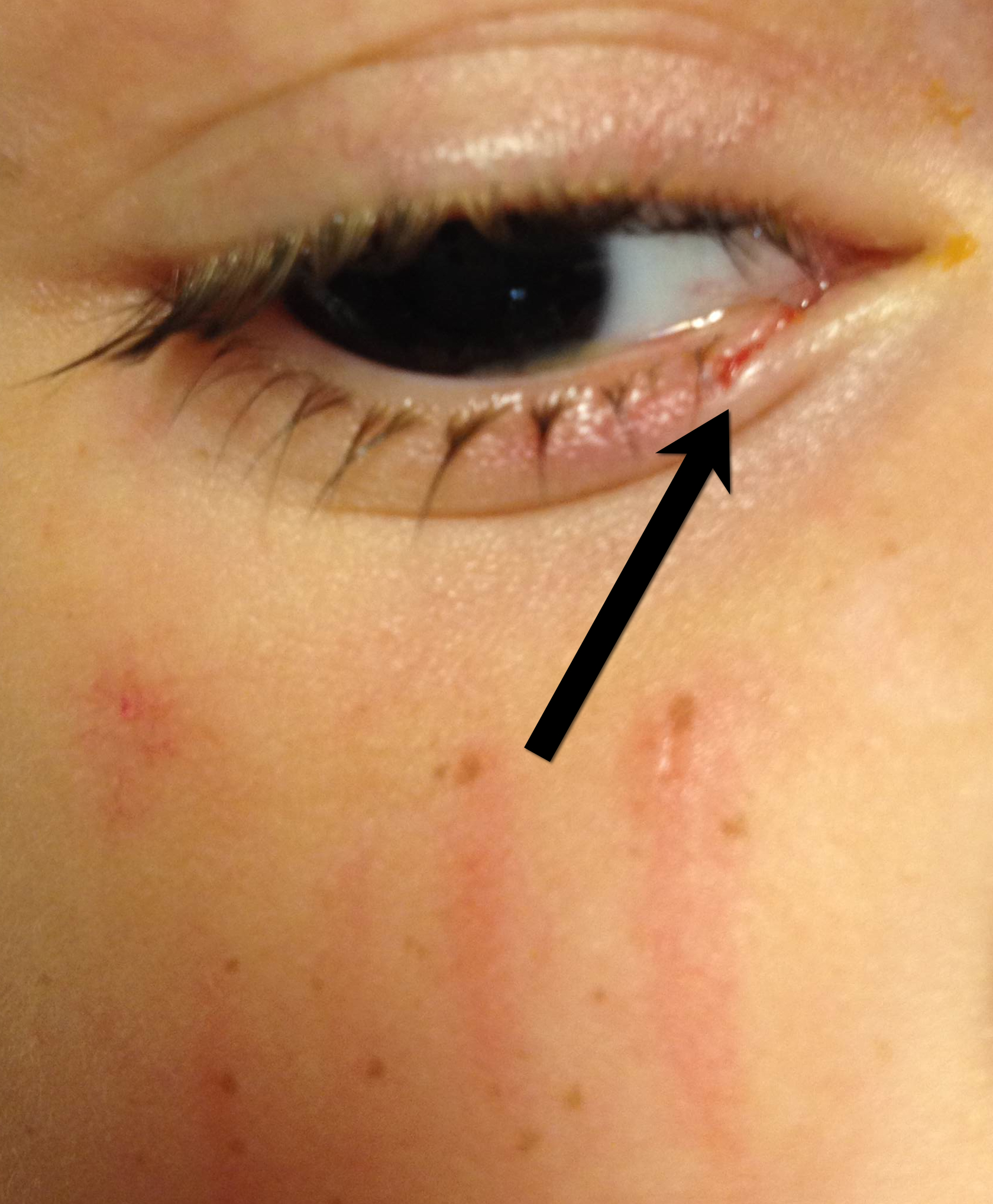
Important injuries to consider (image below):

  • Tarsal tear
  • Laceration of the lacrimal duct
  • Corneal abrasion
  • Globe rupture.

This patient had only a corneal abrasion on fluorescein exam.

Bonus Pearl: #Foam4yrDome

  • Global Medial Educational Project (GMEP.org) is a fantastic resource for anyone interested in medical education, especially Emergency and Critical Care. It's the brainchild of Dr. Mike Cadogan (@sandnsurf).
  • After creating your login and profile, you can answer hundreds of clinical questions, browse open-access media, and connect / network with other GMEP members.
  • With your account, you also have the ability to upload your own questions and media to share, or you may keep them all private in your own personal cloud-based folder to retrieve anytime.

Show References

Follow me on Twitter (@criticalcarenow) or Google+ (+criticalcarenow)


Title: What's the Diagnosis? Case by Dr. Jason Brown

Category: Visual Diagnosis

Posted: 7/15/2013 by Haney Mallemat, MD

Question

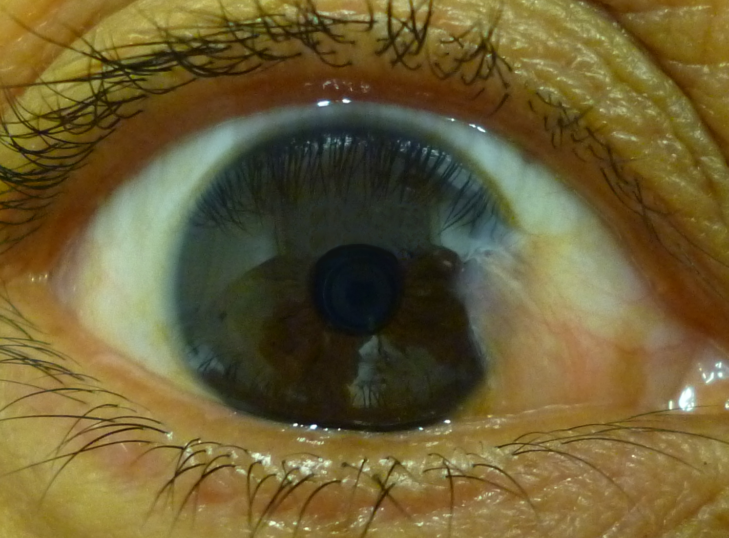
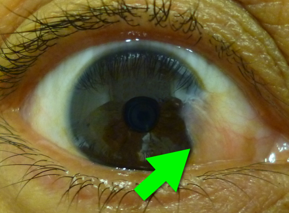
46 year-old female presents with a headache. The following is seen on visual inspection of the eye. What's the diagnosis?
Show Answer

Pterygium

  • Benign wedge-shaped fibrovascular proliferation extending from sclera to nasal portion of cornea
  • Sometimes confused with pinguecula (both are benign)
  • Symptoms range from none, to eye irrigation/redness, to visual impairment (if crosses visual field)
  • Risk factors (UV-light exposure, male gender, and genetic predisposition)
  • Treatment
    • Supportive (artificial tears or short-course topical steroids)
    • Surgical excision, if:
      • discomfort
      • crosses visual-field
      • cosmetic reasons
  • Prevent by blocking UV light from eyes (e.g., sunglasses)

Six-word Summary: Cornea, benign, UV, supportive, surgery, and sunglasses  


Bonus Pearl

As a new academic year begins, I will be sharing some amazing free educational online resources/links. These free materials are known as Free Open Access Meducation (or FOAMed) and for those familiar with FOAMed this is an emerging educational revolution. If you don't know what FOAMed is, read about it here and then read this. Updates will happen every Monday and will be known as #FOAM4yourDome  

Show References

Follow me on Twitter (@criticalcarenow) or Google+ (+criticalcarenow)


Title: What's the Diagnosis? Case by Dr. Zachary Dezman

Category: Visual Diagnosis

Posted: 7/8/2013 by Haney Mallemat, MD

Question

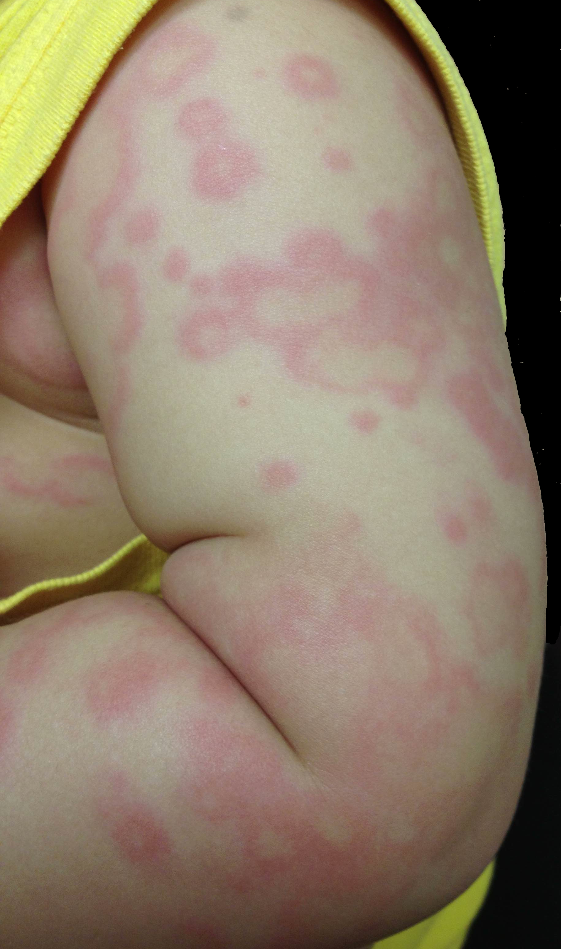
3 year-old male develops rash 5 days after starting amoxicillin for acute otitis media. What's the diagnosis? 
Show Answer

Erythema Multiforme

Erythema multiforme (EM) is a pruritic, erythematous, and blanchable maculopapular rash; it is serpiginous or targetoid in shape, with central clearing or pallor.

EM is generally symmetric, appearing on hands, feet, groin, and extensor aspects of legs and forearms.

It is classically associated with upper respiratory infections, medications, connective tissue diseases, and malignancies.

Treatment includes:

  • stopping offending agent
  • symptomatic treatment (e.g. Benadryl for pruritis)
  • local wound care, especially if mucosal involvement

Show References

Habif, et al, Skin Disease, 3rd Ed. 2011

Follow me on Twitter (@criticalcarenow) or Google+ (+criticalcarenow)


Title: What's the Diagnosis? Case by Dr. Zachary Dezman

Category: Visual Diagnosis

Posted: 7/1/2013 by Haney Mallemat, MD

Question

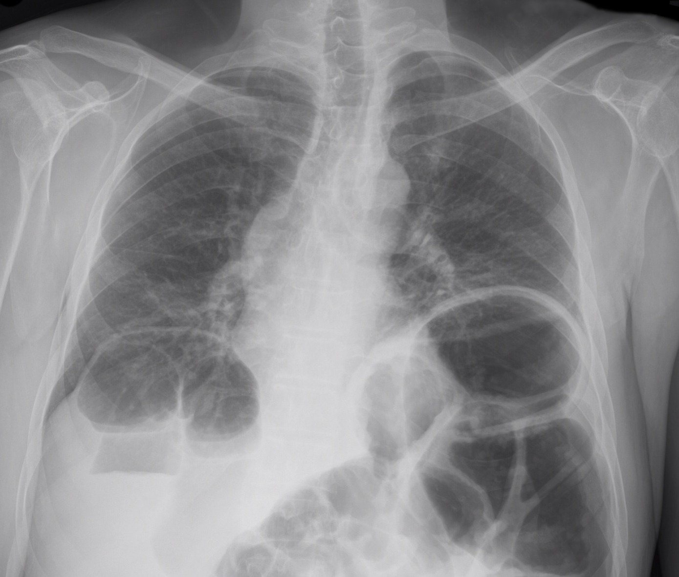
65 year-old male presents with nausea and diffuse abdominal pain, 3 days after knee replacement surgery. What's the diagnosis?
Show Answer

Adynamic ileus

Functional impairment in the transit of intestinal material, in the absence of mechanical obstruction
Common complaints are 
  • Nausea, vomiting, constipation, and obstipation
  • Diffuse abdominal swelling
  • Diffuse abdominal pain

Risk factors include

  • Post-operative state (#1 cause, typically 1-3 days post-op)
  • Pharmacological agents (e.g., opioids, anti-cholinergics, antihistamines, etc.)
  • Infections (e.g., pneumonia, pancreatitis, etc.)
  • Neurological disorders (e.g. diabetic neuropathy)
  • Abdominal trauma

Imaging reveals distention of both large and small bowel without a transition zone, which differs from a small or large bowel obstruction. Such cases can be difficult to differentiate clinically from one another, so physicians often rely on imaging, specifically CT scanning to define a discrete obstruction versus an ileus.


References

1. Hayden and Sprouse, Bowel Obstruction and Hernia, Med Clin N Amer 29 (2011) 319-345

2. American College of Radiology, Suspected Small Bowel Obstruction, ACR Appropriateness Guidelines, rev. 2010

Show References

Follow me on Twitter (@criticalcarenow) or Google+ (+criticalcarenow)


Title: What's the diagnosis? Case submitted by Dr. Jennifer Guyther

Category: Visual Diagnosis

Posted: 6/24/2013 by Haney Mallemat, MD (Updated: 6/24/2013)

Question

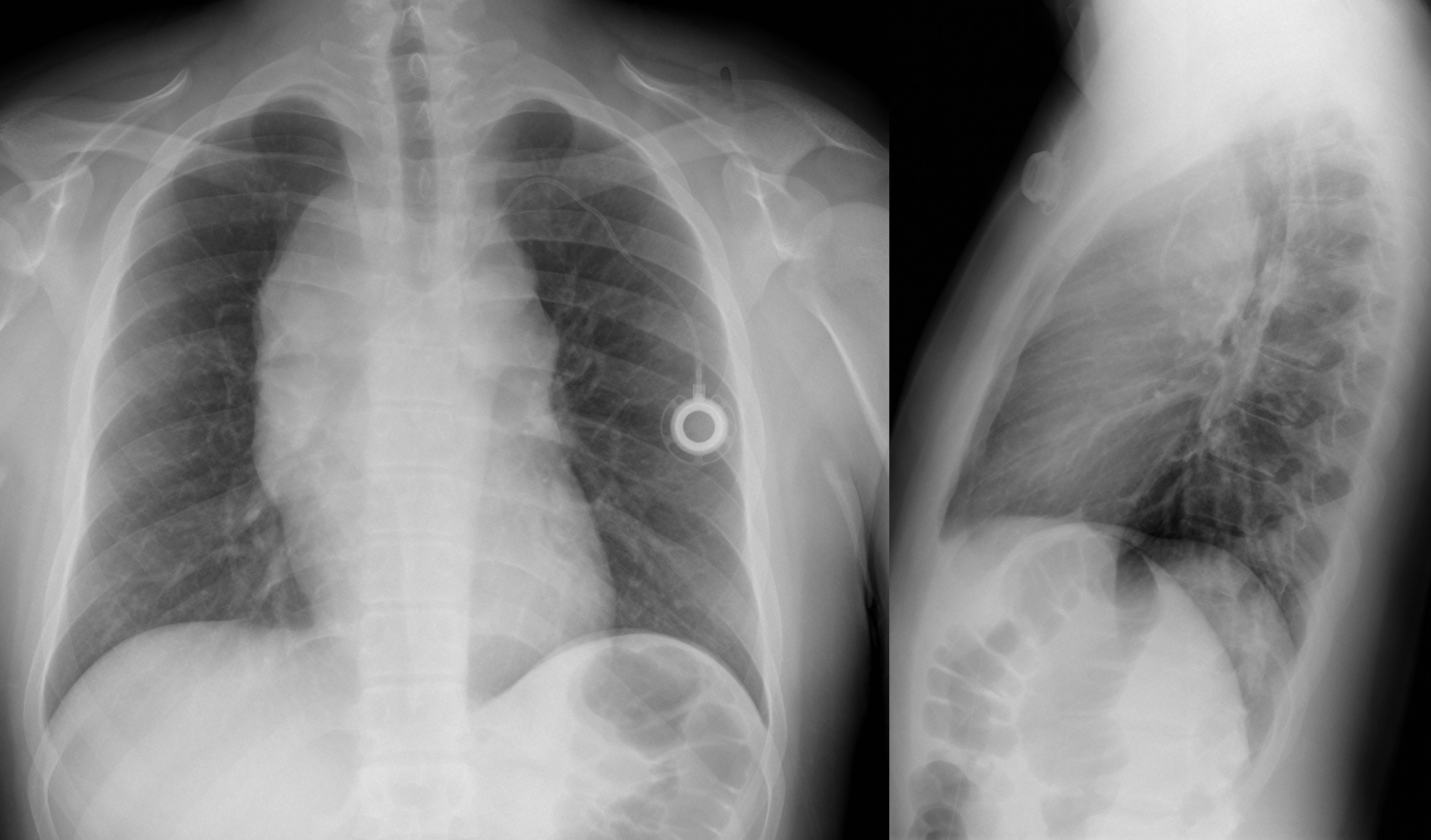
Name three differential diagnoses based on the CXR below.


Show Answer

The diagnosis in this case is Non-Hodgkin lymphoma, but read below for more differentials.

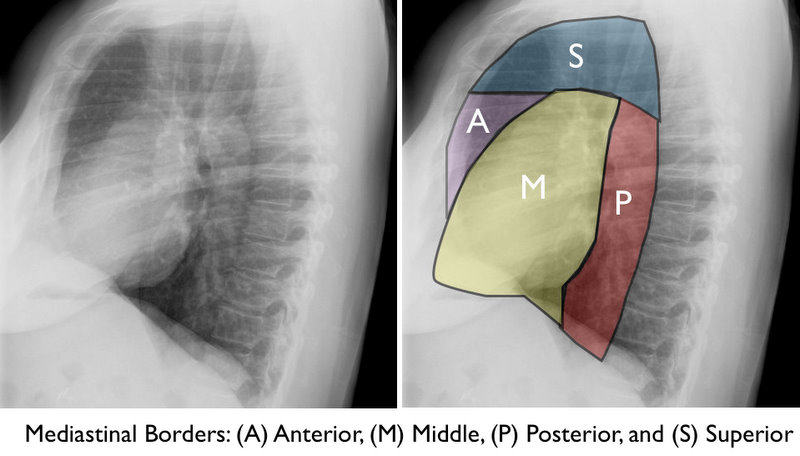
Mediastinal Masses

The mediastinum is subdivided into the regions shown below. Here are some differential diagnoses based on region.

Anterior / Superior Mediastinum

  • Superior vena cava syndrome
  • Ascending Aortic aneurysm
  • Lymphoma
  • Thyroid tumor
  • Parathyroid tumor
  • Thymic masses / tumors
  • Germ-Cell tumor
  • Metastatic disease

Middle Mediastinum

  • Lymphadenopathy
  • Metastatic Disease
  • Lymphoma
  • Developmental cysts
  • Pericardial effusion
  • Foregut duplication
  • Bronchogenic cyst
  • Vascular enlargement
  • Aortic aneurysm

Posterior Mediastinum

  • Neurogenic tumors
    • Peripheral nerves
    • Sympathetic ganglia
    • Paraganglionic tissue
  • Esophageal tumors
  • Carcinoma
  • Diverticula
  • Diaphragmatic (Bochdalek's) hernia

Congratulations to all the graduating residents, especially our own at the University of Maryland. I wish you all the best as you start your phenomenal careers!

Show References

Follow me on Twitter (@criticalcarenow) or Google+ (+criticalcarenow)


Title: What's the Diagnosis? Case written by Dr. Michael Santiago

Category: Visual Diagnosis

Posted: 6/10/2013 by Haney Mallemat, MD

Question

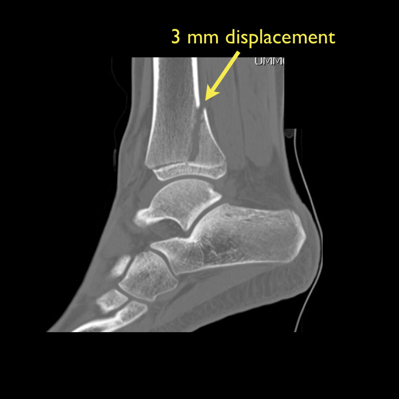
13 year-old female with ankle pain following fall down escalator. What's the diagnosis?
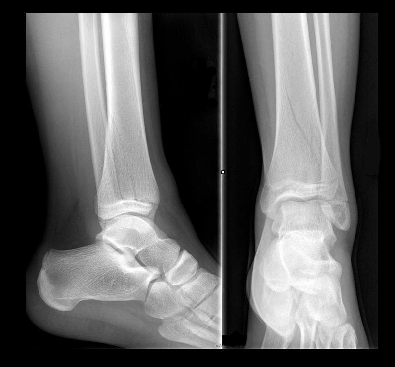


Show Answer
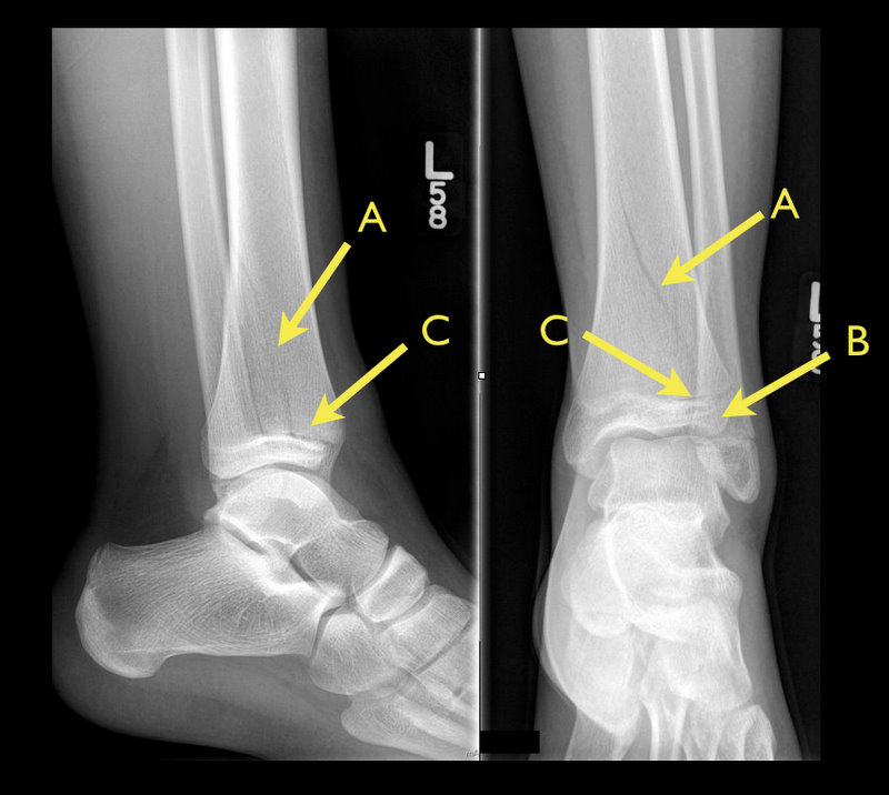
Answer: Tri-plane ankle fracture

Tri-plane Fractures

  • Multi-planar ankle fracture in older children and adolescents during an 18 month window prior to distal tibial-physis closure.
  • 3 planes (see Xray below): coronal (A: tibal metaphysis), sagital (B: epiphysis), and transverse (C: growth plate)
  • Associated fibular spiral fracture (50% cases)
  • Commonly due to external rotational or "twisting" forces.
  • Xrays are helpfully, but CT scan may be indicated as fractures may be more displaced than radiographs suggest.
  • Non-operative treatment for non-displaced fractures:
    • Closed reduction with long-leg splint/cast
    • Reduction may slip once swelling has subsided
  • Indications for operative repair:
    • >2mm displacement of fracture segments
    • Intra-articular fracture

Show References

Follow me on Twitter (@criticalcarenow) or Google+ (+criticalcarenow)


Title: Here's a Solution, for that Pleural Effusion

Category: Visual Diagnosis

Posted: 6/3/2013 by Haney Mallemat, MD

Ever wonder how to place a pigtail catheter?

Check out this video to learn how, click here

Show References

Follow me on Twitter (@criticalcarenow) or Google+ (+criticalcarenow)


Title: What's the Diagnosis? Case submitted by Bethany Radin

Category: Visual Diagnosis

Posted: 5/27/2013 by Haney Mallemat, MD

Question

9 month-old presents with wheezing and the CXR is shown below. What's the diagnosis?
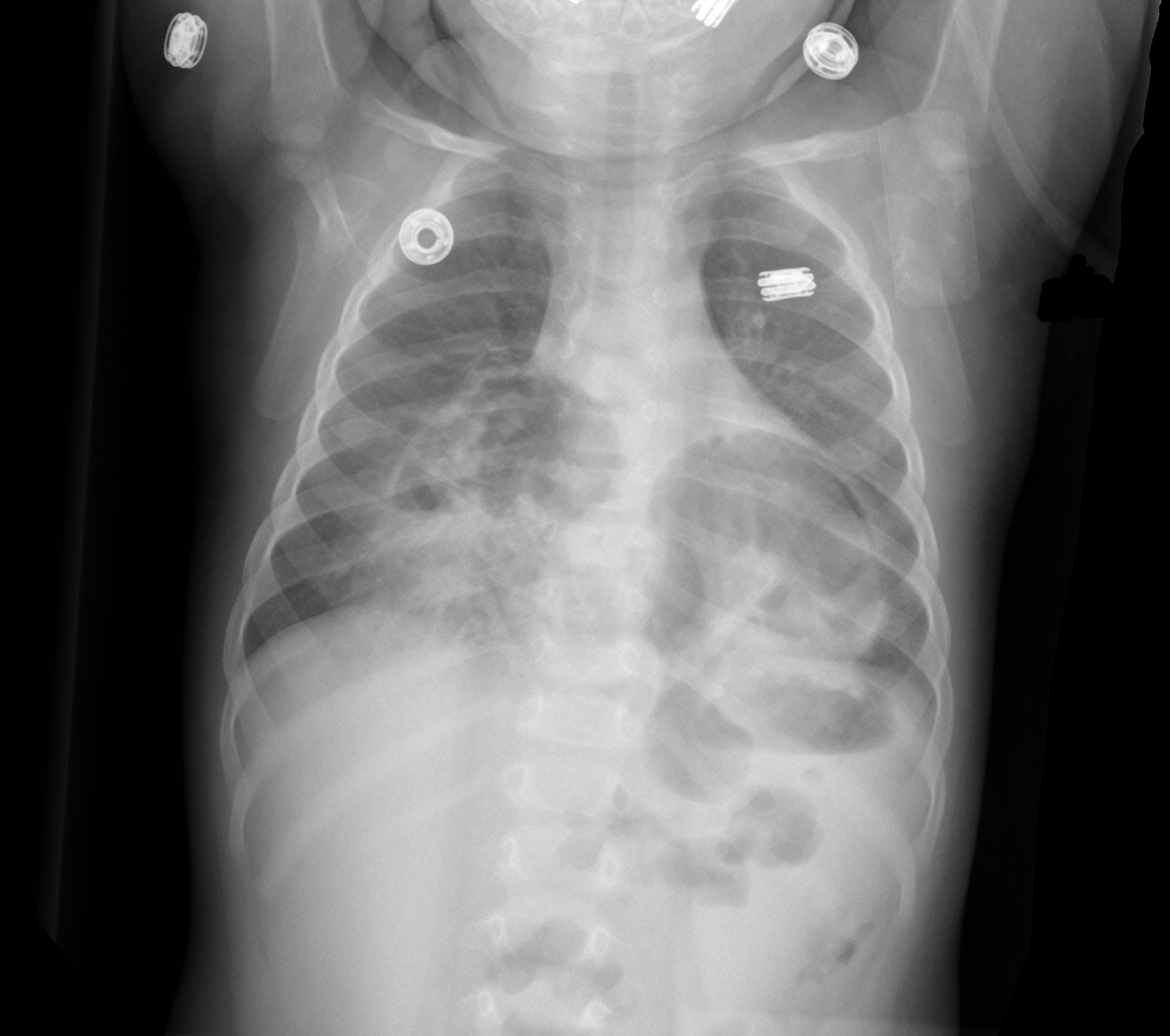


Show Answer
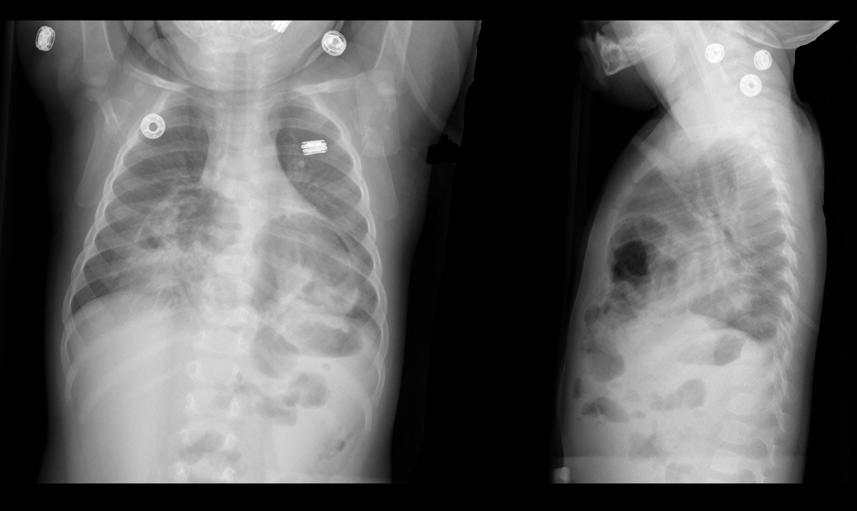
Congenital anterior diaphragmatic defects with herniated bowel.

Show References

Follow me on Twiter (@criticalcarenow) or Google+ (+criticalcarenow)


Title: What's the Diagnosis? Case submitted by Dr. Ryan Spangler

Category: Visual Diagnosis

Posted: 5/21/2013 by Haney Mallemat, MD

Question

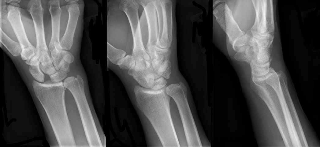
30 year-old male presents with right wrist pain after falling off his bicycle. What's the diagnosis?
Show Answer
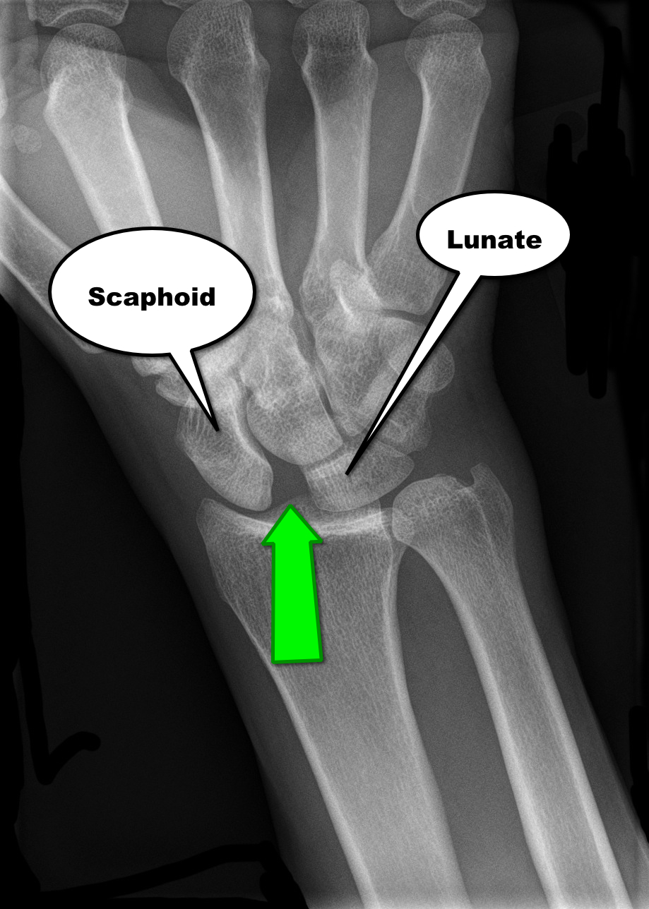

Scapholunate dislocation


Scapholunate Dislocation (a.k.a. “Terry Thomas” or “David Letterman” sign)

  • Increased distance between the scaphoid and lunate bones on AP wrist X-ray; the distance between the two bones should normally be less than 2mm.
  • A widened scapholunate distance suggests ligamentous injury or tear (green arrow below).
  • The widened distance resembles the famous dental diastema (i.e., tooth gap) of Terry Thomas (British comedian) and David Letterman


Show References

Follow me on Twitter (@criticalcarenow) or Google+ (+criticalcarenow)


Title: What's the Diagnosis? Case submitted by Dr. Ali Farzad

Category: Visual Diagnosis

Posted: 5/13/2013 by Haney Mallemat, MD

Question

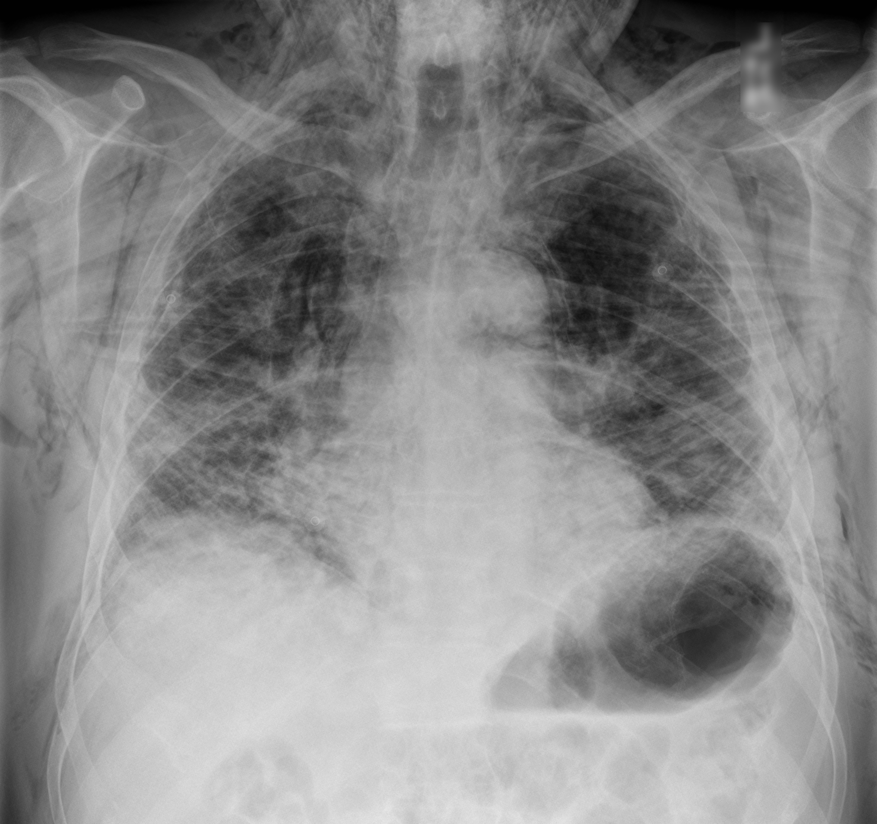
60 year-old male with a history of pulmonary fibrosis presents to the Emergency Department after a lung biopsy. He is complaining of facial swelling and dyspnea. What's the diagnosis?
Show Answer

Answer: Massive subcutaneous emphysema

Click here

Show References

Follow me on Twitter (@criticalcarenow) or Google+ (+criticalcarenow)


Title: What's the Diagnosis? Case by Dr. Siamak Moayedi

Category: Visual Diagnosis

Posted: 5/6/2013 by Haney Mallemat, MD (Updated: 5/6/2013)

Question

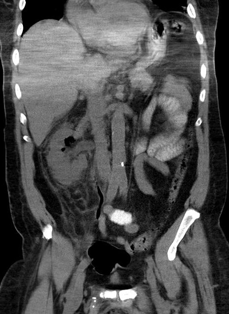
38 year-old male with a past medical history of diabetes presents with back pain and hypotension. CT scan is shown below. What's the diagnosis?
 

Show Answer
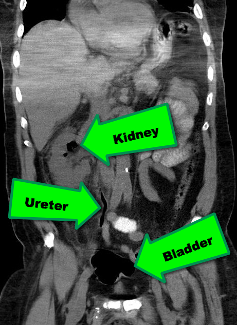
Emphysematous Pyelonephritis (EPN)

EPN is a necrotizing infection of the renal and peri-nephric tissues with accumulation of gas within the tissues and collecting system of the kidney; it is rapidly fatal if not recognized and aggressively treated early.

Risk factors include an immunocompromised host; Diabetics are at particularly increased risk.

The most common etiologic organisms are Escherichia coli, Klebsiella pneumoniae, Proteus mirabilis, and Pseudomonas aeruginosa. Infection with anaerobic species is relatively uncommon.

In addition to the classic signs / symptoms of pyelonephritis, (e.g., flank pain, fever, pyuria, etc.), patients presenting with EPN may experience the passage of air in the urine (pneumaturia) or crepitus over their flank.

Abdominal CT scan is the diagnostic test of choice when clinical presentation and laboratory tests are suggestive (see below)

Treatment should include:

  • Aggressive resuscitation with fluids and vasopressors, as necessary
  • Broad-spectrum antibiotics
  • Percutaneous ureteral drain or stent placement, if obstruction is present

Show References

Follow me on Twitter (@criticalcarenow) or Google+(criticalcarenow)


Title: What's the Diagnosis?

Category: Visual Diagnosis

Posted: 4/29/2013 by Haney Mallemat, MD

Question

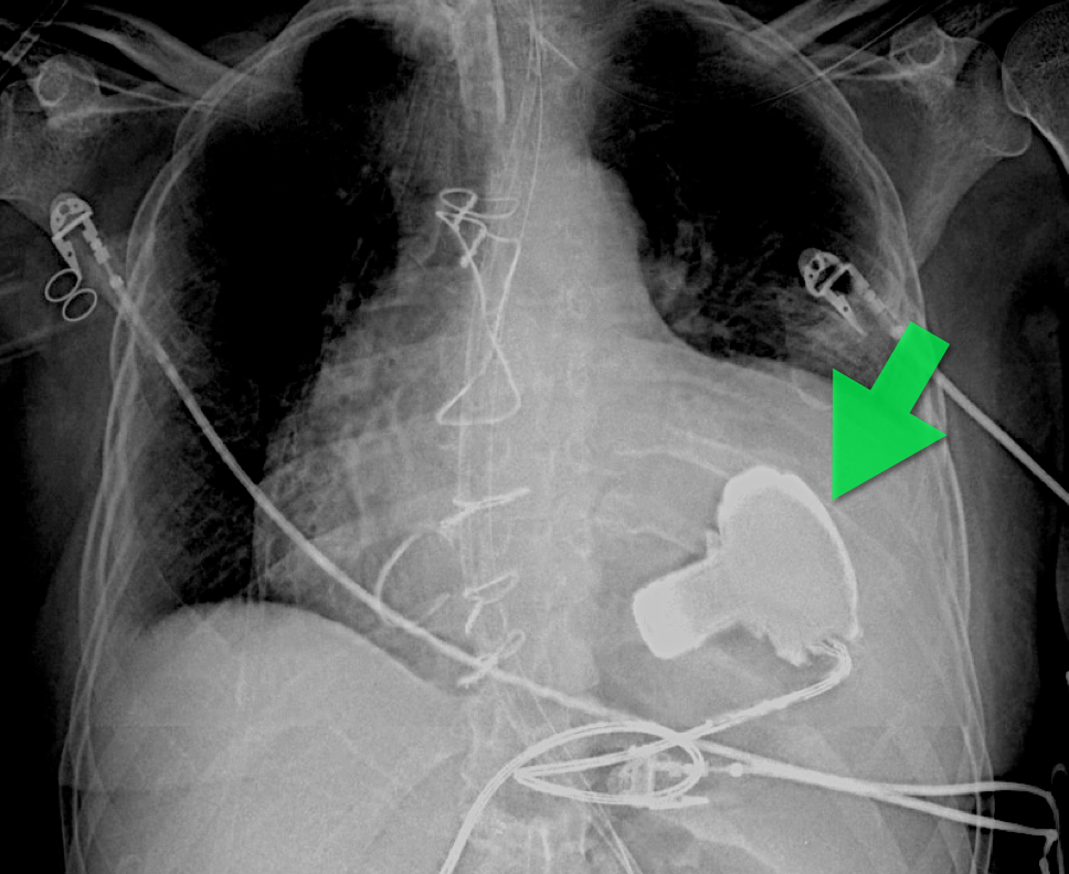
57 year old male presents with a cough. The CXR is shown below. What's the diagnosis?

Show Answer

Answer: Left Ventricular Assist Device (LVAD) 

An LVAD is a mechanical pump that pulls blood from the left ventricle and pumps it to the ascending aorta for systemic delivery; LVADs are increasingly being placed in patients with heart failure so you must be aware of them.

The three indications for an LVAD are:

  • Bridge to cardiac transplant
  • Temporizing measure when a reversible cardiac disease (e.g., myocarditis) exists
  • Destination therapy; patients ineligible for cardiac transplant who would otherwise die from heart failure

There are several models, which have been implanted in people (http://www.mylvad.com/lvad-devices). The main distinction between devices are pulsatile or non-pulsatile (or linear) flow; newer generation devices are non-pulsatile.

Patients with LVAD may develop complications such as infections, arrhythmias, or anemia, but bleeding can be a life-threatening one; bleeding occurs secondary to an acquired von Willebrand factor deficiency, AVMs within the GI tract, or secondary to over anti-coagulation (typically with Warfarin).

Show References

Follow me on Twitter (@criticalcarenow) or Google+ (+criticalcarenow)


Previous  |  1 |  ... |  3 |  4 |  5 |  6 |  7 |  8 |  9 |  10 |  11 |  12 |  13 |  Next
University of Maryland School of Medicine logo
  • Departments
  • Programs
  • Research Centers
  • Institutes
  • Graduate Education
  • CIBR Cores
  • Offices of the Dean
    • University of Maryland, Baltimore
    • University of Maryland Medical Center
    • Medical Alumni Association of the University of Maryland
    • Health Sciences and Human Services Library
University of Maryland School of Medicine
655 W. Baltimore Street
Baltimore, MD 21201
Contact Us

© University of Maryland School of Medicine

    • Webmaster
    • Web Accessibility
    • Site Index
    • UMB Hotline
    • Employment
    • Map