University of Maryland School of Medicine logo
Emergency Medicine
All Departments
UMSOM Assistant
UMEM Educational Pearls
  • Home
  • Departments
  • Emergency Medicine
  • CME
  • UMEM Educational Pearls
  • CME
  • Courses and Conferences
  • Free Open Access Medical Education
  • UMEM Educational Pearls

Search

81-100 of 245 results with category "Visual Diagnosis"

Previous  |  1 |  2 |  3 |  4 |  5 |  6 |  7 |  8 |  9 |  10 |  ... |  13 |  Next

Title: What's the Diagnosis? Case by Dr. R. Gentry Wilkerson

Category: Visual Diagnosis

Posted: 12/1/2014 by Haney Mallemat, MD

Question

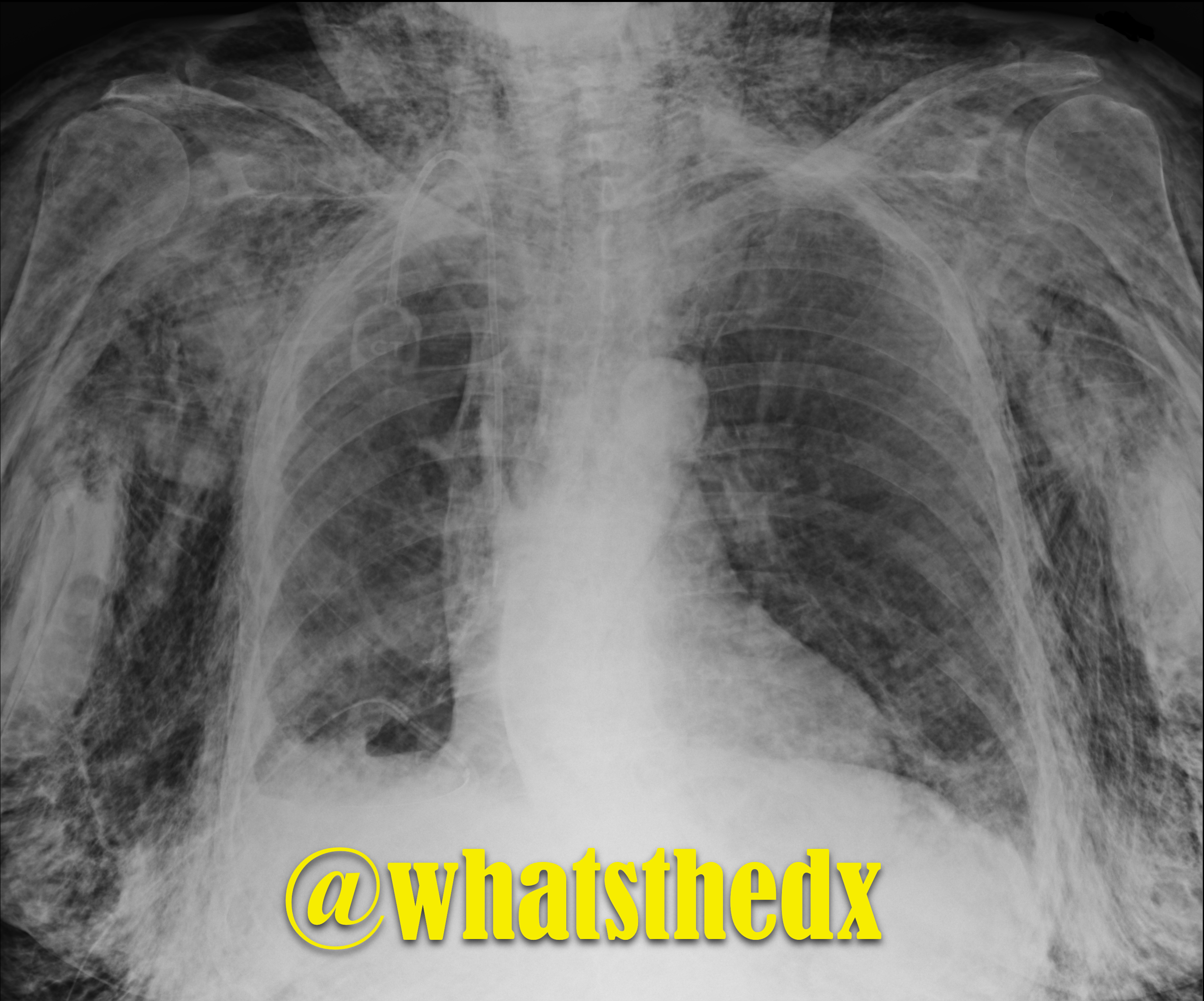
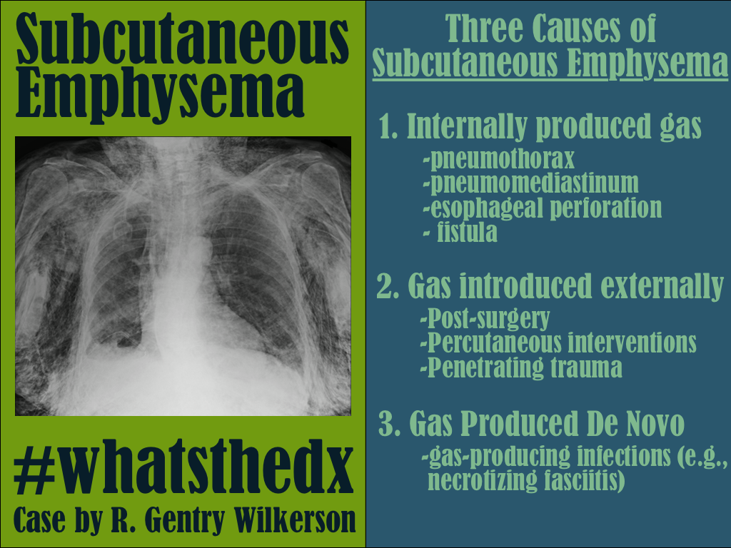
Patient presents with dyspnea. What's the diagnosis and name three potential causes (can be specific to the case or in general)?
Show Answer
Show References

Follow me on Twitter (@criticalcarenow) or Google+ (+criticalcarenow)


Title: What's the Diagnosis? Image by Dr. Yitschok Applebaum, Dr. Jonathan Hoover, and Dr. Semhar Tewelde

Category: Visual Diagnosis

Posted: 11/24/2014 by Haney Mallemat, MD (Updated: 11/25/2014)

Question

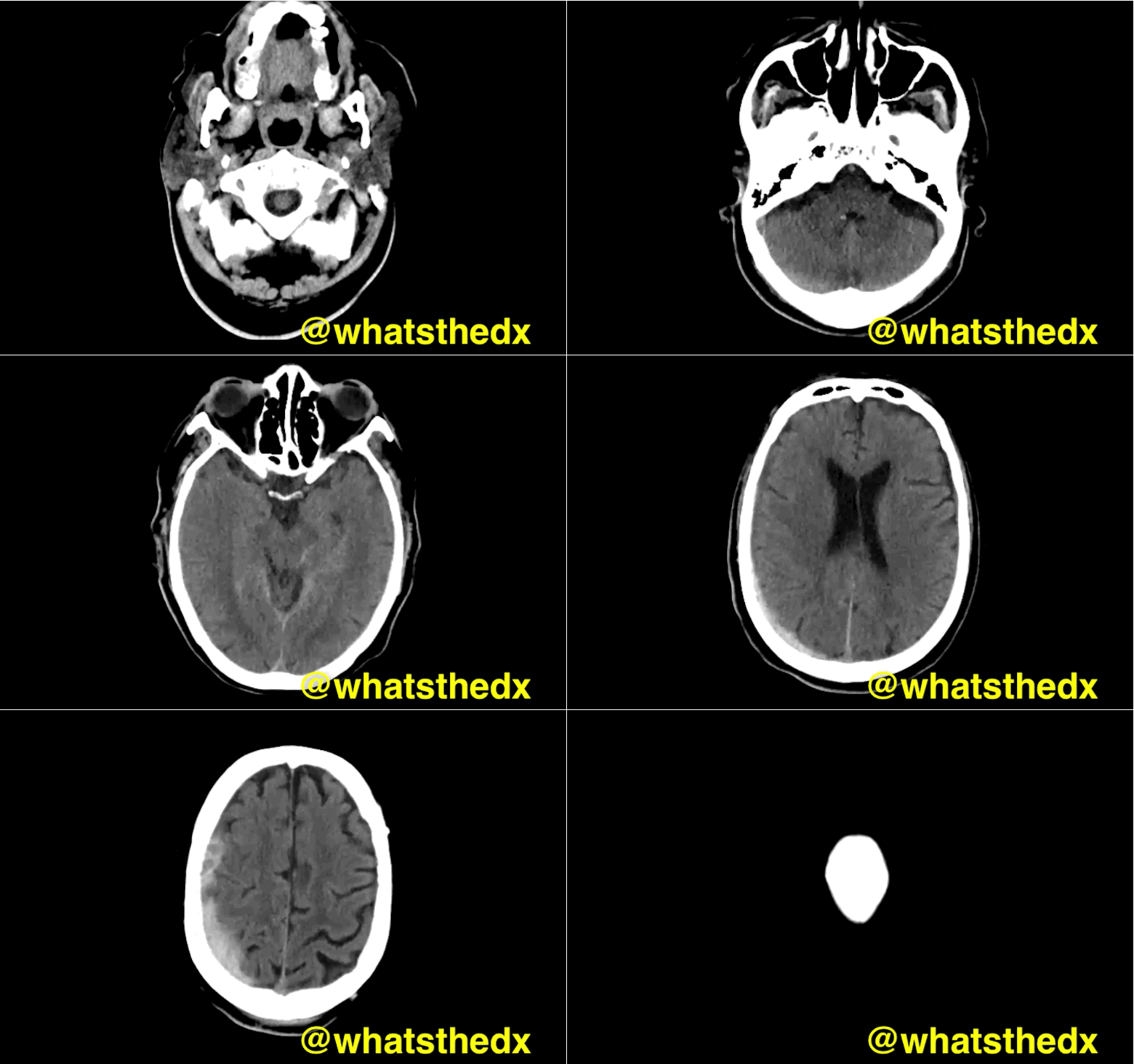
Patient with syncope and then falls down the stairs. What's the diagnosis? (hint: be very,very careful)
Show Answer
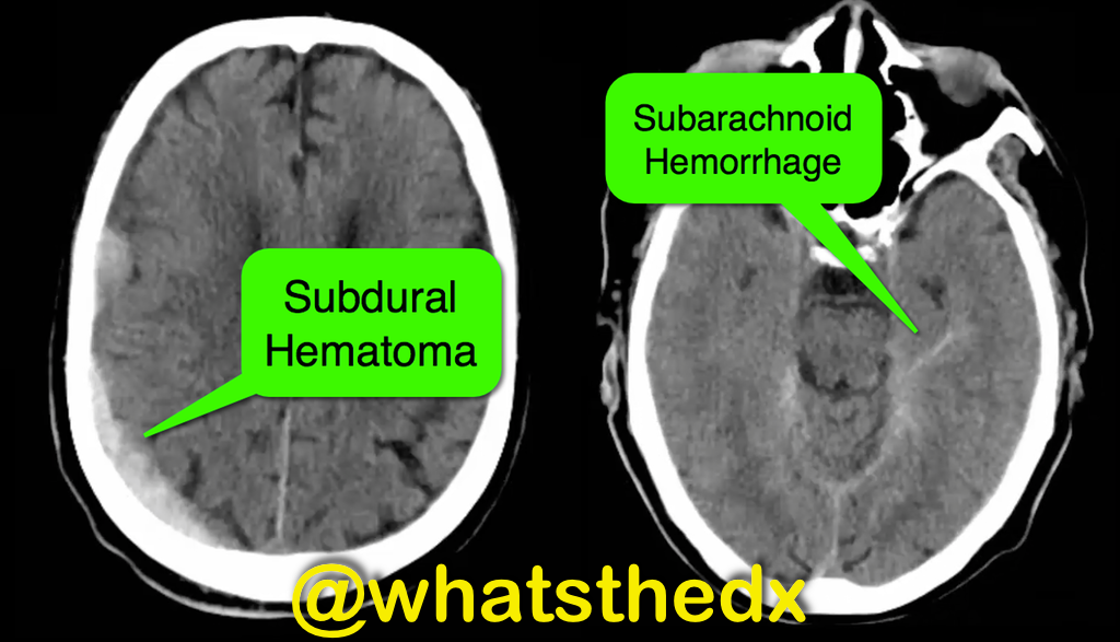
Right subdural hematoma and left subarachnoid hemorrhage


Show References

Follow me on Twitter (@criticalcarenow) or Google+ (+criticalcarenow)


Title: What's the Diagnosis? Images by Dr. Denise Fraga and Dr. Nicole Cimino-Fiallos

Category: Visual Diagnosis

Posted: 11/17/2014 by Haney Mallemat, MD

Question

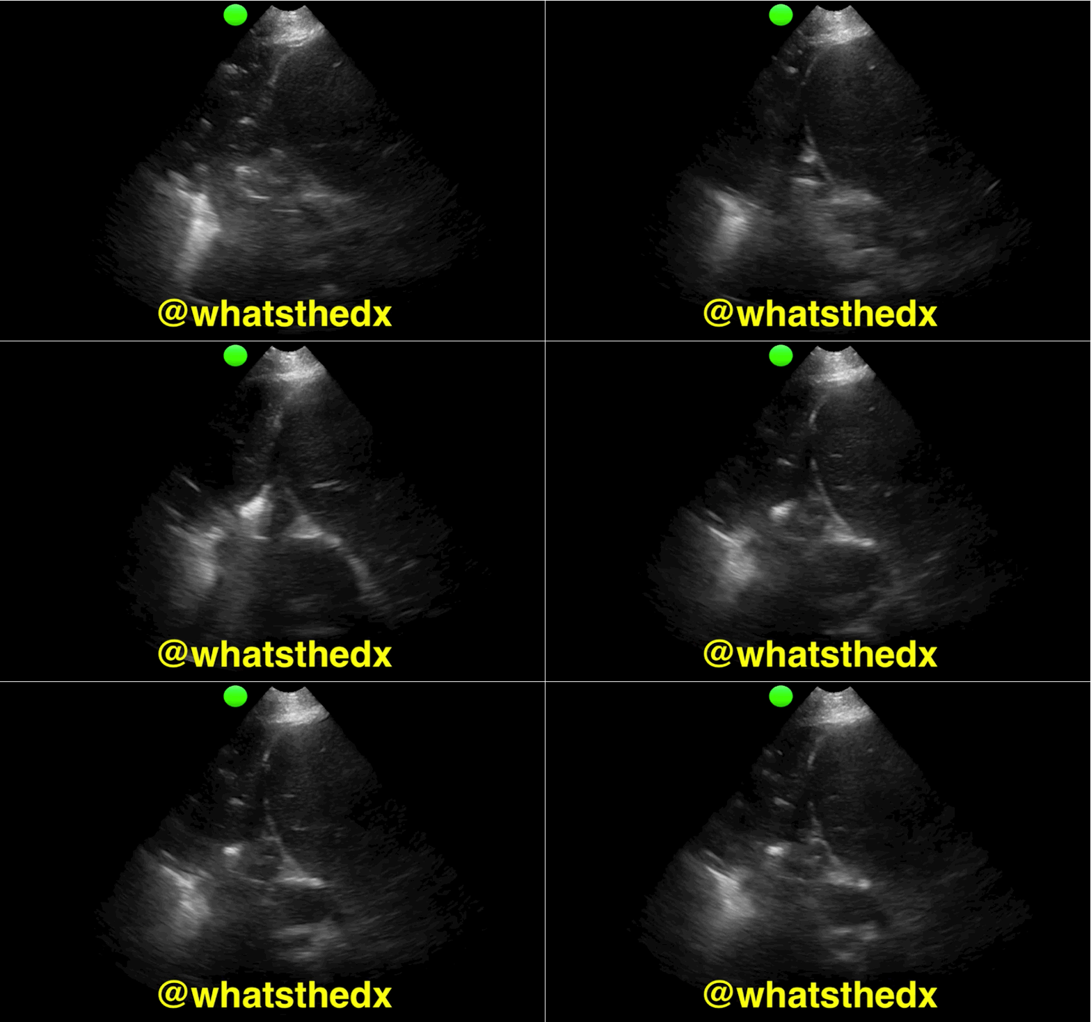
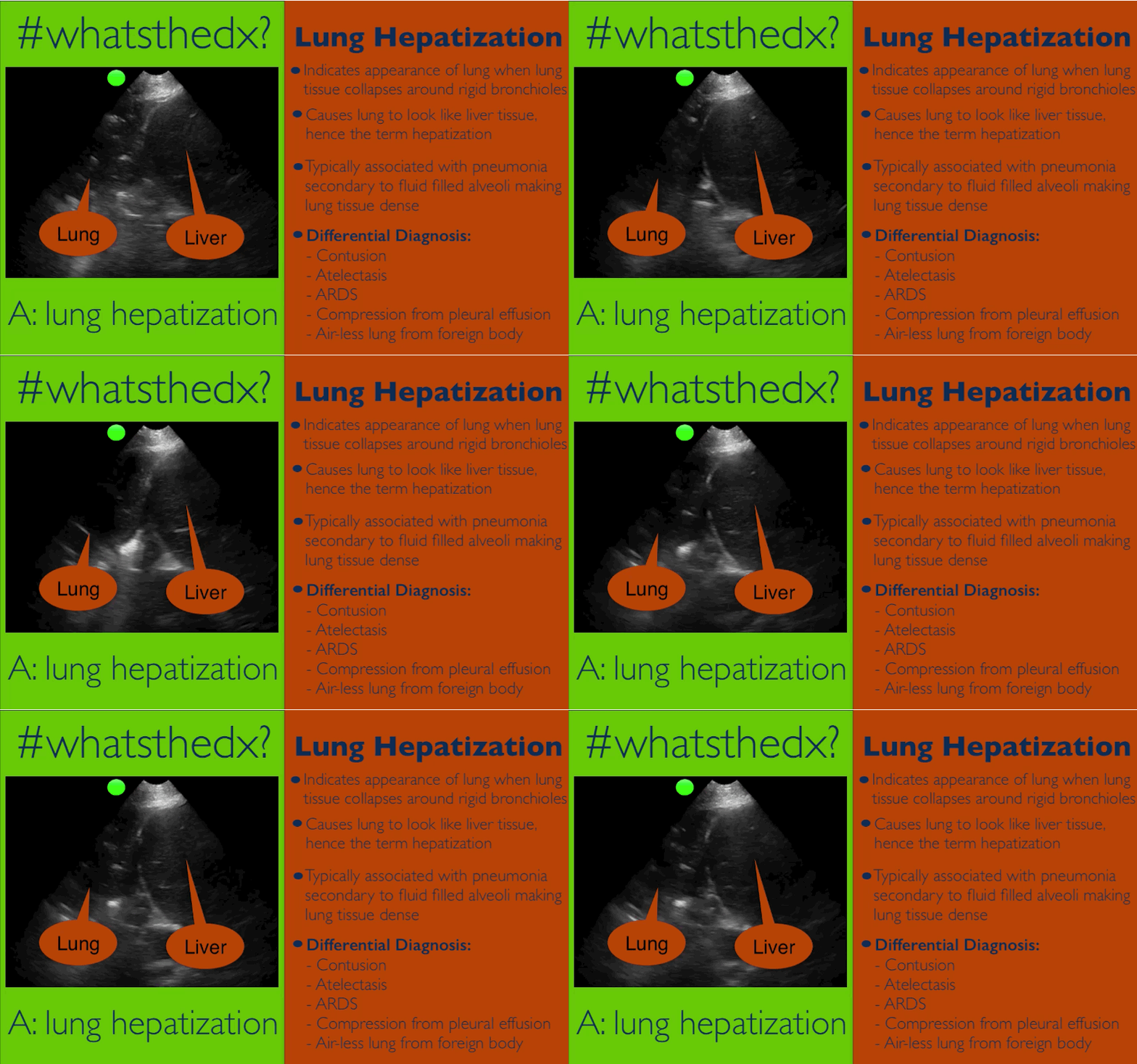
Patient presents with dyspnea & hypoxemia (pulse oximeter is 80%). The "stat" CXR is delayed, but ultrasound is not. What's the diagnosis and what are some differential diagnoses?
Show Answer
Show References

Follow me on Twitter (@criticalcarenow) or Google+ (+criticalcarenow)


Title: What's the Diagnosis?

Category: Visual Diagnosis

Posted: 11/11/2014 by Haney Mallemat, MD

Question

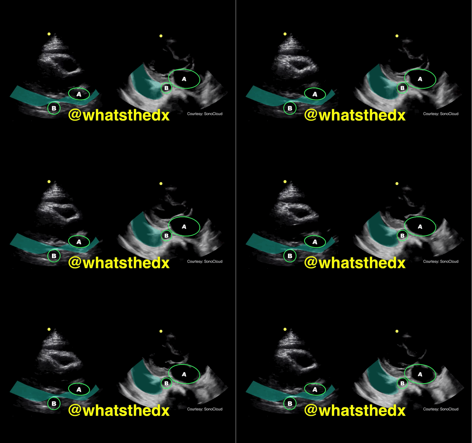
Parasternal long-axis of two different patients. What is the:

  1. structure labeled “A”
  2. structure labeled “B”
  3. diagnosis on the left?
  4. diagnosis on the right?

Show Answer

Answer: 

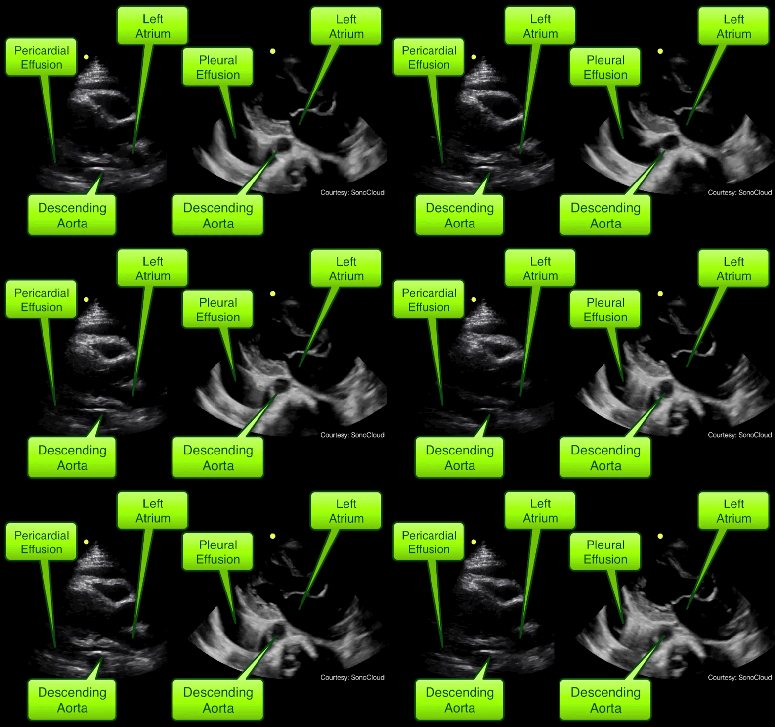
  1. A = Left atrium
  2. B = Descending aorta
  3. Left clip = Pericardial effusion
  4. Right clip = Pleural effusion

Take home pearl: when there is fluid behind the heart, the parasternal long-axis view of the heart is helpful to distinguish between a pleural effusion and a pericardial effusion.

  • Step 1: Find the descending aorta and left atrium
  • Step 2: Is there fluid that crosses in-between the two?
    • If there is, then there is a pericardial effusion
    • If there is no fluid between, then it is a left pleural effusion

Show References

Follow me on Twitter (@criticalcarenow) or Google+ (+criticalcarenow)


Title: What's the Diagnosis? Clip by Dr. Mike Stone (@bedsidesono)

Category: Visual Diagnosis

Posted: 11/4/2014 by Haney Mallemat, MD (Updated: 11/5/2014)

Question

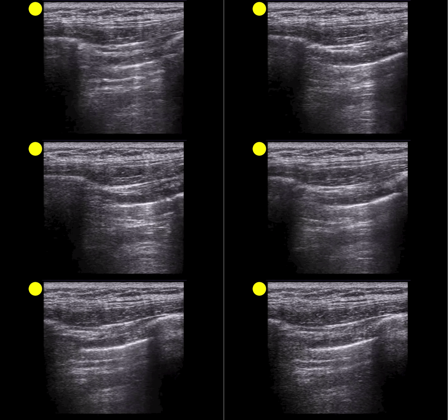
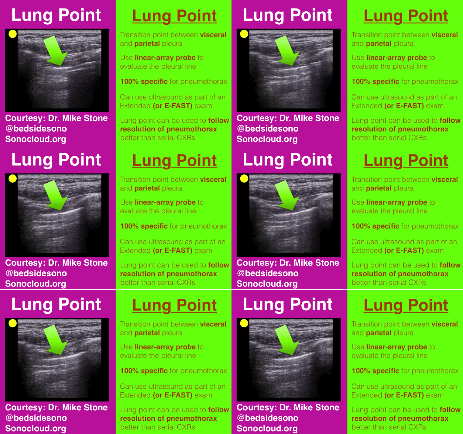
A patient presents post-motor vehicle crash with chest pain and dyspnea. The lung ultrasound is shown below. What's the diagnosis?
Show Answer
Show References

Follow me on Twitter (@criticalcarenow) or Google+ (+criticalcarenow)


Title: What's the Diagnosis?

Category: Visual Diagnosis

Posted: 10/20/2014 by Haney Mallemat, MD (Updated: 11/4/2014)

Question

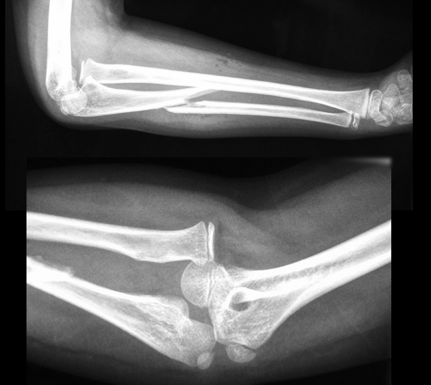
13 year-old right-hand dominant following assault with blunt object. What’s the diagnosis?
Show Answer

Monteggia Fracture

  • Ulnar fracture with dislocation of proximal radioulnar joint (often subtle); do not confuse with Galeazzi fracture (radial-shaft fracture with distal radioulnar dislocation)
  • Interosseous membrane (between radius and ulna) transmits forces to radioulnar joints and causes associated dislocations.
  • Secondary to fall on outstretched hand with arm in hyper-pronation or with direct trauma to forearm as in defensive wounds (e.g., nightstick injury).Suspected forearm fractures should include Xray of the wrist, forearm, and elbow
  • Children may be treated with closed reduction and immobilization; adults usually require open reduction and internal fixation. Radial head dislocations should be reduced within 6-8 hours because of associated articular damage and nerve injury.
  • Radial, ulnar, and median nerve neuropraxias (motor / sensory deficits) may complicate injury.

Show References

Follow me on Twitter (@criticalcarenow) or Google+ (+criticalcarenow)


Title: What's the Diagnosis?

Category: Visual Diagnosis

Posted: 10/13/2014 by Haney Mallemat, MD

Question

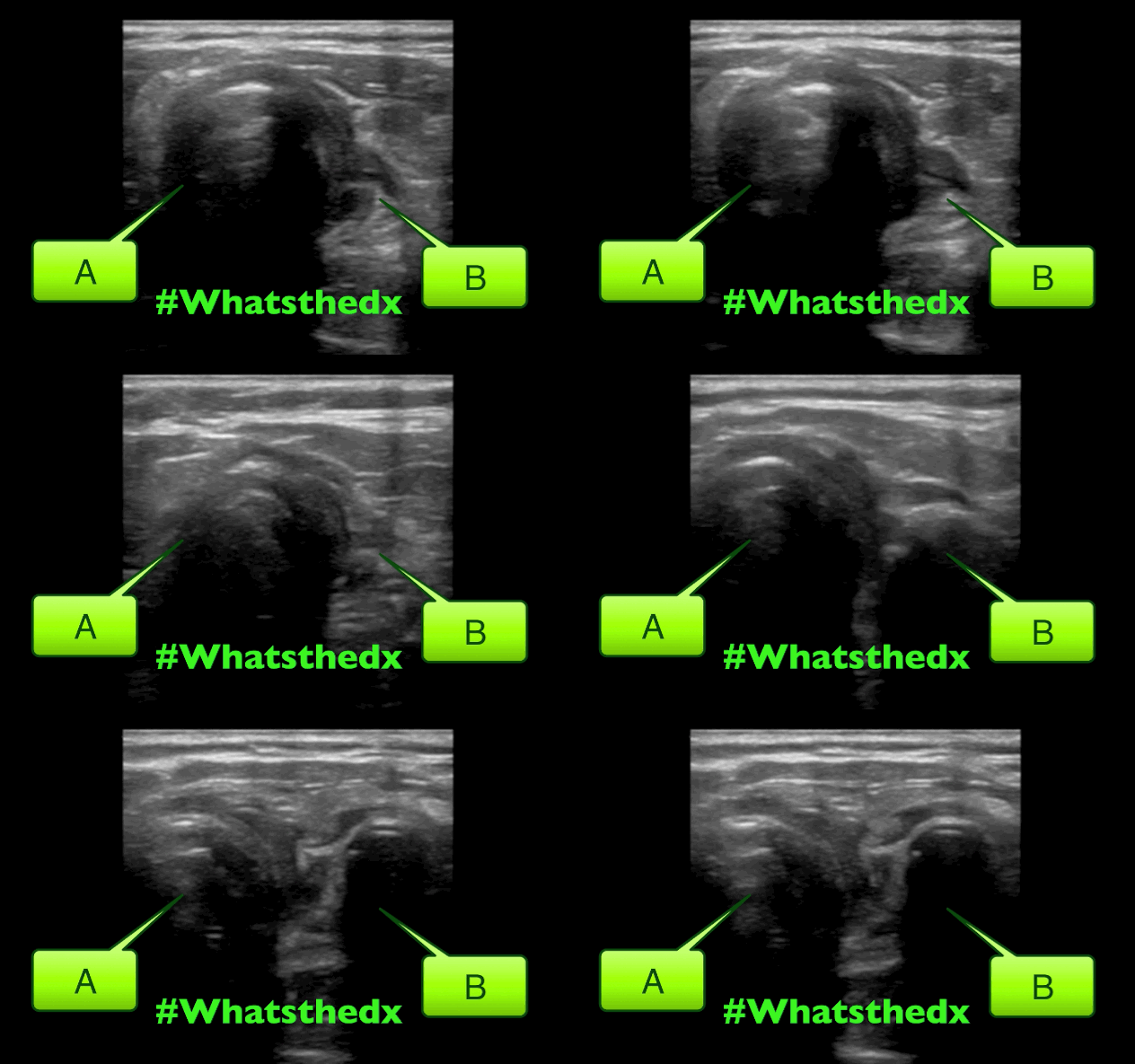
A neck ultrasound is performed during endotracheal intubation. What is labeled "A", what is labeled "B" and what's the diagnosis?
Show Answer
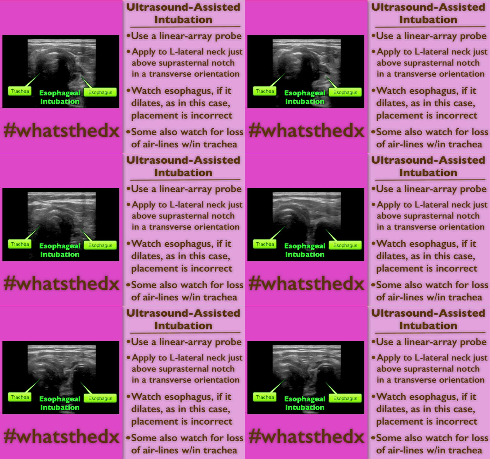
Answer:

A: Trachea

B: Esophagus

This is an esophageal intubation! Take out the tube and try again. Check out this podcast for more on the technique from my good friends at the ultrasound podcast

Here's a summary:

Show References

Follow me on Twitter (@criticalcarenow) or Google+ (+criticalcarenow)


Title: What's the Diagnosis? By Dr. Ari Kestler and Dr. Ashley Strobel

Category: Visual Diagnosis

Posted: 10/6/2014 by Haney Mallemat, MD

Question

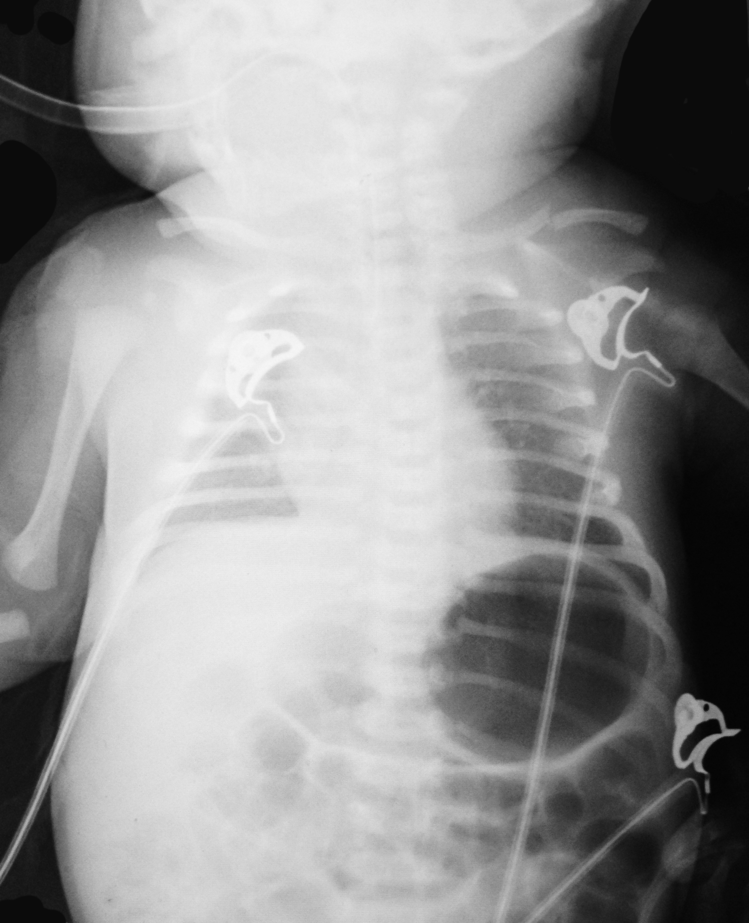
7d old child presents with difficulty feeding,vomiting one time, and now with intermittent apneic episodes. What's the diagnosis? (Careful....this one is tricky!)
Show Answer
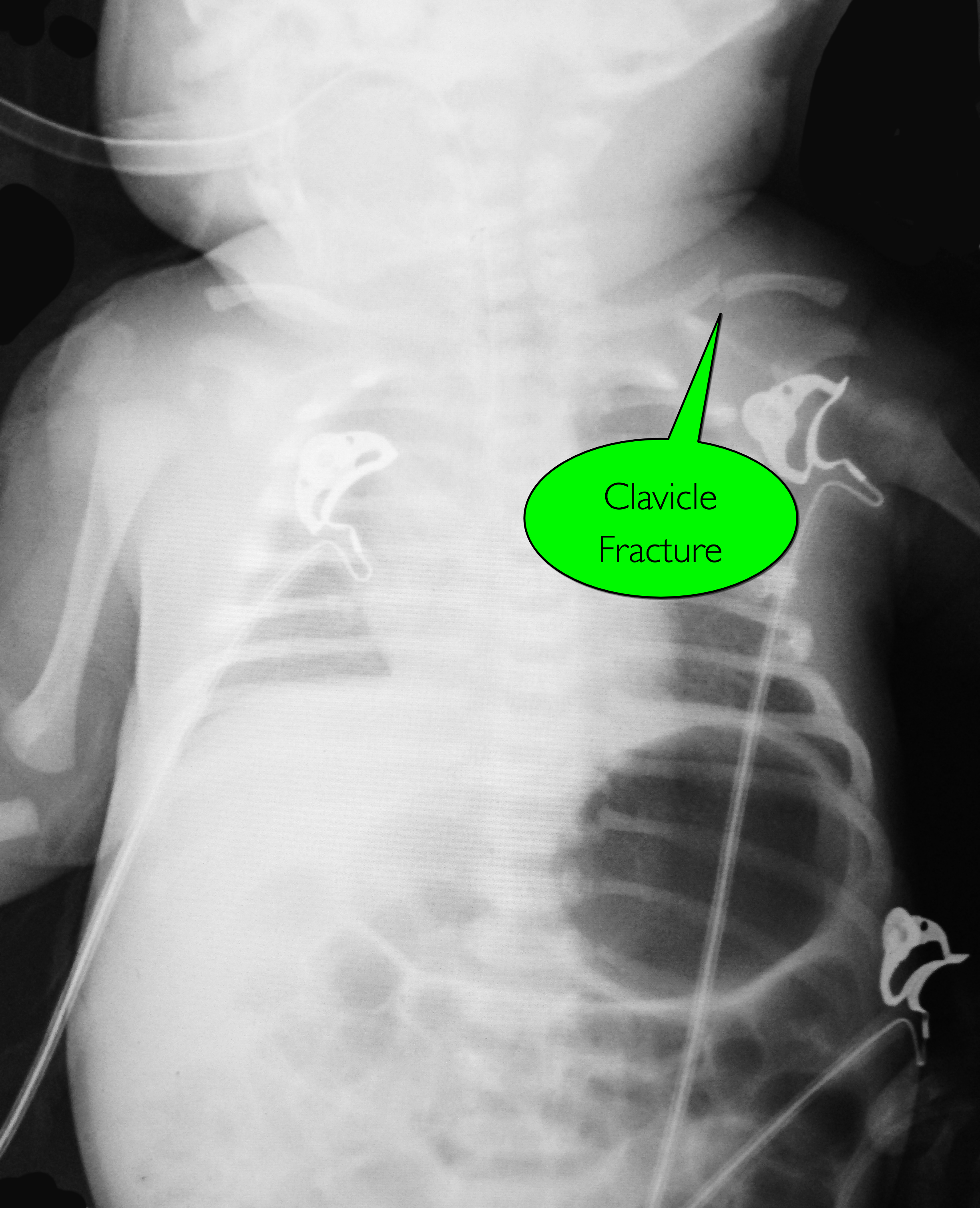
Answer: Workup should include an ALTE and sepsis evaluation PLUS evaluation for non-accidental trauma

Non-accidental trauma (NAT)

NAT is most prevalent in children 0-3 months of age.

Classic metaphyseal lesions, rib fractures, and fractures in various stages of healing are most commonly described in children.

How do we know this is not just birth trauma from a shoulder dystocia, large for gestational age, or difficult vaginal delivery?

  • Key is dating the fracture (see referenced article for more info)
  • Callus not present prior to 8 days of life and callus thickness decreases inversely with fracture age.
  • Subperiosteal new bone formation is highly unlikely in fractures less than 7 days old

Subperiosteal new bone formation appears as a:

  • hazy cortical margin or a thin layer of bone separated from the original cortex by a discrete lucent interval
  • linear hyperdensity parallel to the cortex of the bone
  • see referenced article for images

NAT Work-up:

  1. CT head without contrast if 2 yo
  2. Skeletal survey if 2 yo
  3. AST, ALT, amylase, lipase, CBC with manual diff, BMP, urinalysis, urine toxicology
  4. Consults (Ophthalmology, Social Work, Child Protection, etc.)

Show References

Follow me on Twitter (@criticalcarenow) or Google+ (+criticalcarenow)

Walters MM, Forbes PW, Buonomo C, and Kleinman PK. Healing Patterns of Clavicular Birth Injuries as a guide to fracture dating in cases of possible infant abuse. Pediatric Radiology. October 2014; 44: 1224-1229


Title: What's the Diagnosis? Case by Dr. Maite Huis in 't Veld

Category: Visual Diagnosis

Posted: 9/29/2014 by Haney Mallemat, MD (Updated: 10/1/2014)

Question

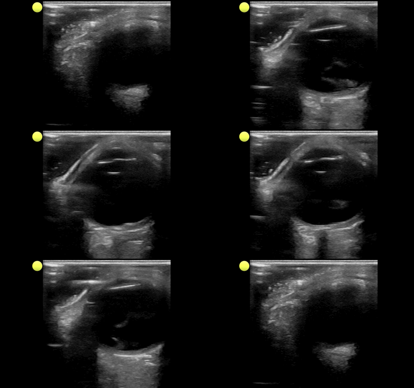
Person presents with painless vision loss after seeing flashes of light. Ultrasound is below. What's the diagnosis?
Show Answer
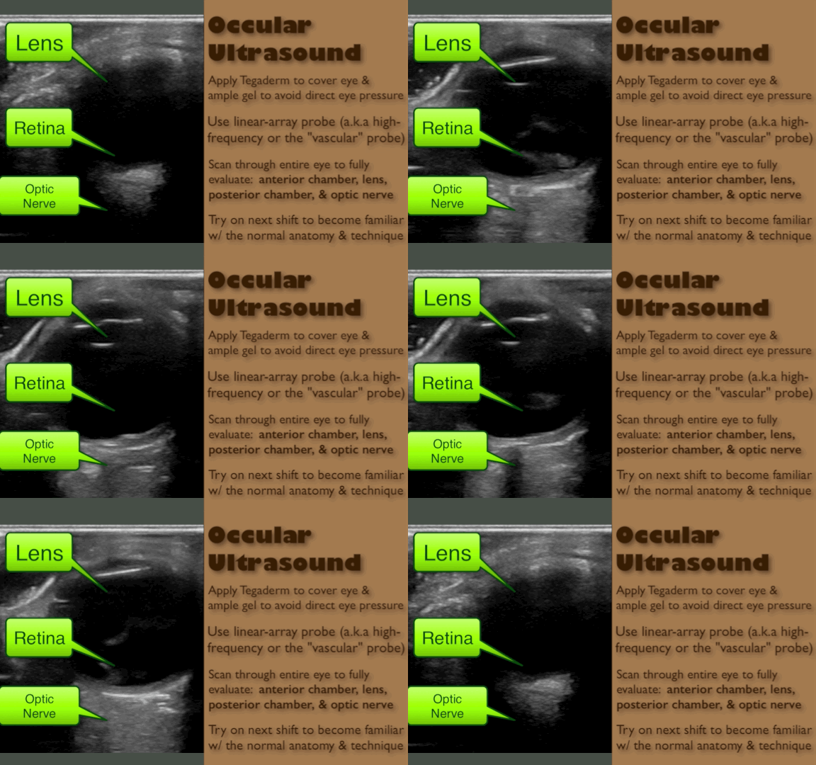
Answer: Vitreous hemorrhage with retinal detachment

Show References

Follow me on Twitter (@criticalcarenow) or Google+ (+criticalcarenow)


Title: What's the Diagnosis? Case by Dr. Leen Alblaihed

Category: Visual Diagnosis

Posted: 9/15/2014 by Haney Mallemat, MD (Updated: 10/1/2014)

Question

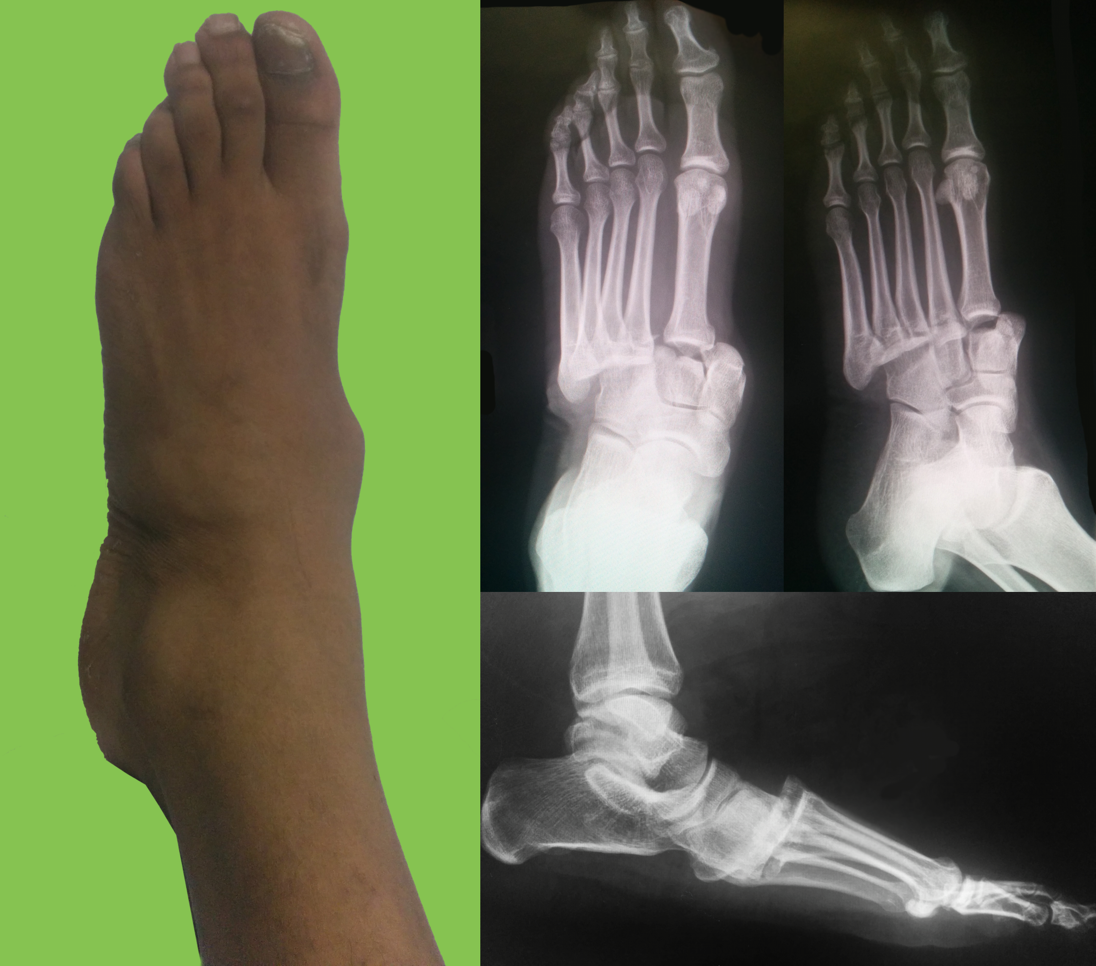
Football player complains of sudden foot pain after begin tackled. What’s the diagnosis? 
Show Answer

Lisfranc fracture-dislocation

  • Lisfranc joint consists of the tarsometatarsal joint (i.e., mid-foot) complex
  • Injuries occur when bones are fractured/avulsed or there is ligamentous disruption, secondary to a direct force or sudden rotation on a downward pointing forefoot; classic injury was fall from a horse while foot is stuck in the stirrup
  • Examine the Lisfranc joint:
    • Hold heel and twist mid-foot; assess for tenderness
    • Piano-key test: move toes up or down (increases stress in mid-foot)
    • Ask patient to stand on one foot with tip-toes; if not painful, then Lisfranc joint is intact
  • X-ray:
    • Fleck sign: fleck of bone between the first and second metatarsal bases, representing avulsion fracture of the Lisfranc ligament
    • >2 mm displacement between medial cuniform and base of 2nd metatarsal
    • If suspicion remains with normal x-ray, obtain weight bearing x-rays
    • No pain but Lisfranc on x-ray, consider Charcot’s joint
  • Treatment is usually surgical. If fracture is stable and non-displaced (<2 mm) use a short leg cast and non-weight bearing for 6 weeks.

Show References

Follow me on Twitter (@criticalcarenow) or Google+ (+criticalcarenow)


Title: What's the Diagnosis? Image by Dr. Michael Abraham

Category: Visual Diagnosis

Posted: 9/8/2014 by Haney Mallemat, MD

Question

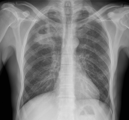
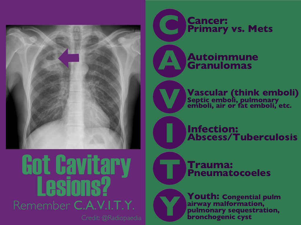
CXR shown below, what's the diagnosis? ...and name 3 differential diagnoses.


Show Answer
Show References

Follow me on Twitter (@criticalcarenow) or Google+ (+criticalcarenow)


Title: What's the Diagnosis?

Category: Visual Diagnosis

Posted: 9/1/2014 by Haney Mallemat, MD

Question

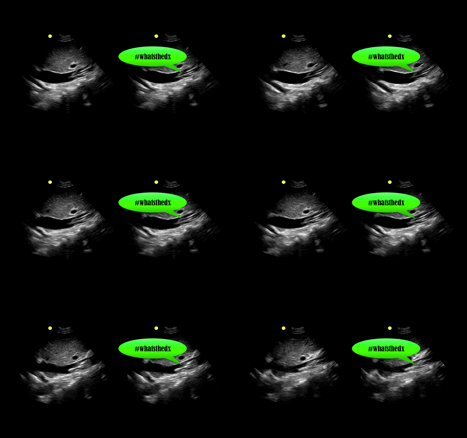
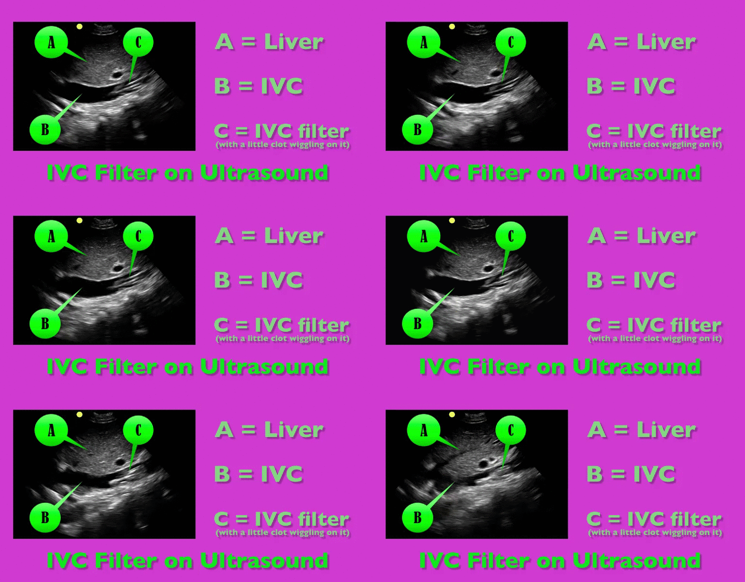
You are scaning the liver with ultrasound and you see this. What's the diagnosis?


Show Answer
Show References

Follow me on Twitter (@criticalcarenow) or Google+ (+criticalcarenow)


Title: What's the Diagnosis?

Category: Visual Diagnosis

Posted: 8/25/2014 by Haney Mallemat, MD

Question

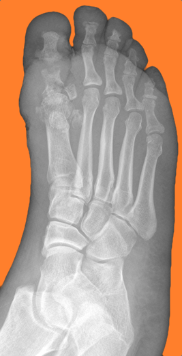
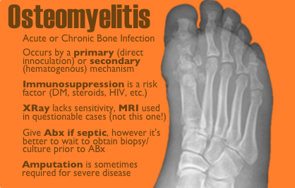
50 year-old female with diabetes complains of pain and discharge from a poorly healing wound. XRay below. What's the diagnosis?
Show Answer
Show References

Follow me on Twitter (@criticalcarenow) or Google+ (+criticalcarenow)


Title: What's the Diagnosis?

Category: Visual Diagnosis

Posted: 8/18/2014 by Haney Mallemat, MD

Question

A critically-ill patient requires fluid resuscitation. Someone hands you a bag of this. What’s the pH of this fluid? 
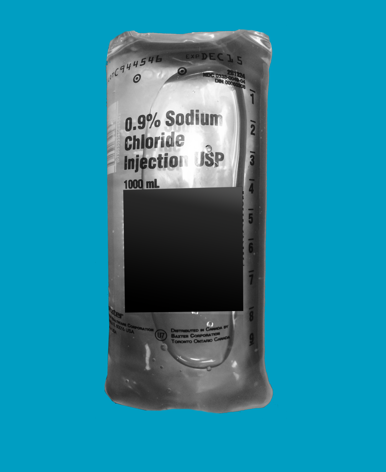
Show Answer
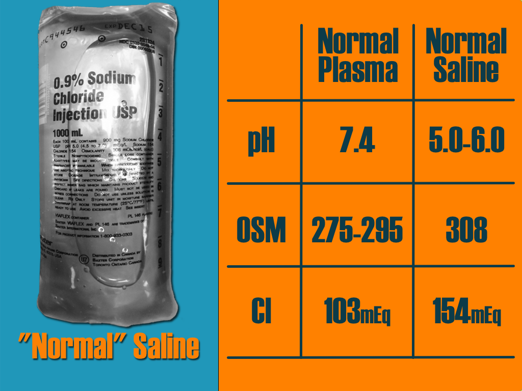
Show References

Follow me on Twitter (@criticalcarenow) or Google+ (+criticalcarenow)


Title: What's the Diagnosis? Image by Dr. Ari Kestler

Category: Visual Diagnosis

Posted: 8/11/2014 by Haney Mallemat, MD (Updated: 8/12/2014)

Question

30 year-old female with complains of night sweats and painful lesions on her fingertips. What’s the diagnosis and list some things to have in the differential diagnosis?

Show Answer

Answer: Osler Nodes

Osler notes are painful, palpable, and erythematous lesions secondary to the deposition of immune-complexes within the pads of digits.

Although classically associated with infective endocarditis, some authors claim that it may be seen in only 10-23% of confirmed cases. Therefore the differential diagnosis should also include:

  • Bacteremia
  • Disseminated gonococcal infection
  • Lupus
  • Vasculitis
  • Drug-induced

Show References

Follow me on Twitter (@criticalcarenow) or Google+ (+criticalcarenow)


Title: What's the Diagnosis? Image by Dr. Andrew Crouter

Category: Visual Diagnosis

Posted: 8/4/2014 by Haney Mallemat, MD (Updated: 8/12/2014)

Question

40 year-old female presents with painful lesions and ulcers on lower extremities. She has had this before, but never to this extent. She also has a history of DVT. What’s the diagnosis?
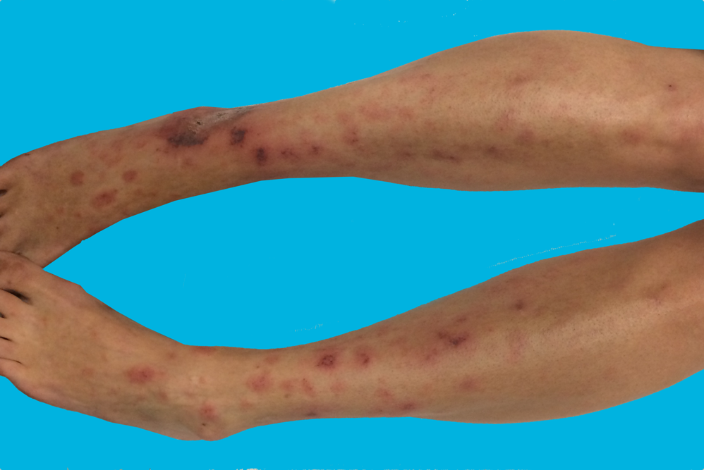
Show Answer

Answer: Livedoid vasculopathy or Livedoid vasculitis (LV)

  • Vascular disease resulting in chronic thrombosis and ulceration of bilateral lower extremities usually in young to middle-aged females. Lesions are painful purpuric macules or papules.
  • A vasoocclusive-type process not an inflammatory one occurring secondary to hyalinization and focal thrombosis of blood vessels. Diagnosis via biopsy.
  • Patients are prothrombotic with an increased risk for VTE’s (altered protein C, increased homocystine, Factor V Leiden mutation, etc.).
  • Treatment goals are to reduce pain, thrombosis (e.g., anticoagulants, antiplatelets, etc.), and superinfected ulcers should be treated with antibiotics covering skin flora. No increased risk of loss of life or limb with the disease.
  • Fantastic differential diagnoses from folks following on Twitter:
    • Polyarteritis nodosum and other vasculitis diseases
    • SLE and anti-phosholipid syndrome
    • Levamisole toxicity
    • Venous stasis ulcers
    • Warfarin-induced skin necrosis

Show References

Follow me on Twitter (@criticalcarenow) or Google+ (+criticalcarenow)


Title: What's the Diagnosis? Case by Dr. Swati Singh

Category: Visual Diagnosis

Posted: 7/28/2014 by Haney Mallemat, MD (Updated: 7/28/2014)

Question

2-day old baby boy presents with forceful vomiting of entire feeds, bloated belly, and has not passed stools since birth. What's the diagnosis?
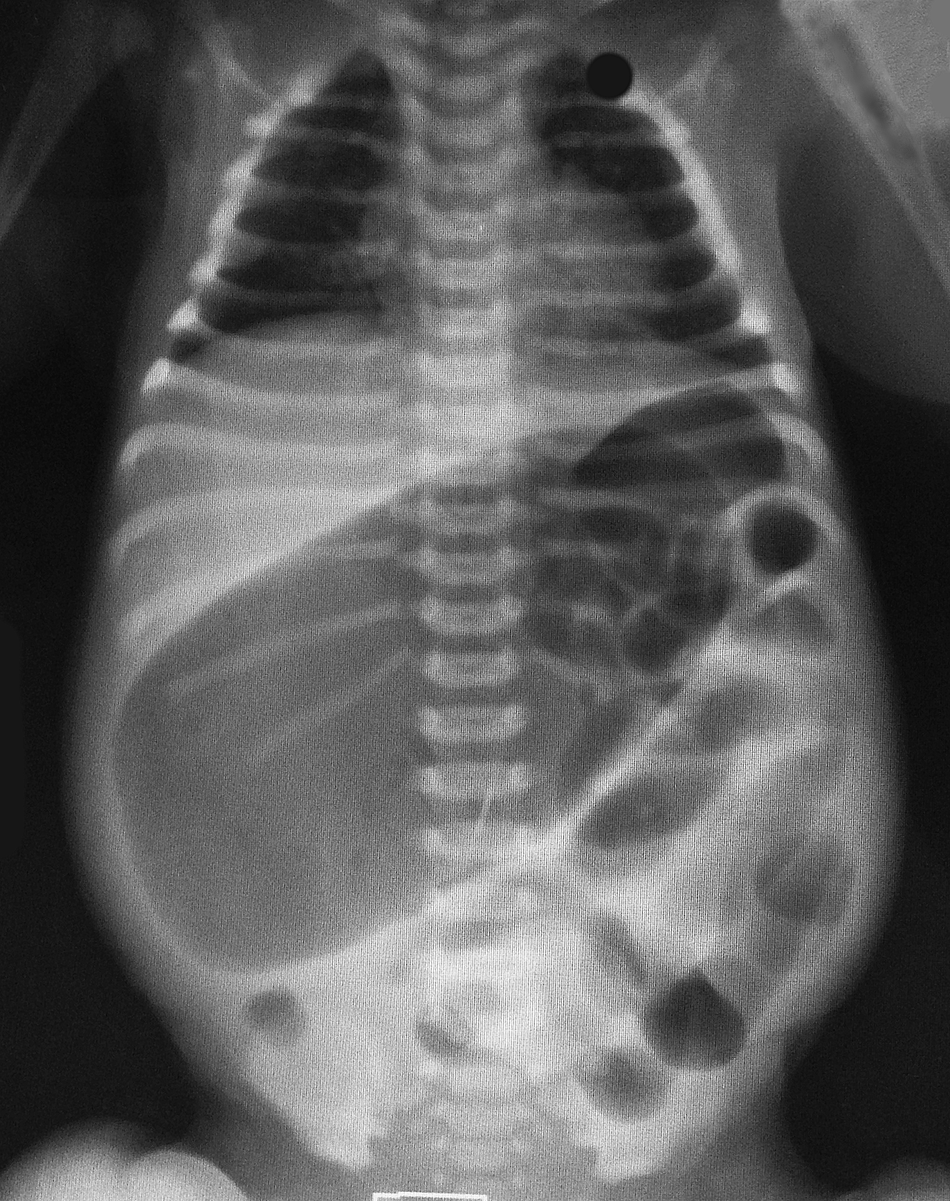
Show Answer

Colonic atresia

Colonic Atresia

  • Condition in which a part of the colon has not formed correctly; colon is completely blocked or missing altogether. The colon is the rarest site of atresia in the GI tract.
  • Diagnosis may be suggested in prenatal sonograms and is usually revealed in affected newborns shortly after birth.
  • Patients usually present with vomiting, abdominal distention, and failure to pass meconium.
  • Differential includes:
    • Small bowel atresia

    • Meconium plug syndrome

    • Hirschsprung's disease 

  • Treatment is supportive and ultimately surgical correction

Show References

Follow me on Twitter (@criticalcarenow) or Google+ (+criticalcarenow)

http://emedicine.medscape.com/article/934014-overview


Title: What's the Diagnosis? Case by Dr. Bennett Myers

Category: Visual Diagnosis

Posted: 7/21/2014 by Haney Mallemat, MD (Updated: 7/22/2014)

Question

45 year-old right-hand dominant patient presents with right hand pain from a prior injury to hand. Patient has also been injecting subcutaneous heroin into hand for relief. What's the diagnosis?
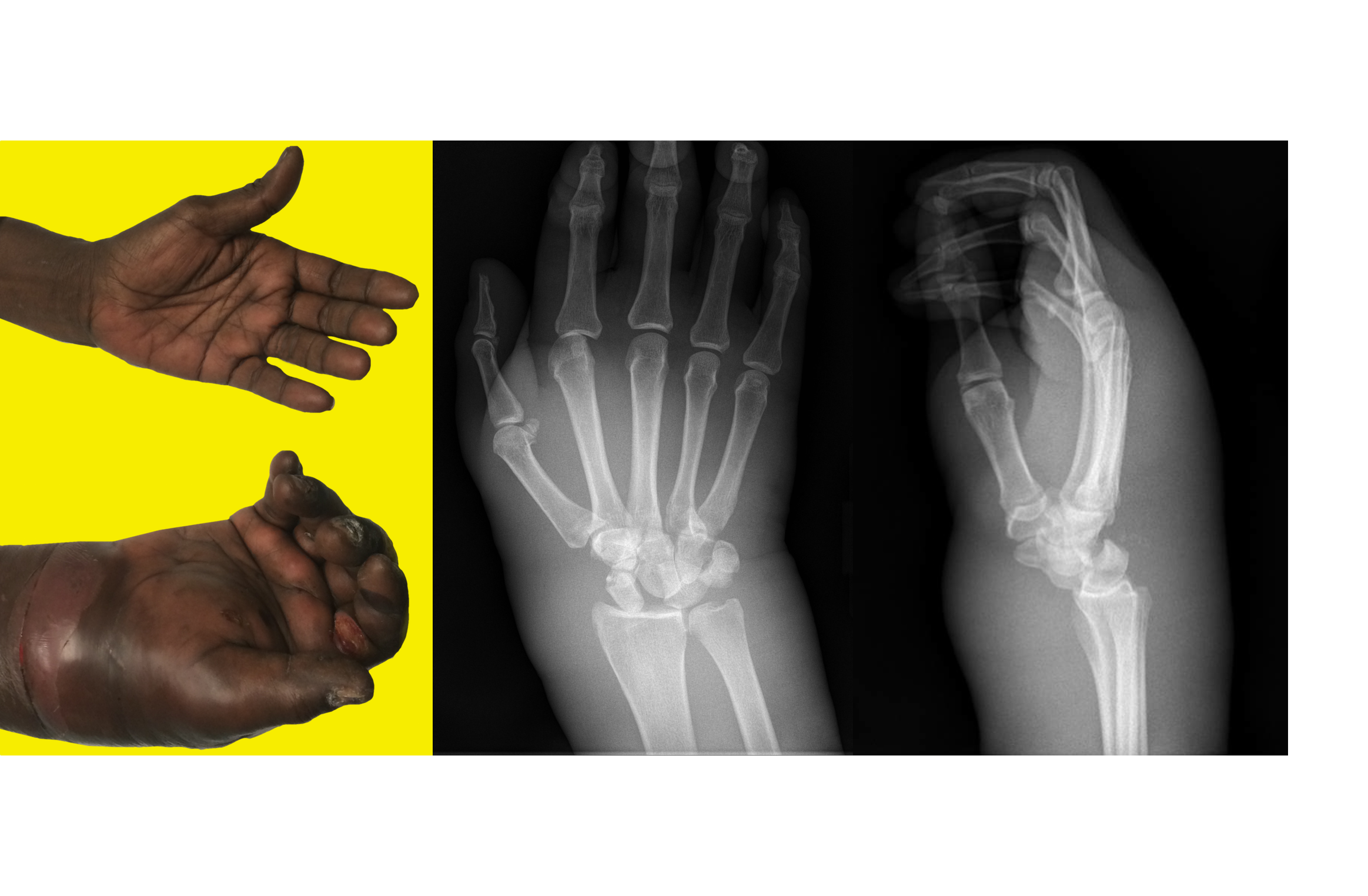
 

Show Answer
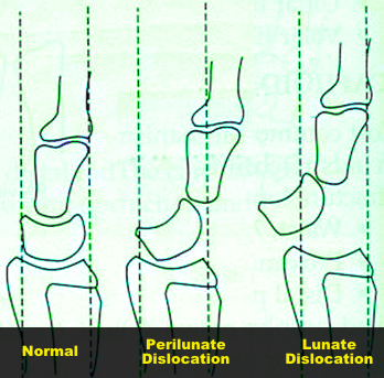
Perilunate dislocation (volar displacement)...and severe hand cellulitis.

Perilunate and Lunate dislocations

  • Lunate dislocation: normal alignment of the distal radius, carpels, and metacarpals; the lunate is displaced 
  • Perilunate dislocation: lunate will demonstrate normal alignment and the remainder of the carpels and metacarpals will typically be displaced in a dorsal direction (occasionally in a volar direction as in this case)
  • These dislocations occur following high-energy trauma, typically with a hyperextended and ulnar deviated hand
  • Plain films are usually enough to make diagnosis, but CT may be used to assess for associated occult fractures (e.g., diagnose co-existing scaphoid fracture


Show References

Follow me on Twitter (@criticalcarenow) or Google+ (+criticalcarenow)


Title: What's the Diagnosis?

Category: Visual Diagnosis

Posted: 7/14/2014 by Haney Mallemat, MD

Question

30 year-old presents with cough & fever. CXR shows mild right lower lobe pneumonia. The lung ultrasound of the right lower lobe is shown below. What's the diagnosis? 
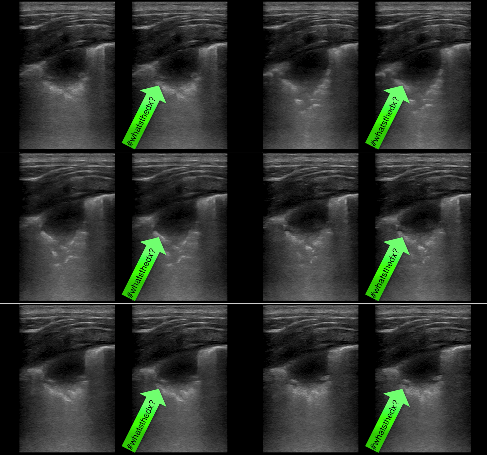
Show Answer

Answer: Pulmonary abscess

  • Patient initially denied IV drugs of abuse, but later disclosed use when this was found
  • Chest CT demonstrated several pulmonary abscesses and the patient was admitted for a workup of endocarditis 

Bottom-line: Not only is ultrasound faster than a CXR in diagnosing pneumonia, unexpected diagnoses may be found that change management

Show References

Follow me on Twitter (@criticalcarenow) or Google+ (+criticalcarenow)


Title: What's the Diagnosis? Image by UMEM alumni Dr. Adam Friedlander

Category: Visual Diagnosis

Posted: 7/7/2014 by Haney Mallemat, MD (Updated: 7/7/2014)

Question

10 year-old male complains of fever and rash (shown below); no other complaints. He went camping 10-days ago. What’s the diagnosis...and what medication(s) should he receive?
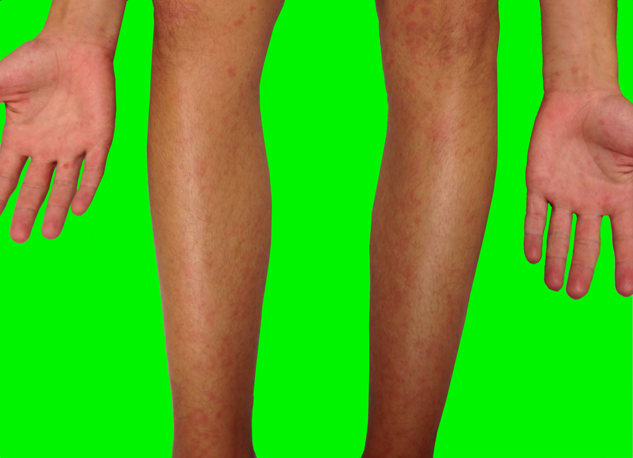
Show Answer

Rocky Mountain spotted fever (RMSF)

  • Tick-borne disease caused by the organism Rickettsia rickettsia; endemic in North, Central, and South America; especially prevalent in southeastern and south-central US
  • Consider diagnosis in patients with unexplained fevers, even without history of a tick bite (reported in only 70%)
  • Common symptoms include:
    • Fever
    • Headache
    • Maculopapular rash (see below)
    • GI symptoms (e.g., nausea, vomiting, etc.)
    • Myalgias
    • CNS  (encephalitis, delirium, seizures, etc.)
  • Classic maculopapular rash appears up to 7 days after fever; starts on wrists and ankles then centripetally spreads to involve the trunk and extremities (including the palms and soles); face is typically spared. In children, periorbital edema is a diagnostic clue.
  • Up to 25% mortality if untreated, but is 5% with appropriate antibiotics; doxycycline is the preferred drug for adults and children; chloramphenicol is an alternative agent and used primarily in pregnant women.

Show References

Follow me on Twitter (@criticalcarenow) or Google+ (+criticalcarenow)


Previous  |  1 |  2 |  3 |  4 |  5 |  6 |  7 |  8 |  9 |  10 |  ... |  13 |  Next
University of Maryland School of Medicine logo
  • Departments
  • Programs
  • Research Centers
  • Institutes
  • Graduate Education
  • CIBR Cores
  • Offices of the Dean
    • University of Maryland, Baltimore
    • University of Maryland Medical Center
    • Medical Alumni Association of the University of Maryland
    • Health Sciences and Human Services Library
University of Maryland School of Medicine
655 W. Baltimore Street
Baltimore, MD 21201
Contact Us

© University of Maryland School of Medicine

    • Webmaster
    • Web Accessibility
    • Site Index
    • UMB Hotline
    • Employment
    • Map