University of Maryland School of Medicine logo
Emergency Medicine
All Departments
UMSOM Assistant
UMEM Educational Pearls
  • Home
  • Departments
  • Emergency Medicine
  • CME
  • UMEM Educational Pearls
  • CME
  • Courses and Conferences
  • Free Open Access Medical Education
  • UMEM Educational Pearls

Search

Title: What's the Diagnosis?

Category: Visual Diagnosis

Posted: 11/11/2014 by Haney Mallemat, MD

Question

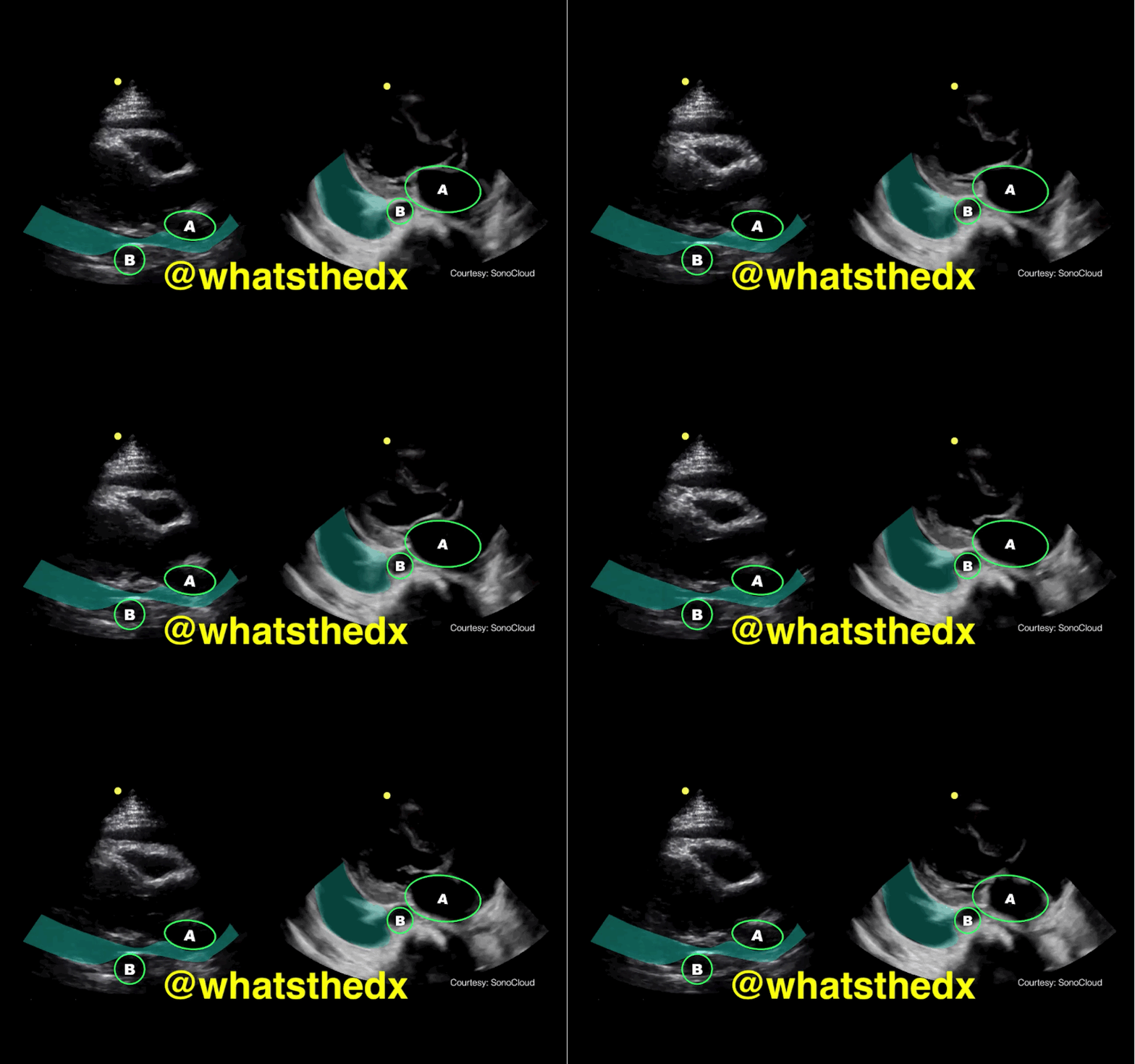
Parasternal long-axis of two different patients. What is the:

  1. structure labeled “A”
  2. structure labeled “B”
  3. diagnosis on the left?
  4. diagnosis on the right?

Answer

Answer: 

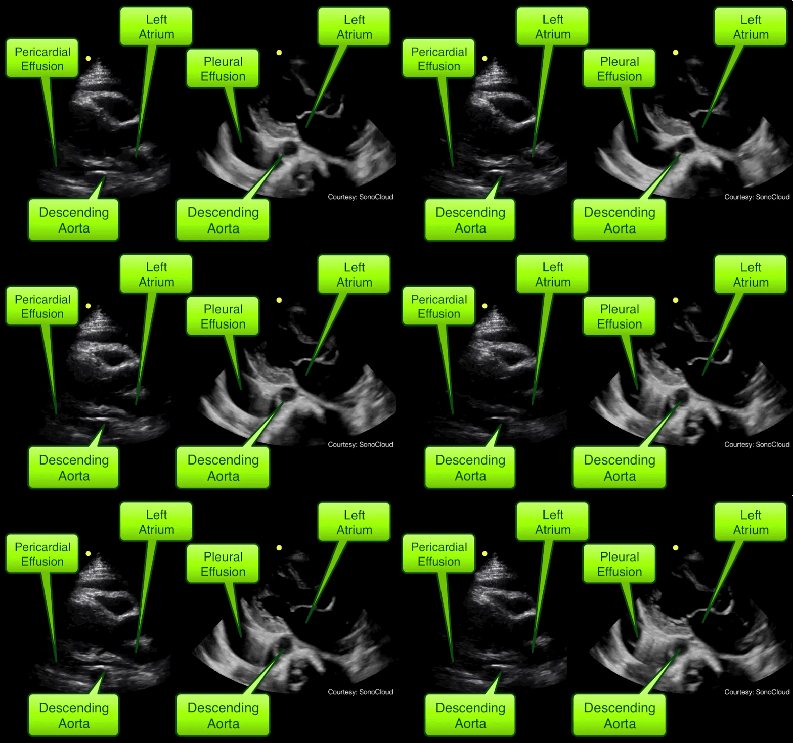
  1. A = Left atrium
  2. B = Descending aorta
  3. Left clip = Pericardial effusion
  4. Right clip = Pleural effusion

Take home pearl: when there is fluid behind the heart, the parasternal long-axis view of the heart is helpful to distinguish between a pleural effusion and a pericardial effusion.

  • Step 1: Find the descending aorta and left atrium
  • Step 2: Is there fluid that crosses in-between the two?
    • If there is, then there is a pericardial effusion
    • If there is no fluid between, then it is a left pleural effusion
References

Follow me on Twitter (@criticalcarenow) or Google+ (+criticalcarenow)


University of Maryland School of Medicine logo
  • Departments
  • Programs
  • Research Centers
  • Institutes
  • Graduate Education
  • CIBR Cores
  • Offices of the Dean
    • University of Maryland, Baltimore
    • University of Maryland Medical Center
    • Medical Alumni Association of the University of Maryland
    • Health Sciences and Human Services Library
University of Maryland School of Medicine
655 W. Baltimore Street
Baltimore, MD 21201
Contact Us

© University of Maryland School of Medicine

    • Webmaster
    • Web Accessibility
    • Site Index
    • UMB Hotline
    • Employment
    • Map