
Search
Subscribe to daily UMEM Pearls (FREE!) via:
561-580 of 889 results with category "Critical Care"
The management of alcohol withdrawal syndrome (AWS) includes supportive care focusing on the ABC’s and administration of benzodiazepines (BDZ).
While BDZ are effective in the treatment of AWS, some patients may require very high doses of BDZ to control symptoms (tachycardia, hypertension, diaphoresis, etc.); unfortunately, high-doses of BDZ may lead to suppression of the respiratory drive and endotracheal intubation.
Dexmedetomidine (DEX) is a sedative agent that is an intravenous alpha2-agonist (it's like clonidine); it reduces sympathetic outflow from the central nervous system and it may help treat withdrawal syndromes. The major benefit of DEX is that it does not suppress the respiratory drive, thus intubation is not required.
Smaller trials and case series have shown that patients with AWS who were treated with BDZ in addition to DEX had better symptom control, lower overall BDZ doses, and less respiratory depression/intubation.
Bottom-line: While more trials are needed, consider adding DEX for patients with AWS who require high-doses of BDZ.
Show References
Ottawa Rules for Subarachnoid Hemmorhage (SAH)
Background
- Headache is a common reason for ER visits
- 1-3% of headaches are SAH
- Misdiagnosis of SAH can be fatal
- Lumbar puncture can be a painful/time-consuming procedure
- Goal is to design a decision rule to help guide the clinician
Design
- Multi-center study at ten Canadian emergency departments.
- 2131 adults with a headache peaking within 1 hour and no neurologic deficits
- Non-traumatic headaches only; GCS of 15 required
- SAH defined as: 1. CT evidence of SAH; 2. Xanthochromia in CSF; or 3. RBCs in the final tube of CSF, WITH positive angiography findings.
Results
132 (6.2%) had SAH
Decision rule including any:
- age 40 years or older
- neck pain or stiffness
- witnessed LOC
- onset during exertion
Had 98.5% sensitivity (95% CI, 94.6%-99.6%) and 27.5% specificity (95% CI, 25.6%-29.5%)
Adding “thunder-clap” headache and “limited neck flexion on examination” (inability to touch chin to chest or raise the head 8cm off the bed if supine) resulted in 100% (95% CI, 97.2%-100%) sensitivity.
The rule was then evaluated using a bootstrap analysis on old cohort data to validate the rule.
Conclusion/Limitations
- Exciting new rule for SAH that needs to be validated in a new, independent cohort
- The rule may not decrease the rate of investigation (CT, LP, or both)
- It may decrease the amount of SAH that are missed on first visit to the ER
- Limited by narrow criteria for inclusion in the rule/not meant for other causes of headache
- See the JAMA editorial with the article for a nice discussion of the difficulties with decision making rules.
- The rule:The Ottawa SAH Rule
-
For alert patients older than 15 y with new severe nontraumatic headache reaching maximum intensity within 1 h
-
Not for patients with new neurologic deficits, previous aneurysms, SAH, brain tumors, or history of recurrent headaches (≥3 episodes over the course of ≥6 mo)
-
Investigate if ≥1 high-risk variables present:
-
Age ≥40 y
-
Neck pain or stiffness
-
Witnessed loss of consciousness
-
Onset during exertion
-
Thunderclap headache (instantly peaking pain)
-
Limited neck flexion on examination
-
Show References
Attachments
Acalculous Cholecystitis in the Critically Ill
- Acute acalculous cholecystitis (AAC) accounts for almost 50% of cases of acute cholecystitis in the critically ill ICU patient.
- Importantly, the mortality rate for AAC can be as high as 50%.
- Risk factors for AAC include:
- CHF
- Cardiac arrest
- DM
- ESRD on hemodialysis
- Postoperative
- Burns
- Unfortunately, the physical exam is unreliable, especially in intubated and sedated patients.
- Furthermore, less than half of patients with AAC are febrile or have a leukocytosis. LFTs can also be normal in up to 20% of patients.
- Ultrasound remains the most common imaging modality for diagnosis.
- Take Home Point: Consider AAC in the septic critically ill patient without a source.
Show References
Ineffective triggering is the most common type of ventilator dyssynchrony. The differential diagnosis includes:
- Auto peep (the most common cause)
- Neuromuscular weakness
- Improper ventilator settings
Auto peep is the most common cause of ineffective triggering and will often occur as a patient cannot create enough inspiratory force to overcome their own intrinsic peep (PEEPi). Patients who are severely tachypnic or those with obstructive lung disease are at high risk for auto peep (not enough time to exhale).
Ineffective triggering can also occur if the patient cannot create enough of a negative inspiratory force to trigger the vent to deliver a positive pressure breath. Prolonged period of mechanical ventilation, over sedation, high cervical spine injuries, or diaphragmatic weakness are common causes.
Lastly, improper trigger sensitivities may make it difficulty for the ventilator to sense when the patient is attempting to take a spontaneous breath.
For an example of a patient with ineffective triggering, check out: http://marylandccproject.org/2013/10/28/vent-problems1/
Show References
The pregnant patient normally has increased cardiac output and minute ventilation by the third trimester. Despite this increase, however, these patients have little cardiopulmonary reserve should they become critically-ill.
Remember the mnemonic T.O.L.D.D. for simple tips that should be done for the pregnant patient who presents critically-ill or with the potential for critical illness:
- Tilt: The supine-hypotension syndrome occurs after the 20th week of pregnancy as the gravid uterus compresses the IVC and aorta, reducing cardiac output by up to 30%. Placing a 30-degree right hip-wedge under the patient will relieve this obstruction.
- Oxygen: the growing uterus pushes up on the base of the lungs reducing the functional residual capacity meaning there is less oxygen reserve and rapid oxygen desaturations. Supplemental oxygen may increase the patient's reserve.
- Lines: The circulatory system reserve is reduced, so early and large bore venous access is important. Remember that lines should be placed above the diaphragm because the enlarging uterus compresses pelvic veins, reducing venous return to the heart.
- Dates: Rapidly determine the gestational age of the fetus as 24 weeks is a critical date to remember (e.g., increased risk of supine-hypotension syndrome, fetal viability, etc.)
- Delivery: Call labor and delivery early on, not only for the consultation, but also for the fetal monitoring that this service provides.
Show References
Background
- Acute lung injury that develops within 6 hours after transfusion of 1 or more units of blood or blood components.
- Increased risk with greater number of transfusions
- Incidence is 1 in 4000
Definition
- Acute onset
- Hypoxemia (PaO2/FiO2 < 300 mm Hg)
- Bilateral pulmonary opacities on chest x-ray
- Absence of left atrial hypertension
Pathogenesis
Two-hit hypothesis: first hit is underlying patient factors causing adherence of neutrophils to the pulmonary endothelium; second hit is caused by mediators in the blood transfusion that activate the neutrophils and endothelial cells.
Differential
Can be confused or overlap with TACO or transfusion-associated volume/circulatory overload, which presents similarly but has evidence of increased BNP, CVP, pulmonary wedge pressure, and left sided heart pressures. Patients with TACO tend to improve with diuretic treatment
Supportive tests
- Echocardiogram
- BNP (tends to be low)
- Transient leukopenia
Treatment
- Supportive care
- Lung protective ventilation strategies
- Fluid restrictive strategy
- Aspirin (shown to be helpful in animal studies)
- Pre-washing of stored RBCs prior to transfusion
- Decrease the amount of transfusions!
Show References
There have been so many great talks at ACEP 2013, but Dr. Michael Winters' talk "The ICU is NOT Ready for Your Patient" was chock full of great critical care pearls. Here are just a few:
- Increased mortality for ICU patients boarding in the Emergency Department; the increase is 1.5% per each hour of delayed transfer.
- Intubated patients should receive analgesia BEFORE sedation; fentanyl is recommended because hemodynamically stable, but you can use anything. Good analgesia will also reduce total sedative dosing
- Use continuous capnography for the intubated patient; can detect equipment malfunction and allow titration of ventilation
- Keep an eye out for abdominal compartment syndrome. Physical exam is not always conclusive, should obtain bladder pressures
- Reduce the risk of ventilator-associated pneumonia by keeping endotracheal cuff pressures adequate and keeping the head of bed elevated 30-45 degrees
Show References
Want to improve your chances of success in the resus room? Download a metronome app on your smartphone and set it to a rate of 100-120 beats per minute. There are a number of cheap (usually free) metronome applications for both iOS and Android devices.
A recent review looked at the evidence behind CPR feedback devices and found:
- Compared to baseline, chest compression rates and end-tidal CO2 improved after activation of the metronomes.
- There was a significant improvement in the hands-off time per minute during CPR
- The proportion of intubation attempts taking under 20 seconds improved
- There were Increased survival rates when implemented in the pre-hospital setting
So instead of going to iTunes and downloading the Bee Gees, go over to the App store and download a free metronome. Your resus team will be able to stay on track with their compressions and even better - they won't have to hear you sing!
Show References
-
The efficacy of epinephrine during out-of hospital cardiac arrest has been questioned in recent years, especially with respect to neurologic outcomes (ref#1).
-
A recent study demonstrated both a survival and neurologic benefit to using epinephrine during in-hospital cardiac arrest when used in combination with vasopressin and methylprednisolone.
-
Researchers in Greece randomized 268 consecutive patients with in-hospital cardiac arrest to receive either epinephrine + placebo (control group; n=138) or vasopressin, epinephrine, and methylprednisolone (intervention arm; n=130)
-
Vasopressin (20 IU) was given with epinephrine each CPR cycle for the first 5 cycles; Epinephrine was given alone thereafter (if necessary)
-
Methylprednisolone (40 mg) was only given during the first CPR cycle.
-
If there was return of spontaneous circulation (ROSC) but the patient was in shock, 300 mg of methylprednisolone was given daily for up to 7 days.
-
-
Primary study end-points were ROSC for 20 minutes or more and survival to hospital discharge while monitoring for neurological outcome
-
The results were that patients in the intervention group had a statistically significant:
-
probability of ROSC for > 20 minutes (84% vs. 66%)
-
survival with good neurological outcomes (14% vs. 5%)
-
survival if shock was present post-ROSC (21% vs. 8%)
-
better hemodynamic parameters, less organ dysfunction, and better central venous saturation levels
-
-
Bottom-line: This study may present a promising new therapy for in-hospital cardiac arrest and should be strongly considered.
Show References
Background:
- Antibiotics are prescribed commonly for URIs including acute bronchitis and community acquired pneumonia.
- Antibiotic prescriptions for non-bacterial causes of URIs lead to antibiotic overuse, which can lead to antibiotic resistance and risk of Clostridium difficile.
- Procalcitonin is a biomarker for bacterial infections and is released in response to bacterial toxins during infections.
- Several algorithms using procalcitonin have been developed to help guide antibiotic treatment of URIs based on blood levels and to aid discontinuing antibiotics when procalcitonin levels have returned to normal, leading to decreased use and length of antibiotic treatment courses.
Clinical Question:
- Does measurement of procalcitonin lead to shorter antibiotic exposure without increasing mortality and treatment failure?
Meta-analysis:
- 14 trials; 2004-11; 4211 patients with a variety of URI severity and type including CAP and COPD exacerbations.
- Inpatient and outpatient settings
- Compared to regular antibiotic treatment without procalcitonin level guidance.
- Primary outcomes: All cause mortality and treatment failure within 30 days.
Conclusions:
- No increase in all-cause mortality using procalcitonin algorithms versus standard therapy in any clinical setting or type of URI (5.7% vs. 6.3%, respectively).
- Treatment failure was LOWER for procalcitonin guided patients in the ED [OR 0.76 (95% CI, 0.61-0.95)].
- Lower antibiotic exposure due to lower prescription rate in COPD exacerbations and bronchitis.
Limitations:
- Non-blinded to outcome assessment.
- Adherence to algorithms was variable.
- Immunosuppressed patients and children were excluded.
Bottom Line:
- Another tool to help aid clinical decision making regarding antibiotic treatment
- Test is around $25-30 and takes about 1 hour to run
- Low levels may indicate a non-bacterial cause of infection.
Show References
Peri-Intubation Cardiac Arrest
- Emergency intubation is a common critical care procedure that carries the risk of life-threatening complications.
- Although cardiac arrest (CA) is an established complication, there is scant literature on the actual incidence ad factors associated with CA in the peri-intubation period.
- In a recent retrospective analysis from Carolinas Medical Center, investigators found:
- Peri-intubation CA occurred in 4.2% of patients and was associated with a 14-fold increase in hospital mortality.
- A pre-RSI shock index > 0.9 was indepedently associated with CA.
- Obese patients had a higher incidence of CA; odds of CA increased 1.37 times for every 10 kg increase in weight.
- Take Home Point: Peri-intubation CA may be more common than previously thought and, not suprisingly, is associated with an increased risk of in-hospital death.
Show References
Necrotizing Pneumonia
Necrotizing pneumonia is a rare, but potentially deadly complication of bacterial pneumonia.
It is characterized by the finding of pneumonic consolidation with multiple areas of necrosis within the lung parenchyma. Necrotic foci may coalesce, resulting in a localized lung abscess, or pulmonary gangrene if involving an entire lobe.
Most common pathogens: S. aureus, S. pneumoniae, and Klebsiella pneumonia.
Others include S. epidermidis, E. coli, Acinetobacter baumannii, H. influenzae and Pseudomonas.
Contrast-enhanced chest CT is the diagnostic test of choice and is also helpful in evaluating for parenchymal complications.
Empiric antibiotic therapy should include:
- Broad spectrum coverage for commonly implicated pathogens (vancomycin, pseudomonal-dose piperacillin/tazobactam)
- PLUS either clindamycin or metronidazole to cover possibly involved anaerobes
Consider an early surgical evaluation for the patient with necrotizing pneumonia complicated by septic shock, empyema, bronchopleural fistula, or hemoptysis.
Show References
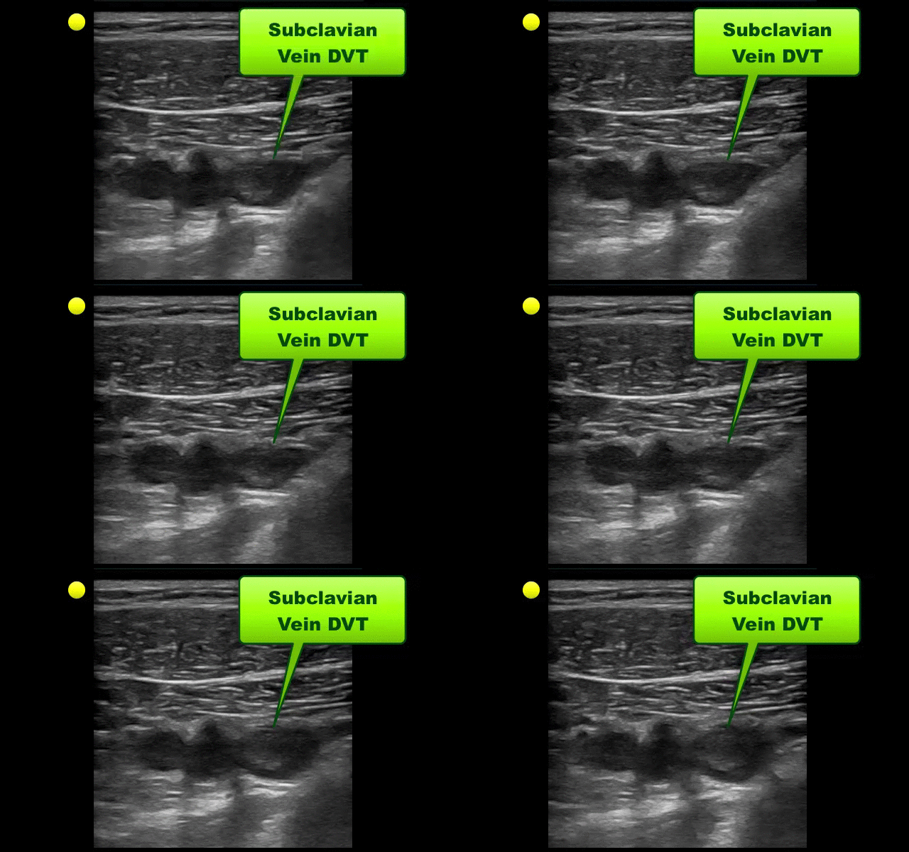
UEDVT comprise 10% of all DVTs (majority are lower extremity), but incidence of UEDVT is rising; UEDVTs are categorized into distal (veins distal to axillary vein) or proximal (from superior vena cava to axillary vein)
Compared to lower extremity DVT, UEDVTs have lower:
- mortality
- risk of pulmonary embolism
- rates of recurrence
75% of UEDVT are secondary (indwelling catheters, pacemakers, malignancy, etc.) and 25% are primary in nature; #1 primary cause of UEDVT is Paget – Schroetter disease
Up to 25% of patients with primary UEDVTs are eventually found to have an underlying malignancy; patients with idiopathic UEDVT should be referred for cancer workup
Treatment includes removal of the catheter (if no longer needed) and:
- anticoagulation (minimum of 3 months)
- consideration of thrombolytics, including catheter-directed administration
- mechanical thrombolysis (clot aspiration, fragmentation, etc.)
- surgical thrombectomy / venous bypass
Show References
Background
- Stroke is common in the first few weeks after a transient ischemic attack (TIA) or minor ischemic stroke.
- Aspirin reduces the risk of recurrent stroke by 12% or so.
- Thus far there is a trend toward no benefit from dual anti-platelet treatment.
Trial
- Randomized, double blind, placebo-controlled trial conducted in China.
- 5170 patients were randomized to either combination therapy with clopidogrel and aspirin (clopidogrel at an initial dose of 300 mg, followed by 75mg per day for 90 days, plus aspirin 75 mg per day for 21 days) or to placebo plus aspirin.
- Primary outcome was stroke during 90 days of follow-up using intention to treat analysis
Results
- Stroke occurred in 8.2% of patients in the aspirin-clopidogrel group as compared with 11.7% in the aspirin group (Hazard ratio 0.68; 95% confidence interval, 0.57-0.81; p<0.001). Rates of hemorrhage were similar in both groups (0.3%).
- Relative risk reduction of stroke at 90 days by 32%.
Conclusions
- Patients with acute TIA or minor stroke may benefit from combination therapy with no increased risk of hemorrhage
Bottom Line:
- 41,561 patients were screened in order to find 5170 appropriate patients!
- Patients with major stroke, who are risk for hemorrhage, and have isolated sensory TIAs, were excluded.
- The trial was conducted in China, so the results may not apply in other countries (A similar trial, the Platelet-Oriented Inhibition in New TIA and Minor Ischemic Stroke (POINT) study is being done in North America).
- Decision to treat should be made with neurology assistance.
Show References
Clostridium Difficile Associated Diarrhea and The Elderly Patient
- Infectious diseases remain the leading cause of mortality in the elderly.
- An infection that is increasing in prevlance among elderly patients is Clostridium difficile-associated diarrhea (CDAD).
- Mortality rates are up to 3.5 times higher in elderly patients with CDAD compared to younger patients.
- Antimicrobial therapy within the previous 6 weeks is the strongest risk factor for CDAD.
- Though any antibiotic may cause CDAD, clindamycin, fluoroquinolones, and cephalosporins have the highest risk.
- Importantly, the diarrhea may not always bloody.
- Metronidazole remains the treatment of choice for uncomplicated infections.
Show References
Bad brain, good lungs.... Right?
A recent retrospective study reviewed the incidence of acute respiratory distress syndrome (ARDS) in patients presenting with spontaneous intracerebral hemorrhage over a 10-year period. After reviewing 1,665 patients, the authors found that:
- The development of ARDS occurred in approximately 27% of patients with spontaneous ICH (similiar to previous literature).
- The incidence ARDS after spontaneous ICH was similiar to other "high-risk" conditions such as sepsis, trauma, & aspiration.
- Modifiable risk factors include: high tidal volume ventilation, higher total fluid balance, & transfusion of PRBCs/FFP.
It's of particular importance to note that high tidal volume ventilation (>8cc/kg) was the single greatest modifiable factor for the development of ARDS.
Bottom line: Try and use lung-protective ventilation strategies (6-8cc/kg ideal body weight) and avoid excessive volume resuscitation in your critically-ill patients whenever possible. Even in cases of isolated intracerebral hemorrhage - where the patient's lungs may appear to be completely normal - traditional tidal volume settings may be harmful.
Show References
Question
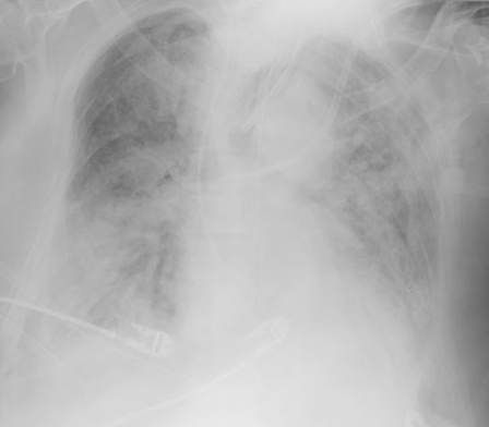
Elderly patient who originally presented for severe pancreatitis now intubated for worsening hypoxemia. CXR is shown below, what's the diagnosis?
Show Answer
Show References
HIV, ART, and the ICU
- Though survival has dramatically improved for patients with HIV, there has been no decrease in the quantity of ICU admissions for this select patient population.
- One of the most common reasons for ICU admission is now adverse effects of antiretroviral therapy (ART).
- When managing a critically ill HIV patient in the ED or ICU, consider the following effects of ART as an etiology:
- Lactic acidosis
- Seen with nucleoside reverse transcriptase inhibitors (NRTIs): greatest risk with didanosine, stavudine, and zidovudine
- Presentation: fatigue, malaise, vomiting, abdominal pain, hepatomegaly
- Lactate often > 10 mmol/L
- Abacavir hypersensitivity
- Usually within first 6 weeks of drug initiation
- Presentation: rash, fever, shortness of breath, vomiting, abdominal pain
- Can rapidly progress to cardiovascular collapse
- Lactic acidosis
Show References
COPD treatment guidelines (e.g., GOLD) recommend 10-14 days of steroid therapy following a COPD exacerbation to prevent recurrences; the supporting data is weak.
A recent noninferiority trial (here) compared patients with a severe COPD exacerbation who received either a 5-day course (n=156) or 14-day course (n=155) of prednisone 40mg.
The results were:
- No significant reduction in time until the next exacerbation (primary end-point)
- No significant difference in mortality, incidence of mechanical ventilation, FEV1, or dyspnea scores (secondary end-points)
What you need to know:
- This was a non-inferiority trial, which has limitations
- All subjects received broad-spectrum antibiotics and an initial dose of IV steroid
- Surprisingly, there were no differences between groups with respect to steroid complications (e.g., hyperglycemia, hypertension, etc.)
Bottom-line: 5 days of prednisone may be as effective as 14-days for COPD exacerbations.
Show References
Hydroxyethyl starch (HES) is a colloid used for volume resuscitation in critically-ill patients.
Previous studies (click here) have compared crystalloids to HES during fluid resuscitation and have demonstrated that HES has an increased cost with more adverse effects. Adverse effects may include:
- Coagulopathy
- Acute kidney injury
- Increased mortality
In the United States, the Federal Drug Administration published a warning on June 24th 2013 with respect to the use of HES in critically ill adult patients. Specifically, it warned about the use of HES in patients,
- with sepsis
- with pre-existing kidney injury
- admitted to the ICU
- undergoing heart surgery with cardiopulmonary bypass
If a decision to use HES is made, the FDA warning advises to:
- discontinue use of HES at the first sign of renal injury or coagulopathy
- continue to monitor renal function for at least 90 days (all patients)
Bottom line: With an increased cost and evidence of harm compared to crystalloids, it appears the indications for use of HES are rapidly declining.