University of Maryland School of Medicine logo
Emergency Medicine
All Departments
UMSOM Assistant
UMEM Educational Pearls
  • Home
  • Departments
  • Emergency Medicine
  • CME
  • UMEM Educational Pearls
  • CME
  • Courses and Conferences
  • Free Open Access Medical Education
  • UMEM Educational Pearls

Search

161-180 of 321 results by Haney Mallemat

Previous  |  1 |  ... |  4 |  5 |  6 |  7 |  8 |  9 |  10 |  11 |  12 |  13 |  14 |  ... |  17 |  Next

Title: Immune Thrombocytopenia Purpura (ITP)

Category: Critical Care

Posted: 6/18/2013 by Haney Mallemat, MD

Keep Immune Thrombocytopenic Purpura (ITP) in your differential for patients with thrombocytopenia and evidence of bleeding. Although ITP has classically been described in children, it can occur in adults; especially between 3rd- 4th decade.

Thrombocytopenia leads to the extravasation of blood from capillaries, leading to skin bruising, mucus membrane petechial bleeding, and intracranial hemorrhage.

ITP occurs from production of auto-antibodies which bind to circulating platelets. This leads to irreversible uptake by macrophages in the spleen. Causes of antibody production include:

  • Medication exposure
  • Infection (usually viral), including HIV and hepatitis
  • Immune disorders (e.g., lupus)
  • Pregnancy
  • Idiopathic

Suspect ITP in patients with isolated thrombocytopenia on a CBC without other blood-line abnormalities. Abnormality in other blood-line warrants consideration of another diagnosis (e.g., leukemia).

ITP cannot be cured; treatments include:

  • Steroid to suppress antibody production (first-line therapy)
  • Intravenous immunoglobulin (IVIG)
  • IV Rho immunoglobulin (for Rh+ patients only)
  • Rituximab +/- dexamethasone
  • Splenectomy (rare cases of massive hemorrhage refractory to pharmacologic treatment)


Title: What's the Diagnosis? Case written by Dr. Michael Santiago

Category: Visual Diagnosis

Posted: 6/10/2013 by Haney Mallemat, MD

Question

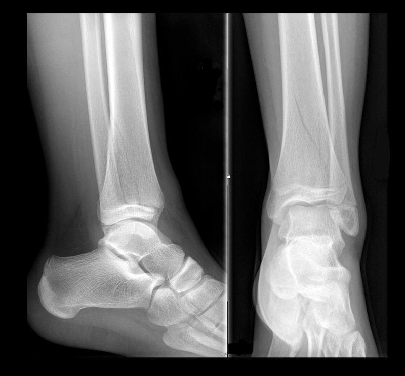
13 year-old female with ankle pain following fall down escalator. What's the diagnosis?


Show Answer
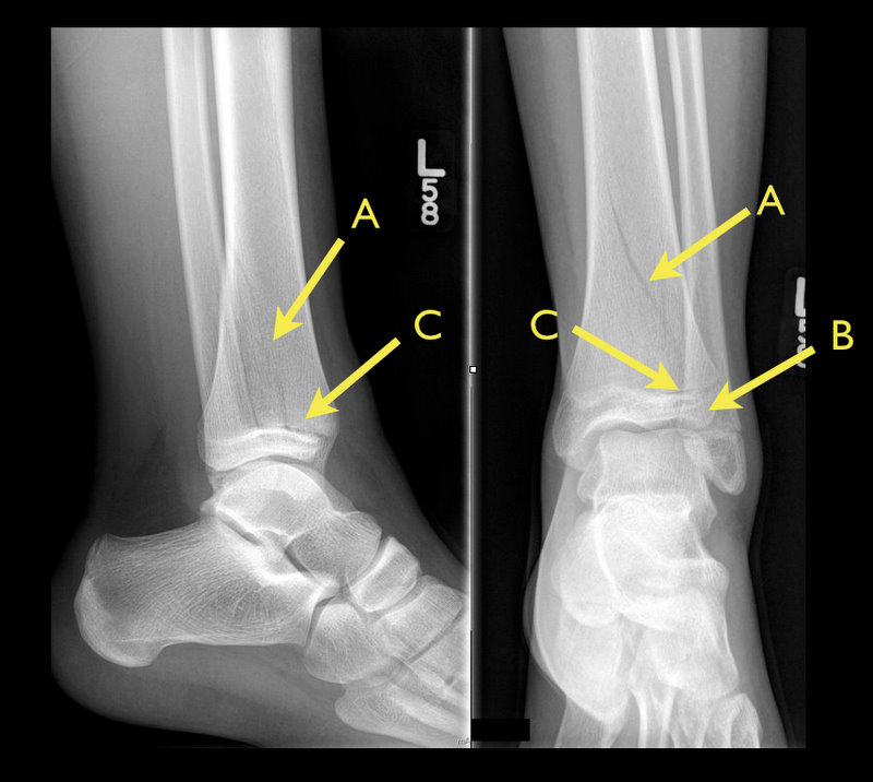
Answer: Tri-plane ankle fracture

Tri-plane Fractures

  • Multi-planar ankle fracture in older children and adolescents during an 18 month window prior to distal tibial-physis closure.
  • 3 planes (see Xray below): coronal (A: tibal metaphysis), sagital (B: epiphysis), and transverse (C: growth plate)
  • Associated fibular spiral fracture (50% cases)
  • Commonly due to external rotational or "twisting" forces.
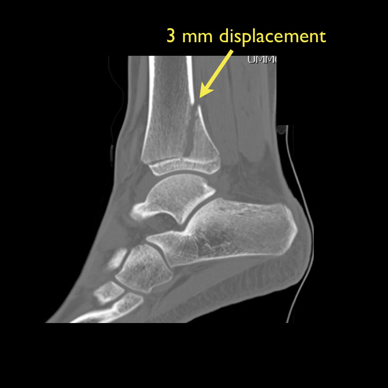
  • Xrays are helpfully, but CT scan may be indicated as fractures may be more displaced than radiographs suggest.
  • Non-operative treatment for non-displaced fractures:
    • Closed reduction with long-leg splint/cast
    • Reduction may slip once swelling has subsided
  • Indications for operative repair:
    • >2mm displacement of fracture segments
    • Intra-articular fracture

Show References

Follow me on Twitter (@criticalcarenow) or Google+ (+criticalcarenow)


Title: Be Bold and Make Inpatients Cold

Category: Critical Care

Posted: 6/4/2013 by Haney Mallemat, MD

Therapeutic hypothermia (TH) following out-of-hospital cardiac arrest (OHCA) has increasingly been utilized since it was first described. TH following in-hospital cardiac arrest (IHCA), on the other hand, is not as commonplace or consistent despite a recommendation by the American Heart Association (AHA).

A recent prospective multi-center cohort-study demonstrated that of 67,498 patients with return of spontaneous circulation (ROSC) following IHCA only 2.0% of patients had TH initiated; of those 44.3% did not even achieve the target temperature (32-34 Celsius). 

The factors found to be most associated with instituting TH were:

  • Younger patients
  • Admission to non-ICU units
  • Arrests occurring Monday through Friday (as compared to weekends)
  • Arrests within teaching hospitals (as compared to non-teaching institutions)

Bottom-line: Hospitals should consider instituting and adhering to local TH protocols for in-house cardiac arrests.

Show References

Mikkelsen, M. et al. Use of Therapeutic Hypothermia After In-Hospital Cardiac Arrest. Crit Care Med 2013 Jun;41(6):1385-1395

Follow me on Twitter (@criticalcarenow) or Google+ (+criticalcarenow)


Title: Here's a Solution, for that Pleural Effusion

Category: Visual Diagnosis

Posted: 6/3/2013 by Haney Mallemat, MD

Ever wonder how to place a pigtail catheter?

Check out this video to learn how, click here

Show References

Follow me on Twitter (@criticalcarenow) or Google+ (+criticalcarenow)


Title: What's the Diagnosis? Case submitted by Bethany Radin

Category: Visual Diagnosis

Posted: 5/27/2013 by Haney Mallemat, MD

Question

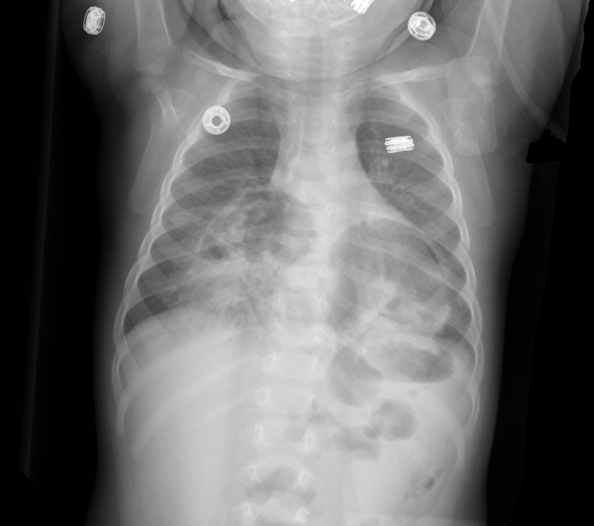
9 month-old presents with wheezing and the CXR is shown below. What's the diagnosis?


Show Answer
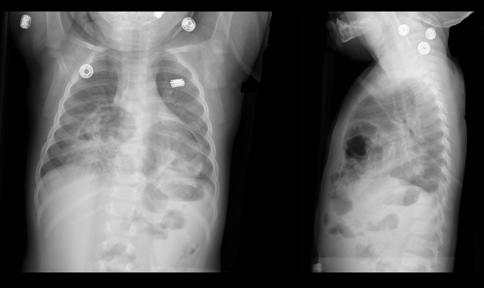
Congenital anterior diaphragmatic defects with herniated bowel.

Show References

Follow me on Twiter (@criticalcarenow) or Google+ (+criticalcarenow)


Title: The Macklin Effect by Dr. Ali Farzad

Category: Critical Care

Posted: 5/21/2013 by Haney Mallemat, MD

The Macklin Effect

Pneumomediastinum (click here for image) may be caused by many things:

  1. Esophageal perforation (e.g., complication from EGD)
  2. Tracheal / Bronchial injury (e.g., trauma, complication of bronchoscopy, etc.)
  3. Abdominal viscus perforation with translocation of air across the diaphragmatic hiatus
  4. Air may reach mediastinum along the fascial planes of the neck.
  5. Alveolar rupture, also known as the "Macklin Effect"

The "Macklin Effect" is typically a self-limiting condition leading to spontaneous pneumomediastinum and massive subcutaneous emphysema after the following:

  1. Alveolar rupture from increased alveolar pressure (e.g., asthma, blunt trauma, positive pressure ventilation, etc.)
  2. Air released from alveoli dissects along broncho-vascular sheaths and enters mediastinum
  3. Air may subsequently track elsewhere (e.g., cervical subcutaneous tissues, face, epidural space, peritoneum, etc.)

Pneumomediastinum secondary to the Macklin effect frequently leads to an extensive workup to search for other causes of mediastinal air. Although, no consensus exists regarding the appropriate workup, the patient's history should guide the workup to avoid unnecessary imaging, needless dietary restriction, unjustified antibiotic administration, and prolonged hospitalization.

Treatment of spontaneous pneumomediastinum includes:

  • Supplemental oxygen and observation for airway obstruction secondary to air expansion within the neck
  • Avoiding positive airway pressure, if possible
  • Avoiding routine chest tubes (unless significant pneumothorax is present)
  • Administering prophylactic antibiotics are typically unnecessary
  • Ordering imaging as needed

Show References

Follow me on Twitter (@criticalcarenow) or Google+ (+criticalcarenow)


Title: What's the Diagnosis? Case submitted by Dr. Ryan Spangler

Category: Visual Diagnosis

Posted: 5/21/2013 by Haney Mallemat, MD

Question

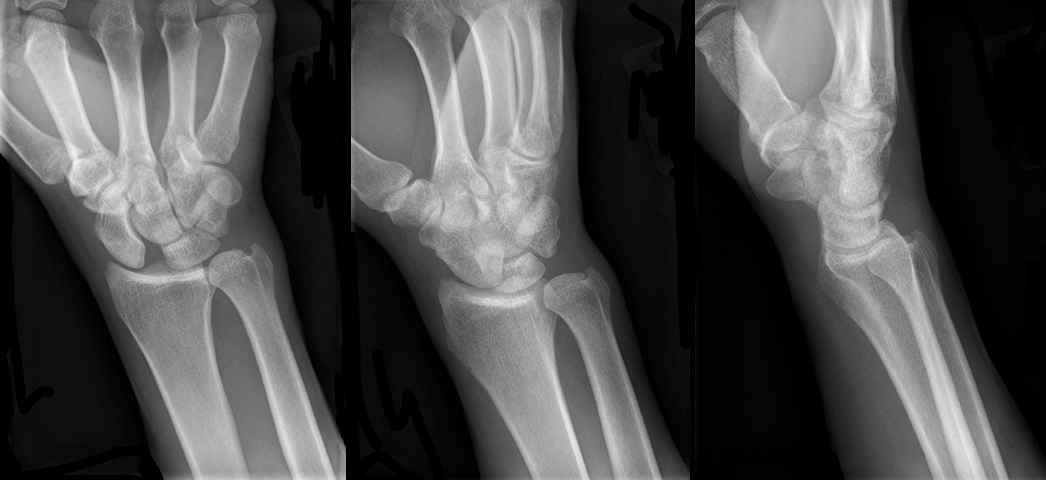
30 year-old male presents with right wrist pain after falling off his bicycle. What's the diagnosis?
Show Answer
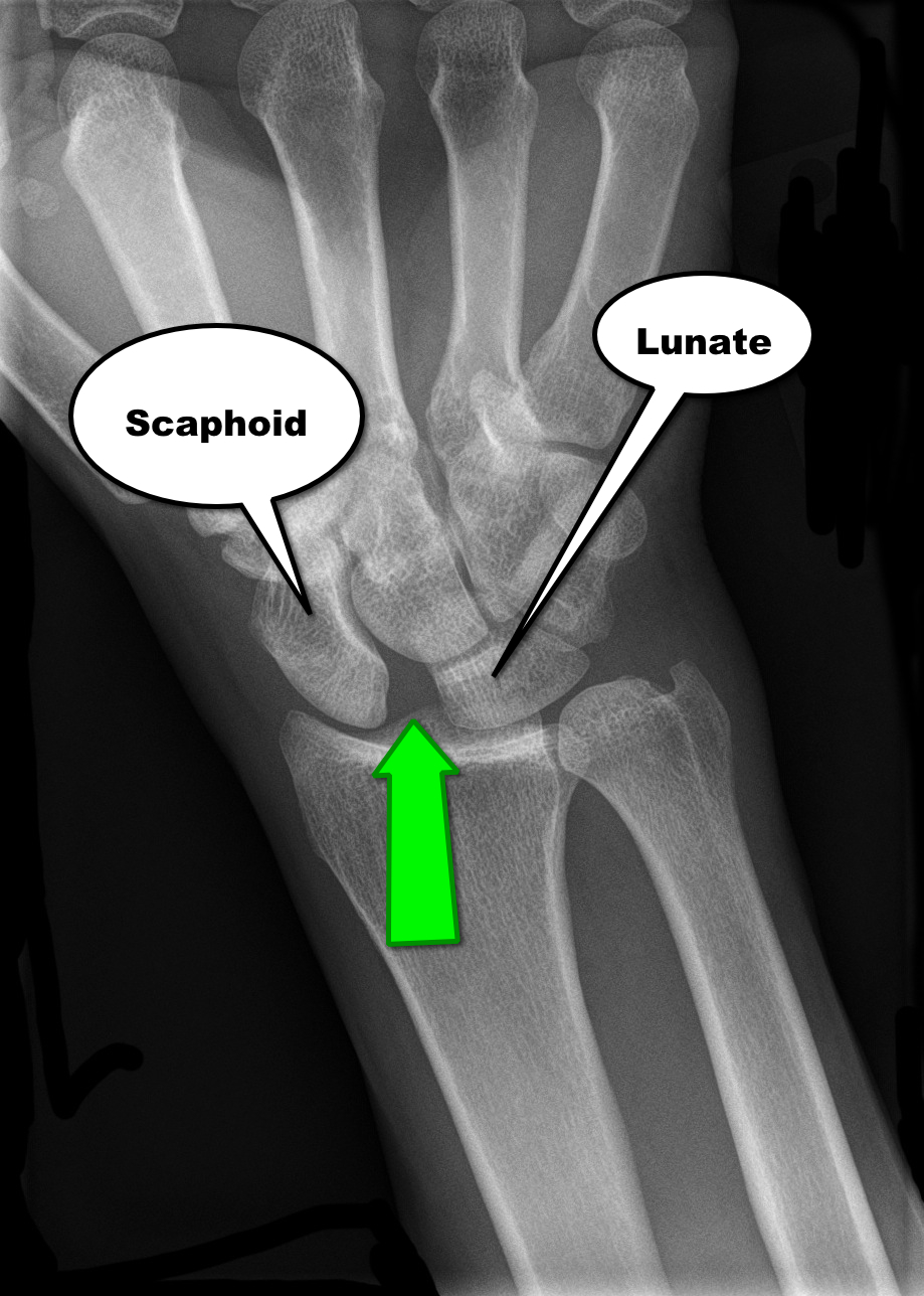

Scapholunate dislocation


Scapholunate Dislocation (a.k.a. “Terry Thomas” or “David Letterman” sign)

  • Increased distance between the scaphoid and lunate bones on AP wrist X-ray; the distance between the two bones should normally be less than 2mm.
  • A widened scapholunate distance suggests ligamentous injury or tear (green arrow below).
  • The widened distance resembles the famous dental diastema (i.e., tooth gap) of Terry Thomas (British comedian) and David Letterman


Show References

Follow me on Twitter (@criticalcarenow) or Google+ (+criticalcarenow)


Title: What's the Diagnosis? Case submitted by Dr. Ali Farzad

Category: Visual Diagnosis

Posted: 5/13/2013 by Haney Mallemat, MD

Question

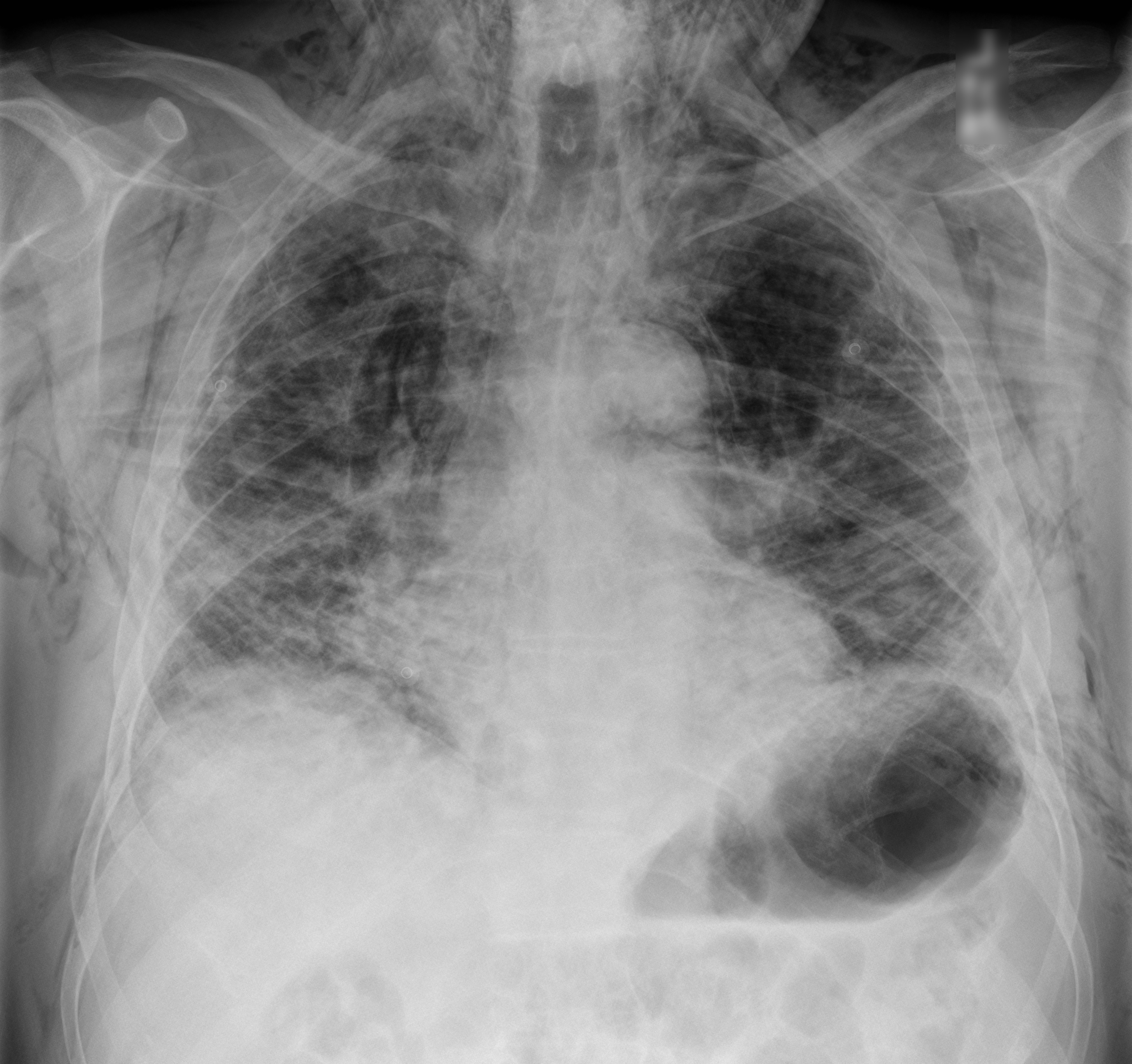
60 year-old male with a history of pulmonary fibrosis presents to the Emergency Department after a lung biopsy. He is complaining of facial swelling and dyspnea. What's the diagnosis?
Show Answer

Answer: Massive subcutaneous emphysema

Click here

Show References

Follow me on Twitter (@criticalcarenow) or Google+ (+criticalcarenow)


Title: Can your breath, predict fluid responsiveness best?

Category: Critical Care

Posted: 5/7/2013 by Haney Mallemat, MD

Fluid boluses are often administered to patients in shock as a first-line intervention to increase cardiac output. Previous literature states, however, that only 50% of patients in shock will respond to a fluid bolus. 

Several validated techniques exist to distinguish which patients will respond to a fluid bolus and which will not; one method is the passive leg raise (PLR) maneuver  (more on PLR here). A drawback to PLR is that it requires direct measurement of cardiac output, either by invasive hemodynamic monitoring or using advanced bedside ultrasound techniques.

Another technique to quantify changes in cardiac output is through measurement of end-tidal CO2 (ETCO2). The benefits of measuring ETCO2 is that it can be continuously measured and can be performed non-invasively on mechanically ventilated patients.

A 5% or greater increase in end-tidal CO2 (ETCO2) following a PLR maneuver has been found to be a good predictor of fluid responsiveness with reliability similar to invasive measures.


Show References

Monnet, X. et al. End-tidal carbon dioxide is better than arterial pressure for predicting volume responsiveness by the passive leg raising test. Intensive Care Med. 2013 Jan;39(1):93-100.

Follow me on Twitter (@criticalcarenow) or Google+ (+criticalcarenow)


Title: What's the Diagnosis? Case by Dr. Siamak Moayedi

Category: Visual Diagnosis

Posted: 5/6/2013 by Haney Mallemat, MD (Updated: 5/6/2013)

Question

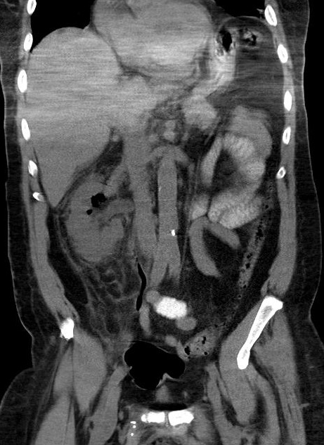
38 year-old male with a past medical history of diabetes presents with back pain and hypotension. CT scan is shown below. What's the diagnosis?
 

Show Answer
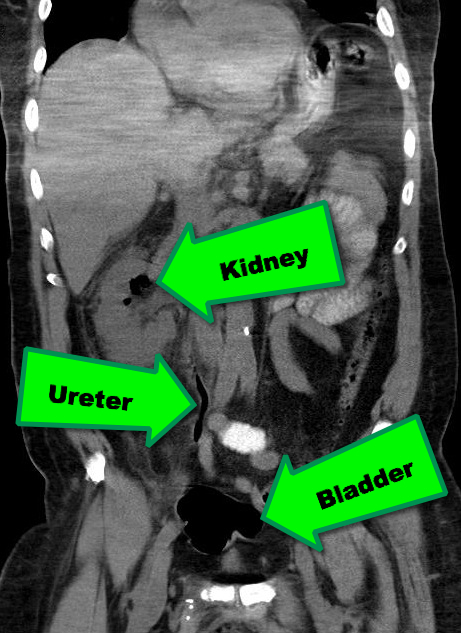
Emphysematous Pyelonephritis (EPN)

EPN is a necrotizing infection of the renal and peri-nephric tissues with accumulation of gas within the tissues and collecting system of the kidney; it is rapidly fatal if not recognized and aggressively treated early.

Risk factors include an immunocompromised host; Diabetics are at particularly increased risk.

The most common etiologic organisms are Escherichia coli, Klebsiella pneumoniae, Proteus mirabilis, and Pseudomonas aeruginosa. Infection with anaerobic species is relatively uncommon.

In addition to the classic signs / symptoms of pyelonephritis, (e.g., flank pain, fever, pyuria, etc.), patients presenting with EPN may experience the passage of air in the urine (pneumaturia) or crepitus over their flank.

Abdominal CT scan is the diagnostic test of choice when clinical presentation and laboratory tests are suggestive (see below)

Treatment should include:

  • Aggressive resuscitation with fluids and vasopressors, as necessary
  • Broad-spectrum antibiotics
  • Percutaneous ureteral drain or stent placement, if obstruction is present

Show References

Follow me on Twitter (@criticalcarenow) or Google+(criticalcarenow)


Title: What's the Diagnosis?

Category: Visual Diagnosis

Posted: 4/29/2013 by Haney Mallemat, MD

Question

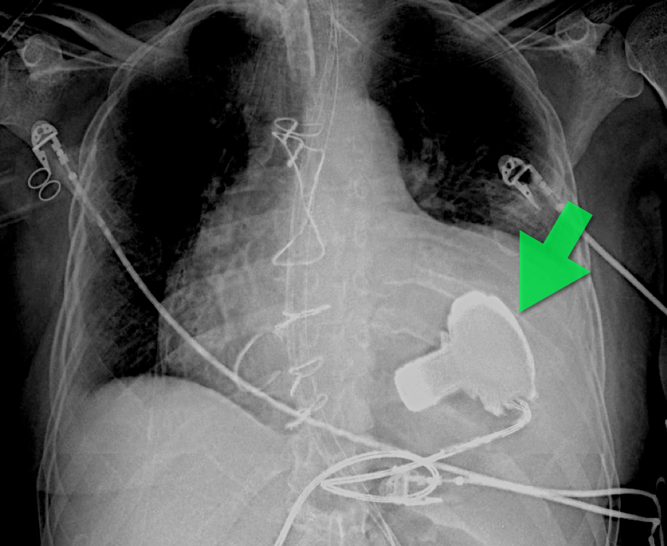
57 year old male presents with a cough. The CXR is shown below. What's the diagnosis?
Show Answer

Answer: Left Ventricular Assist Device (LVAD) 

An LVAD is a mechanical pump that pulls blood from the left ventricle and pumps it to the ascending aorta for systemic delivery; LVADs are increasingly being placed in patients with heart failure so you must be aware of them.

The three indications for an LVAD are:

  • Bridge to cardiac transplant
  • Temporizing measure when a reversible cardiac disease (e.g., myocarditis) exists
  • Destination therapy; patients ineligible for cardiac transplant who would otherwise die from heart failure

There are several models, which have been implanted in people (http://www.mylvad.com/lvad-devices). The main distinction between devices are pulsatile or non-pulsatile (or linear) flow; newer generation devices are non-pulsatile.

Patients with LVAD may develop complications such as infections, arrhythmias, or anemia, but bleeding can be a life-threatening one; bleeding occurs secondary to an acquired von Willebrand factor deficiency, AVMs within the GI tract, or secondary to over anti-coagulation (typically with Warfarin).

Show References

Follow me on Twitter (@criticalcarenow) or Google+ (+criticalcarenow)


Title: Necrotizing Fasciitis

Category: Critical Care

Posted: 4/23/2013 by Haney Mallemat, MD

Necrotizing fasciitis (NF) is a rapidly progressive bacterial infection of the fascia with secondary necrosis of the subcutaneous tissue. In severe cases, the underlying muscle (i.e., myositis) may be affected.

Risk factors for NF include immunosuppression (e.g., transplant patients), HIV/AIDS, diabetes, etc.

There are three categories of NF:

  • Type I (poly-microbial infections)
  • Type II (Group A streptococcus; sometimes referred to as the “flesh-eating bacteria)
  • Type III (Clostridial myonecrosis; known as gas gangrene)

In the early stage of disease, diagnosis may be difficult; the physical exam sometimes does not reflect the severity of disease. Labs may be non-specific, but CT or MRI is important to diagnose and define the extent of the disease when planning surgical debridement.

Treatment should be aggressive and started as soon as the disease is suspected; this includes:

  • Aggressive fluid and/or vasopressor therapy
  • Broad spectrum antibiotics covering for gram-positive, gram-negative, and anaerobic bacteria; clindamycin should be added initially as it suppresses certain bacterial toxin formation
  • Emergent surgical consult for debridement
  • Once the patient is stable, other treatments may include intravenous immunoglobulin and hyperbaric oxygen therapy


Show References

Follow me on Twitter (@criticalcarenow) or Google+ (+criticalcarenow)


Title: What's the Diagnosis?

Category: Visual Diagnosis

Posted: 4/17/2013 by Haney Mallemat, MD

Question

35 year-old female presents with fever and hypotension. Bedside ultrasound is performed and is shown here. What's the diagnosis? 

Show Answer

Answer: Aortic valve vegetation secondary to infective endocarditis. 

Show References

Follow me on Twitter (@criticalcarenow) or Google+ (+criticalcarenow)


Title: Adrenal Insufficiency

Category: Critical Care

Posted: 4/12/2013 by Haney Mallemat, MD (Updated: 7/21/2026)

Adrenal insufficiency (AI) can be a life-threating condition and is classified as primary (failure of the adrenal gland) or secondary (failure of hypothalamic- pituitary axis).

Common causes of primary adrenal insufficiency include autoimmune destruction, infectious causes (TB and CMV), or interactions with drugs (e.g., anti-fungals, Etomidate, etc.). Secondary causes are usually due to abrupt withdrawal of steroids after chronic use, although sepsis and diseases of the hypothalamus or pituitary (e.g., CVA) may occur.

Signs and symptoms include fatigue, weakness, skin pigmentation, dizziness, abdominal pain, and orthostatic hypotension; it should be suspected with any of the following: hyponatremia, hyperkalemia, hypoglycemia, hypercalcemia, low free-cortisol level, and hemodynamic instability despite resuscitation.

Treatment:
• Correct underlying the disorder
• Resuscitation and hemodynamic support
• Correct hypoglycemia and electrolyte abnormalities
• Treat with hydrocortisone, cortisone, prednisone, or dexamethasone +/- fludrocortisone (Note: dexamethasone is attractive choice in the ED because it will not interfere with ACTH stimulation test)


Show References


Neary, N and Nieman, L. Adrenal Insufficiency: Etiology, diagnosis and treatment. Curr Opin Endocrinol Diabetes Obes. 2010 Jun;17(3):217-23.


Title: What s the Diagnosis? Case by Dr. Zachary Dezman

Category: Visual Diagnosis

Posted: 4/8/2013 by Haney Mallemat, MD

Question

64 year-old female presents with chest pain following an argument with her husband. Her echocardiogram (apical four-chamber view) and ECG are shown. Her initial troponin is 10. What's the diagnosis?


Show Answer

Answer: Takotsubo (a.k.a Stress) cardiomyopathy (TC)

TC is left ventricular dysfunction (hypokinesia, akinesia, or dyskinesia) secondary to catecholamine surge (e.g., physical / emotional stress) causing myocardial stunning; it is not due to acute coronary occlusion. TC disproportionately affects postmenopausal women, occurring in up to 90% of cases.

Patients often present with chest pain or dyspnea; 85% of patients will have an abnormal ECG (e.g., ST elevation or T wave inversions), making diagnosis difficult to differentiate between TC and acute coronary syndrome (ACS). TC has been found to be the diagnosis in 2.5% of patients initially worked up for ACS.

Diagnosis can usually be confirmed with echocardiography but cardiac catheterization (with ventriculogram) is sometimes performed if ACS is strongly suspected. Catheterization demonstrates normal coronary arteries with an abnormal ventriculogram (click for video), typically in mid-to apical portion of the left ventricle.

Treatment is symptomatic and similar to congestive heart failure (e.g., diuretics, beta-blockers, etc.); 95% of patients have full recovery within one month. 

Refer to this prior PEARL by Dr. Tewelde for additional information


Reference

T Pilgrima, T Wyss, Takotsubo cardiomyopathy or transient left ventricular apical ballooning syndrome: A systematic review, Int J Card 2008 Mar 14;124(3):283-92

Show References

Follow me on Twitter (@criticalcarenow) or Google+ (+criticalcarenow)


Title: What's the Diagnosis?

Category: Visual Diagnosis

Posted: 4/1/2013 by Haney Mallemat, MD

Question

What's the Diagnosis?

Show Answer

Answer: Normal CXR...April Fool's Day ;) 

Our international friends can learn more about April Fool'sDay here

Show References

Follow me on Twitter (@criticalcarenow) or Google+ (+criticalcarenow)


Title: "D.O.P.E.S. like D.O.T.T.S."

Category: Critical Care

Posted: 3/26/2013 by Haney Mallemat, MD

There are several reasons why a mechanically ventilated patient may decompensate post-intubation. Immediate action is often needed to reverse the problem, but it can be difficult to remember where to start as the vent alarm is sounding and the patient is decompensating.

Consider using the mnemonic “D.O.P.E.S. like D.O.T.T.S.” to assist you in first diagnosing the problem (D.O.P.E.S.) and then fixing the problem (D.O.T.T.S.). You can view an entire lecture on the Crashing Ventilated Patient here.

Step 1: Could this decompensation be secondary to D.O.P.E.S.?

  • Displaced ET tube / ET tube cuff not inflated or has a leak
  • Obstruction of ET tube
  • Pneumothorax
  • Equipment malfunction (disconnection of the ventilator, incorrect vent settings, etc.)
  • Stacking (breath stacking / Auto- PEEP; click here for a review)

Step 2: Fix the problem with D.O.T.T.S.

  • Disconnect – Disconnect patient from the ventilator
  • Oxygen – Oxygenate patient with a BVM and feel for resistance as you bag
  • Tube position / function – Did the ET tube migrate? Is it kinked or is there a mucus plug?
  • Tweak the vent – Are the settings correct for this patient?
  • Sonogram (ultrasound) – Sonogram to look for pneumothorax, mainstem intubation, etc. 

Show References

Follow me on Twitter (@criticalcarenow) or Google+ (+criticalcarenow)


Title: What's the Diagnosis? Case by Dr. Zachary Dezman and Dr. Phillip Stafford

Category: Visual Diagnosis

Posted: 3/25/2013 by Haney Mallemat, MD

Question

35 year-old male presents after a motor vehicle crash. No blood seen at the meatus of the penis and a Foley catheter is placed (see photo below). What's the next diagnostic step?

Show Answer

Answer: Retrograde cystogram

Traumatic bladder rupture

Gross or microscopic hematuria (>50 RBCs per high-power field) following blunt trauma requires a retrograde cystogram to evaluate for bladder injury. If blood is present at the meatus, however, urethral injury (not discussed) should be suspected and retrograde urethrogram should be performed before passing a Foley catheter.

A retrograde cystogram is performed by infusing diluted contrast into the bladder (200-400 mL) to gently distend the bladder and allow visualization of potential rupture(s). A CT scan of the abdomen and pelvis is then performed to determine if any contrast has leaked from the bladder; alternatively an AP pelvis can also be used, but is not as sensitive as CT.

Traumatic bladder ruptures are categorized as either intra-peritoneal or extra-peritoneal (note: X-ray below demonstrates extra-peritoneal rupture).

Treatment for bladder rupture:

  • Extra-peritoneal bladder rupture typically requires only 1-3 weeks of Foley catheter; the catheter can be removed after a successful voiding trial.  
  • Intra-peritoneal bladder rupture requires an exploratory laparotomy because this injury does not heal on its own. Urine draining into the abdominal cavity may cause complications such as urinary ascites and electrolyte imbalances.  

Prognosis is typically good for either injury.

Show References

Follow me on Twitter (@criticalcarenow) or Google+ (+criticalcarenow)


Title: What's the Diagnosis? Case written by Dr. Zachary Dezman

Category: Visual Diagnosis

Posted: 3/19/2013 by Haney Mallemat, MD

Question

A 56-year-old woman with a history of psoriasis presents with fever, nausea, and painful pin-point pustules on an erythematous base. Her dermatologist recently reduced her prednisone dose. What's the diagnosis?

Show Answer

Answer: Pustular psoriasis

Pustular psoriasis

Occurs in patients with psoriasis, classically occurring after a decrease in dose or cessation of systemic steroids

Symptoms include:

  • Acute eruption of painful small pustules (2-3 mm in diameter) that rapidly become confluent into large “lakes of pus”
  • Systemic symptoms may also occur (e.g., fever, malaise, nausea, or arthritis).

These lesions can eventually drain and desquamate, leaving large patches of exposed dermis.

Complications include:

  • Super-infection (most common)
  • ARDS
  • AKI
  • Hypothermia secondary to loss of epidermis
  • Hypovolemia secondary to fluid losses from the skin

Patients may be admitted for supportive care and treatment with disease-modifying antirheumatic drugs (DMARDs) such as cyclophosphamide or methotrexate.


REFERENCE

L Naldi, D Gambini, The clinical spectrum of psoriasis, Clinics in Dermatology, Volume 25, Issue 6, November–December 2007, Pages 510-518

Show References

Follow me on Twitter (@criticalcarenow) or Google+ (+criticalcarenow)


Title: Don't Fall Asleep on Auto-PEEP

Category: Critical Care

Posted: 3/12/2013 by Haney Mallemat, MD

Mechanically ventilated patients can develop a condition in which air becomes trapped within the alveoli at end-expiration; this is called auto-PEEP.

Auto-peep has several adverse effects:

  • Barotrauma from positive pressure trapped within the alveoli 
  • Increased work of breathing
  • Worsening pulmonary gas exchange
  • Hemodynamic compromise secondary to increased intra-thoraic pressure

Auto-PEEP classically occurs in intubated patients with asthma or emphysema, but it may also occur in the absence of such disease. The risk of auto-PEEP is increased in patients with:

  • Short expiration times (i.e., inadequate time for the evacuation of alveolar air at end-expiration)
  • Bronchoconstriction
  • Plugging of the bronchi (e.g., mucus or foreign body) creating a one-way valve and air-trapping

Auto-PEEP may be treated by:

  • Reducing tidal volume
  • Reducing the respiratory rate
  • Decreasing inspiratory time
  • Increasing PEEP

Patients may need to be heavily sedated to accomplish the above ventilator maneuvers.

Show References

Follow me on Twtitter (@criticalcarenow) or Google+ (+criticalcarenow)


Previous  |  1 |  ... |  4 |  5 |  6 |  7 |  8 |  9 |  10 |  11 |  12 |  13 |  14 |  ... |  17 |  Next
University of Maryland School of Medicine logo
  • Departments
  • Programs
  • Research Centers
  • Institutes
  • Graduate Education
  • CIBR Cores
  • Offices of the Dean
    • University of Maryland, Baltimore
    • University of Maryland Medical Center
    • Medical Alumni Association of the University of Maryland
    • Health Sciences and Human Services Library
University of Maryland School of Medicine
655 W. Baltimore Street
Baltimore, MD 21201
Contact Us

© University of Maryland School of Medicine

    • Webmaster
    • Web Accessibility
    • Site Index
    • UMB Hotline
    • Employment
    • Map