
University of Maryland School of Medicine logo
Emergency Medicine
All Departments
UMSOM Assistant
UMEM Educational Pearls
  • Home
  • Departments
  • Emergency Medicine
  • CME
  • UMEM Educational Pearls
  • CME
  • Courses and Conferences
  • Free Open Access Medical Education
  • UMEM Educational Pearls

Search

101-120 of 321 results by Haney Mallemat

Previous  |  1 |  2 |  3 |  4 |  5 |  6 |  7 |  8 |  9 |  10 |  11 |  ... |  17 |  Next

Title: What's the Diagnosis? Case by Dr. Ashley Strobel

Category: Visual Diagnosis

Posted: 5/19/2014 by Haney Mallemat, MD

Question

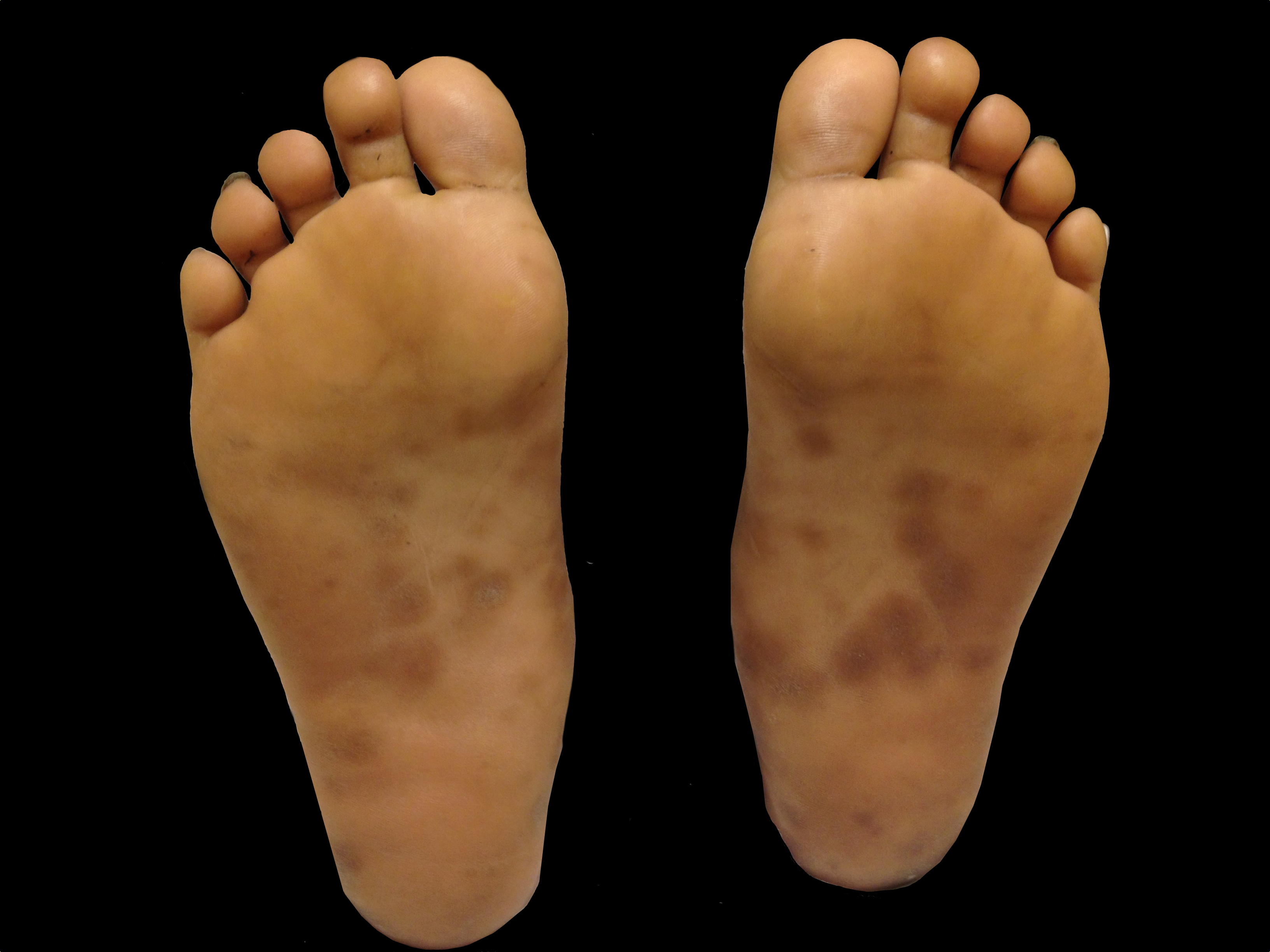
A patient presents with the rash shown below and is treated with penicillin. Fever, headache, and myalgia develop four hours later. What’s the diagnosis?
Show Answer

Answer: Jarisch-Herxheimer reaction

  • Reaction observed 2-8 hours following antibiotic treatment of syphilis as a result of endotoxin released from dying from organisms; it is not an allergy to penicillin, nor a dose related phenomenon.
  • Symptoms may include:
    • Fever / chills / rigors
    • Headache
    • Flushing / Vasodilation
    • Worsening rash
    • Hypotension
  • Can occur when following treatment of any stage of syphilis although most commonly occurs following treatment of primary syphilis; also with other illnesses caused by spirochetes (Lyme, Leptospirosis, etc.)
  • Reaction is typically self-limiting and treatment is supportive care

Show References

Follow me on Twitter (@criticalcarenow) or Google+ (+criticalcarenow)


Title: Above or Below the Clavicle? What's the Best Approach for Ultrasound Cannulation

Category: Critical Care

Posted: 5/13/2014 by Haney Mallemat, MD

Prior literature has demonstrated the safety and feasibility of placing subclavian lines with ultrasound guidance; here's a link to a short educational video describing the technique. 

The literature has been varied, however, as to which approach is best for venous cannulation with ultrasound; the supraclavicular (SC) or infraclavicular (IC) approach (see references below)

A recent study evaluated both approaches in healthy volunteers in order to determine which approach is superior for cannulation using ultrasound.

98 patients were prospective evaluated by Emergency Medicine physicians with training in ultrasound. In each patient, both SC and IC views were evaluated on both the left and right sides; each view was given a grade for ease of favorability (no patients were actually cannulated)

Overall, it was found that the SC view was significantly more favorable compared to the IC view; the right SC was non-significantly preferred compared to the left SC.

Show References

Follow me on Twitter (@criticalcarenow) or Google+ (+criticalcarenow)

Stachura, M. et al. A Comparison of the Supraclavicular and Infraclavicular Views for Imaging the Subclavian Vein with Ultrasound. The American Journal of Emergency Medicine (in press) 

Fragou, M., Gravvanis, A., and Vasilios, D. Real-time ultrasound-guided subclavian vein cannulation versus the landmark method in critical care patients: A prospective randomized study. Crit Care Med. 2011; 39: 1607–1612

Mallin, M., Louis, H., and Madsen, T. A novel technique for ultrasound-guided supraclavicular subclavian cannulation. Am J Emerg Med. 2010; 28: 966–969

Czarnik, T., Gawda, R., Perkowski, T. et al. Supraclavicular approach is an easy and safe method of subclavian vein catheterization even in mechanically ventilated patients. analysis of 370 attempts. Anesthesiology. 2009; 111:334–339


Title: What's the Diagnosis?

Category: Visual Diagnosis

Posted: 5/5/2014 by Haney Mallemat, MD

Question

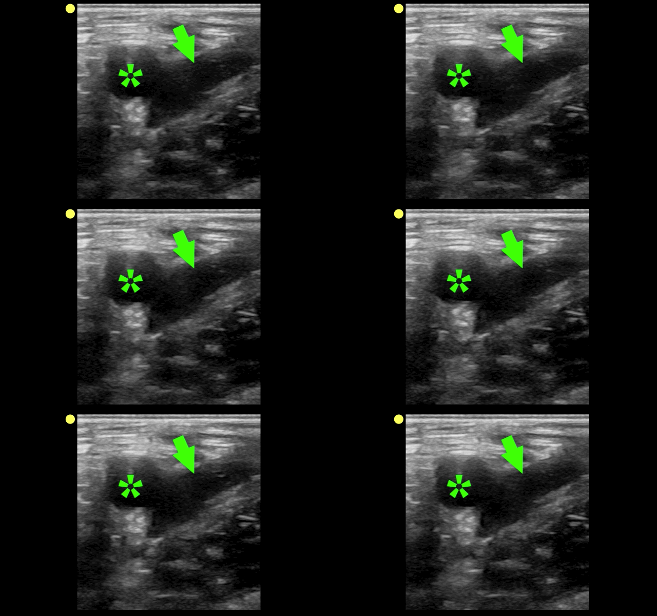
The clip below demonstrates normal right femoral anatomy. The structure with the asterisk is the right common femoral vein and the arrow is pointing to a branch of the right femoral vein. What is the name of the branch and what is its importance during lower extremity ultrasound?
Show Answer

Answer: Greater Saphenous Vein; it is one of the two regions that should be compressed when evaluating for a lower extremity DVT in the Emergency Department (the other is at the trifurcation of the popliteal vein).

Here is a podcast from the Ultrasound Podcast describing the entire bedisde DVT exam http://www.ultrasoundpodcast.com/2011/08/dvt/

Show References

Follow me on Twitter (@criticalcarenow) or Google+ (+criticalcarenow)a


Title: What's the Diagnosis? Case by Dr. Christina Tupe

Category: Visual Diagnosis

Posted: 4/28/2014 by Haney Mallemat, MD

Question

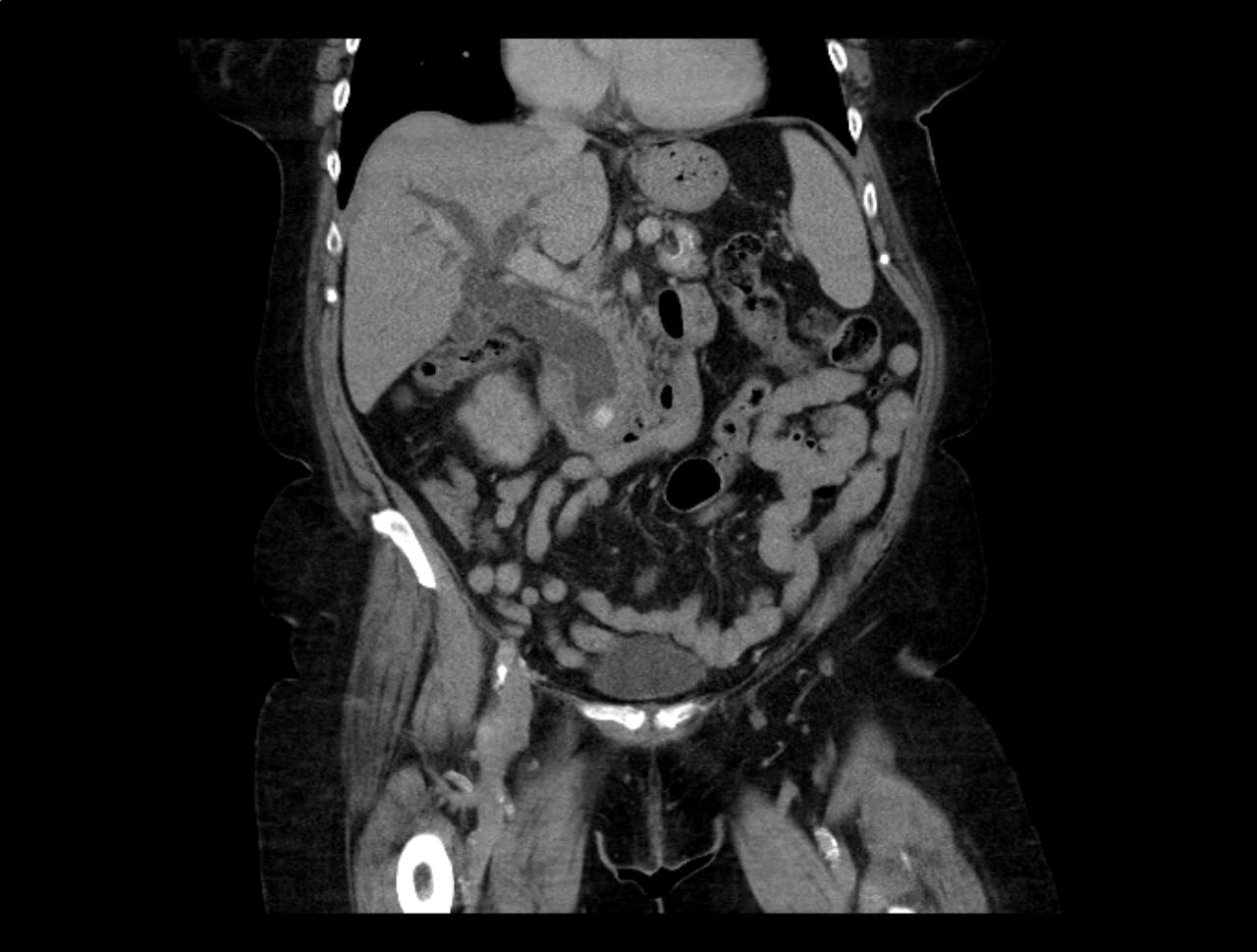
66 year-old female presents with one week of epigastric and right flank pain. Urinalysis was normal. What’s the diagnosis?
Show Answer
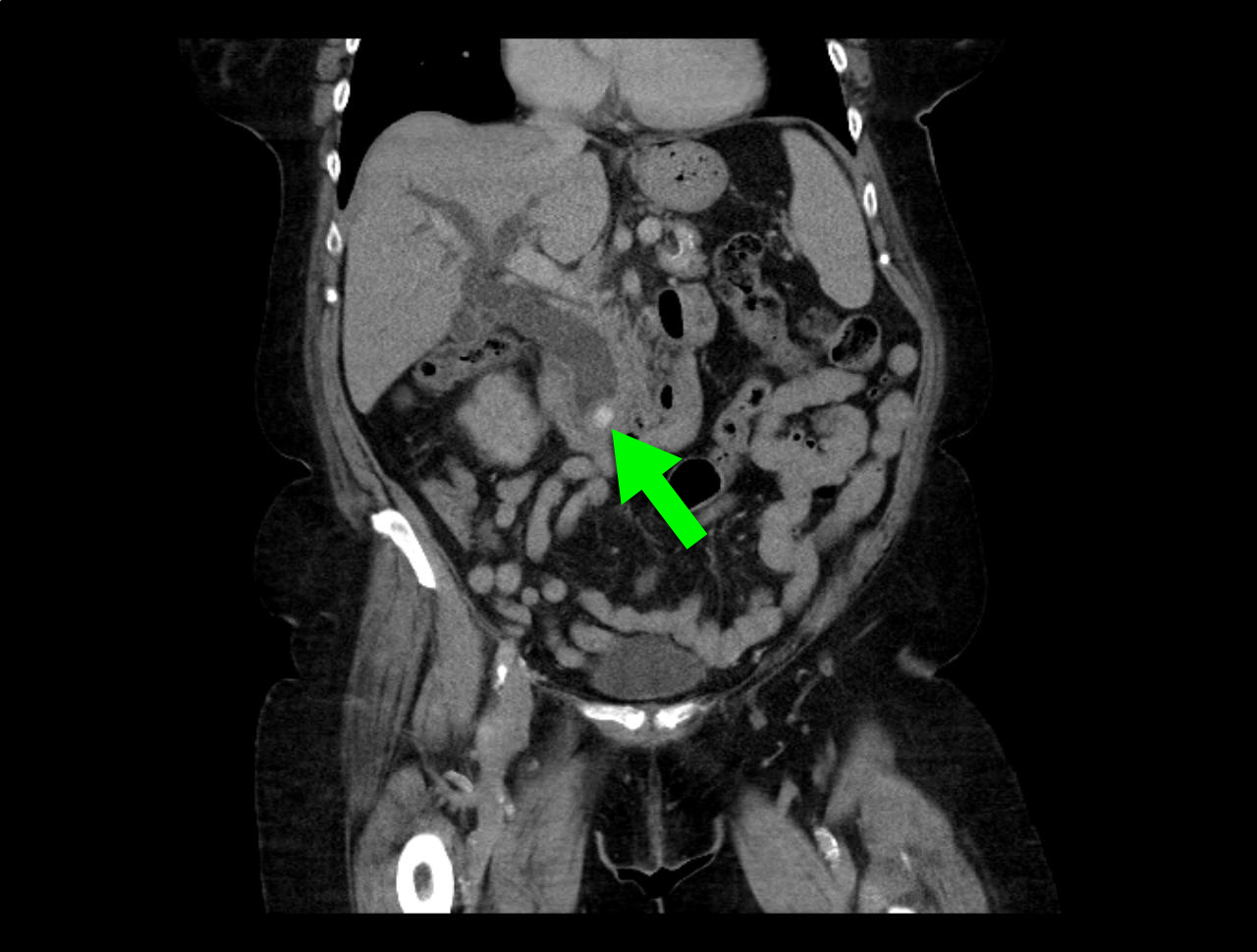
Answer: Choledocholithiasis with pancreatitis secondary to 2.2 cm common bile duct stone

Show References

Follow me on Twitter (@criticalcarenow) or Google+ (+criticalcarenow)


Title: Does My Patient Need More Fluids?

Category: Critical Care

Posted: 4/15/2014 by Haney Mallemat, MD

  • Only 50% of hemodynamically unstable patients will improve their hemodynamics in response to a fluid bolus. However, because excessive fluid administration can lead to organ edema and dysfunction, it is important to give hemodynamically unstable patients only the necessary amount of fluids to improve their hemodynamics.

  • There are two general categories of assessing a patient's response to volume administration; static and dynamic assessments (see referenced article below):

    • Static assessment (generally unreliable, but traditionally used):

      • Physical exam (dry mucus membranes, cool extremities, etc.)

      • Urine output

      • Blood pressure

      • Central venous pressure via central-line

    • Dynamic assessment (more reliable but more labor intensive)

      • Pulse Pressure Variation

      • IVC Distensibility Index

      • End-expiratory occlusion test

      • Passive Leg-Raise

  • There is no simple way to accurately determine the need for a fluid bolus however the integration of the techniques above can help the clinician make better decisions.

Show References

Napoli,A.Physiologic and Clinical Principles behind Noninvasive Resuscitation Techniques and Cardiac Output Monitoring. Cardiol Res Pract. 2012

Follow me on Twitter (@criticalcarenow) or Google+ (+criticalcarenow)


Title: What's theDiagnosis? Images by Dr. Ari Kestler

Category: Visual Diagnosis

Posted: 4/14/2014 by Haney Mallemat, MD

Question

25 year-old female (G1P1) presents with 3 weeks of vaginal bleeding. Her serum beta-HCG is 65,000. Her bedside ultrasound is below; what's the diagnosis? 
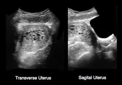
Show Answer
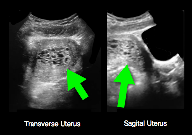
Answer: Hydatidiform mole (molar pregnancy)

Show References

Follow me on Twitter (@criticalcarenow) or Google+ (+criticalcarenow)


Title: What's the Diagnosis? Case by Dr. Ali Farzad

Category: Visual Diagnosis

Posted: 4/7/2014 by Haney Mallemat, MD

Question

23 year-old female presents complaining of progressive right lower quadrant pain after doing "vigorous" pushups. CT abdomen/pelvis below. What’s the diagnosis? (Hint: it’s not appendicitis)
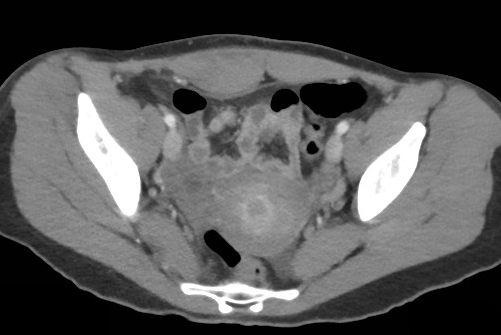
 

Show Answer
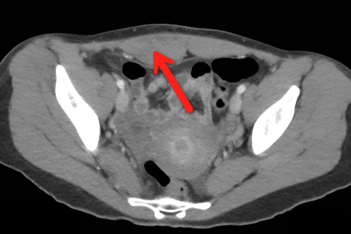
Answer: Rectus sheath hematoma

Rectus Sheath Hematoma (RSH)

Rectus muscle tear causing damage to the superior or inferior epigastric arteries with subsequent bleeding into the rectus sheath; uncommon cause of abdominal pain but mimics almost any abdominal condition.

May occur spontaneously, but suspect with the following risk factors:

  • Coagulopathy (#1 cause); acquired (e.g., warfarin) or inherited disorder
  • Rectus muscle trauma
  • Vigorous or sudden contraction of rectus muscle
  • Increased intra-abdominal pressure from vigorous coughingPregnancy (gestation, labor, or post-partum)

Typically a self-limiting condition, but hypovolemic shock may result from significant hematoma expansion.

  • Hemodynamically stable (non-expanding hematoma): conservative treatment (rest, analgesia, and ice)
  • Hemodynamically unstable (expanding hematoma): treat with fluid resuscitation, reversal of coagulopathy, and transfusion of blood products.

Show References

Follow me on Twitter (@criticalcarenow) or Google+ (+criticalcarenow)


Title: What's the Diagnosis? Case by Dr. Ali Farzad

Category: Visual Diagnosis

Posted: 4/7/2014 by Haney Mallemat, MD (Updated: 7/21/2026)

Question

23 year-old female presents complaining of progressive right lower quadrant pain after doing "vigorous" pushups. CT abdomen/pelvis below. What’s the diagnosis? (Hint: it’s not appendicitis)


Show Answer

Answer: Rectus sheath hematoma

Rectus Sheath Hematoma (RSH)

Rectus muscle tear causing damage to the superior or inferior epigastric arteries with subsequent bleeding into the rectus sheath; uncommon cause of abdominal pain but mimics almost any abdominal condition.

May occur spontaneously, but suspect with the following risk factors:

  • Coagulopathy (#1 cause); acquired (e.g., warfarin) or inherited disorder
  • Rectus muscle trauma
  • Vigorous or sudden contraction of rectus muscle
  • Increased intra-abdominal pressure from vigorous coughingPregnancy (gestation, labor, or post-partum)

Typically a self-limiting condition, but hypovolemic shock may result from significant hematoma expansion.

  • Hemodynamically stable (non-expanding hematoma): conservative treatment (rest, analgesia, and ice)
  • Hemodynamically unstable (expanding hematoma): treat with fluid resuscitation, reversal of coagulopathy, and transfusion of blood products.


Title: What's the Diagnosis? Image by Dr. Ashley Strobel

Category: Visual Diagnosis

Posted: 3/31/2014 by Haney Mallemat, MD (Updated: 4/1/2014)

Question

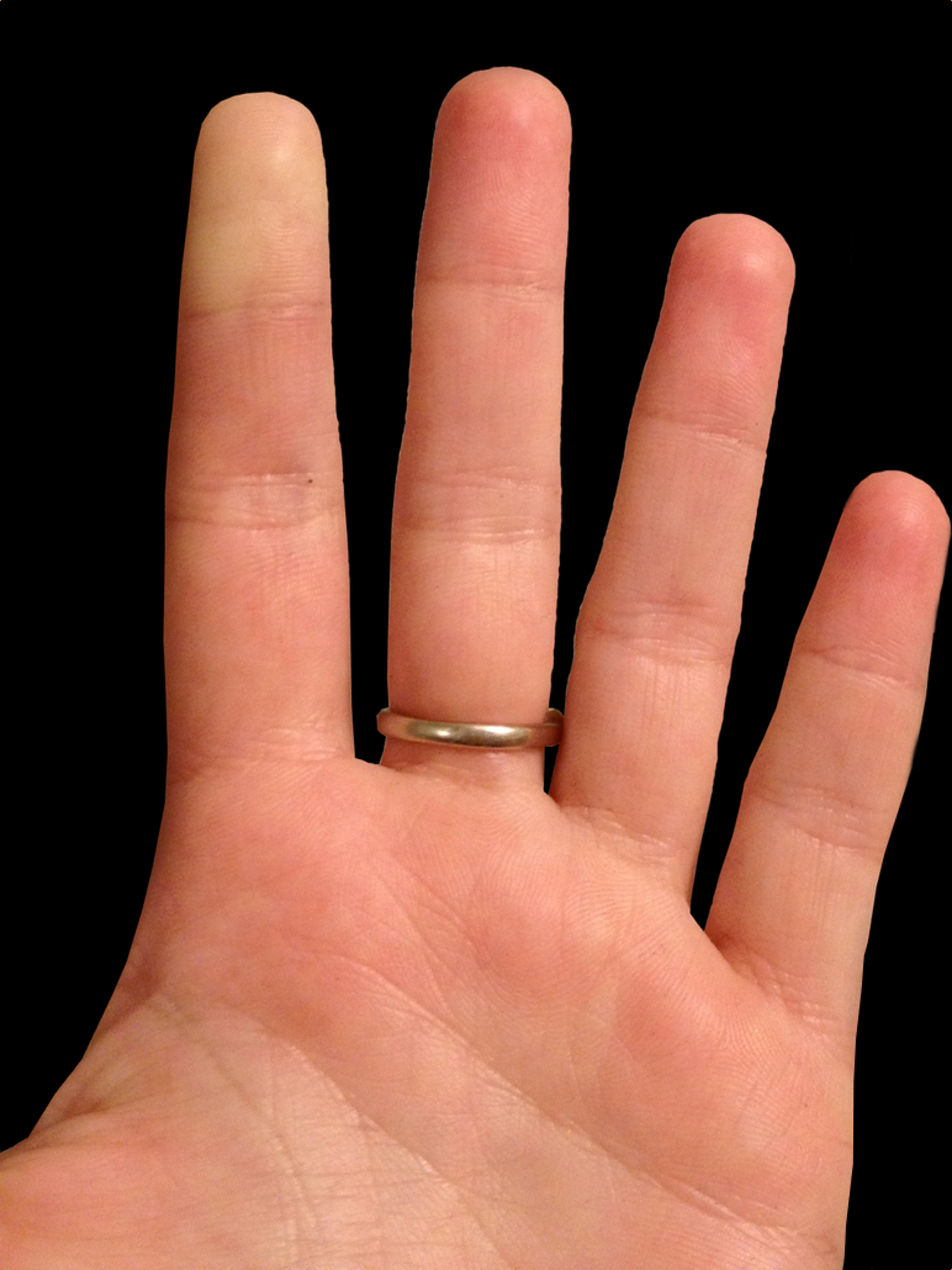
25 year-old female presents with the following. It seems to have occurred spontaneously and spontaneously resolves during her ED evaluation.
 

Show Answer
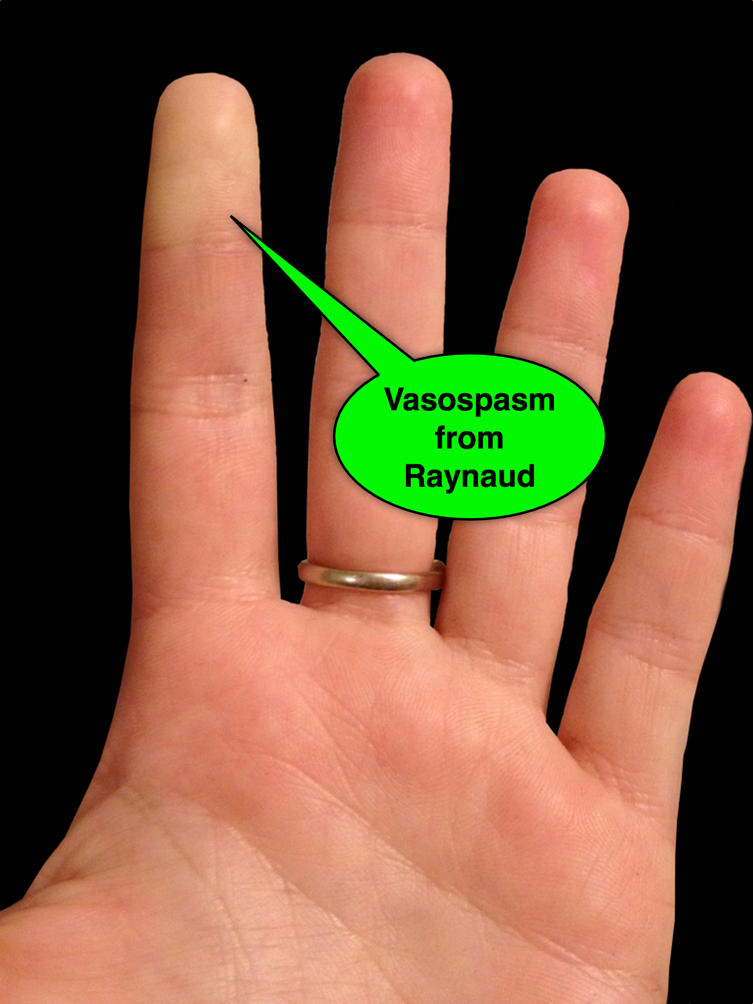
Answer: Raynaud phenomenon

  • Raynaud phenomenon is recurrent vasospasm of fingers or toes
  • Causes
    • Primary (e.g., external factors; such as cold temperature or stress)
    • Secondary to underlying illness, typically autoimmune disease (e.g., progressive systemic sclerosis or SLE)
  • Treatment
    • Avoid known triggers
    • Low-dose calcium channel blockers (e.g., nifedipine)
    • Topical nitroglycerine applied to areas of vasoconstriction
    • Identify and treat secondary disorders

Show References

Follow me on Twitter (@criticalcarenow) or Google+ (+criticalcarenow)


Title: A New Era in Management of the Septic Patient?

Category: Critical Care

Posted: 3/19/2014 by Haney Mallemat, MD

In 2001, Rivers et al. published a landmark article demonstrating an early-goal directed protocol of resuscitation that reduced mortality in septic Emergency Department patients.

Many questions have arisen throughout the years with respect to that trial; critics have complained about the overwhelming change in clinical practice based on this one single-center randomized trial.

Challenging Rivers data are the ProCESS (Protocolized Care for Early Septic Shock) investigators, who released the results from a multi-center randomized control trial of 1351 septic Emergency Department patients; the primary end-point was 60-day mortality. Click here for NEJM article.

Patients in this trial were randomized to one of three groups:

  • Protocol-based EGDT

  • Protocol-based standard (did not require central lines, inotropes, or blood transfusions

  • Usual care (no specific protocol; care was left to the bedside clinicians)

Bottom-line: The investigators did not find any difference in mortality between patients in the three groups and comment that the most important aspects of managing the septic patient may be prompt recognition and early treatment with IV fluids and antibiotics.

Show References

Follow me on Twitter (@criticalcarenow) or Google+ (+criticalcarenow)


Title: What's the Diagnosis? Case by Dr. Ali Farzad

Category: Visual Diagnosis

Posted: 3/17/2014 by Haney Mallemat, MD

Question

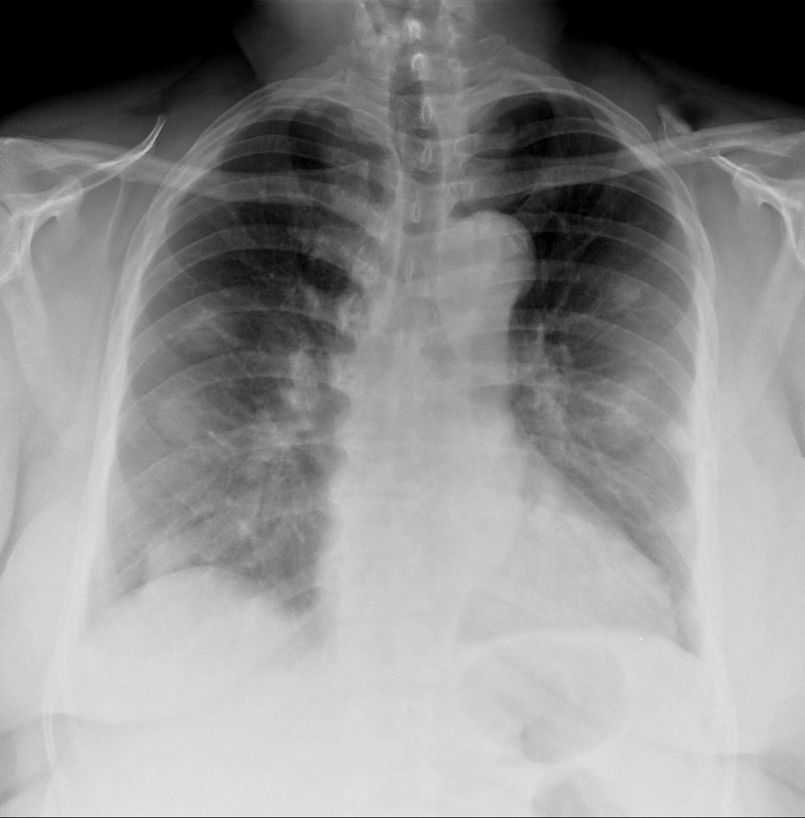
62 year-old male presents with weakness, chills, cough, and malaise. Recently, he had four teeth extracted but felt fine immediately after the extraction. Past medical history includes diabetes and hypertension; CXR is below. What’s the diagnosis?
 

Show Answer
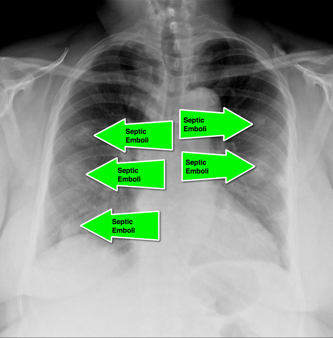
Answer: Septic pulmonary emboli (a.k.a. pulmonary puffballs); he was eventually diagnosed with bacterial endocarditis.

  • This history with the presence of "puffballs" on CXR, should clue the practitioner to the diagnosis of septic pulmonary emboli secondary to endocarditis
  • CT of the chest will confirm the diagnosis and possibly demonstrate cavitary lesions.
  • Blood cultures should be promptly obtained and the patient should be started on broad-spectrum antibiotics.

Show References

Follow me on Twitter (@criticalcarenow) or Google+ (+criticalcarenow)


Title: What's the Diagnosis? Image by Dr. Kami Hu

Category: Visual Diagnosis

Posted: 3/14/2014 by Haney Mallemat, MD

Question

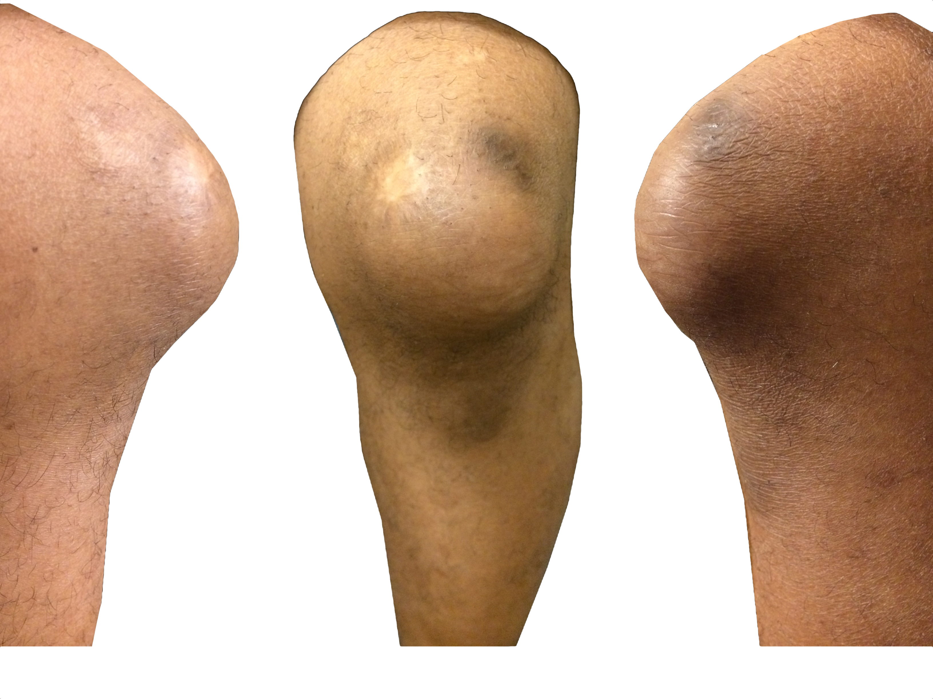
35 year-old carpet-layer presents with swelling of the superior portion of his knee that has progressively gotten worse over one week. He has no fever and has full range of motion (although pain is worse with movement). The knee is not tender to touch and the area is not erythematous or warm. What's the diagnosis?
 

Show Answer

Answer: Pre-patellar bursitis

  • The pre-patellar bursa is a superficial and synovial-lined space that reduces friction at the knee, separating the patella from the skin and the patellar tendon.
  • Inflammation to the bursa occurs secondary to direct knee trauma (either acute or chronic) and is typically benign.
  • It is associated with occupations that have a high incidence of trauma to the knee or repeated kneeling; hence the condition’s many names (carpet-layer's knee, coal-miner's knee, housemaid's knee, nun's knee)
  • Caution must be taken not to miss patients with septic bursitis (e.g., bacterial), especially in immunocompromised patients. If septic bursitis is suspected, fluid should be aspirated from the bursa and sent for analysis
  • Treatment for non-septic bursitis includes rest, (i.e., reduce trauma), NSAIDs, and physical therapy. If it remains persistent, incision and drainage by a qualified clinicians may be performed.

Show References

Follow me on Twitter (@criticalcarenow) or Google+ (+criticalcarenow)


Title: What's the Diagnosis? Image by Dr. Thuy Pham

Category: Visual Diagnosis

Posted: 3/3/2014 by Haney Mallemat, MD

Question

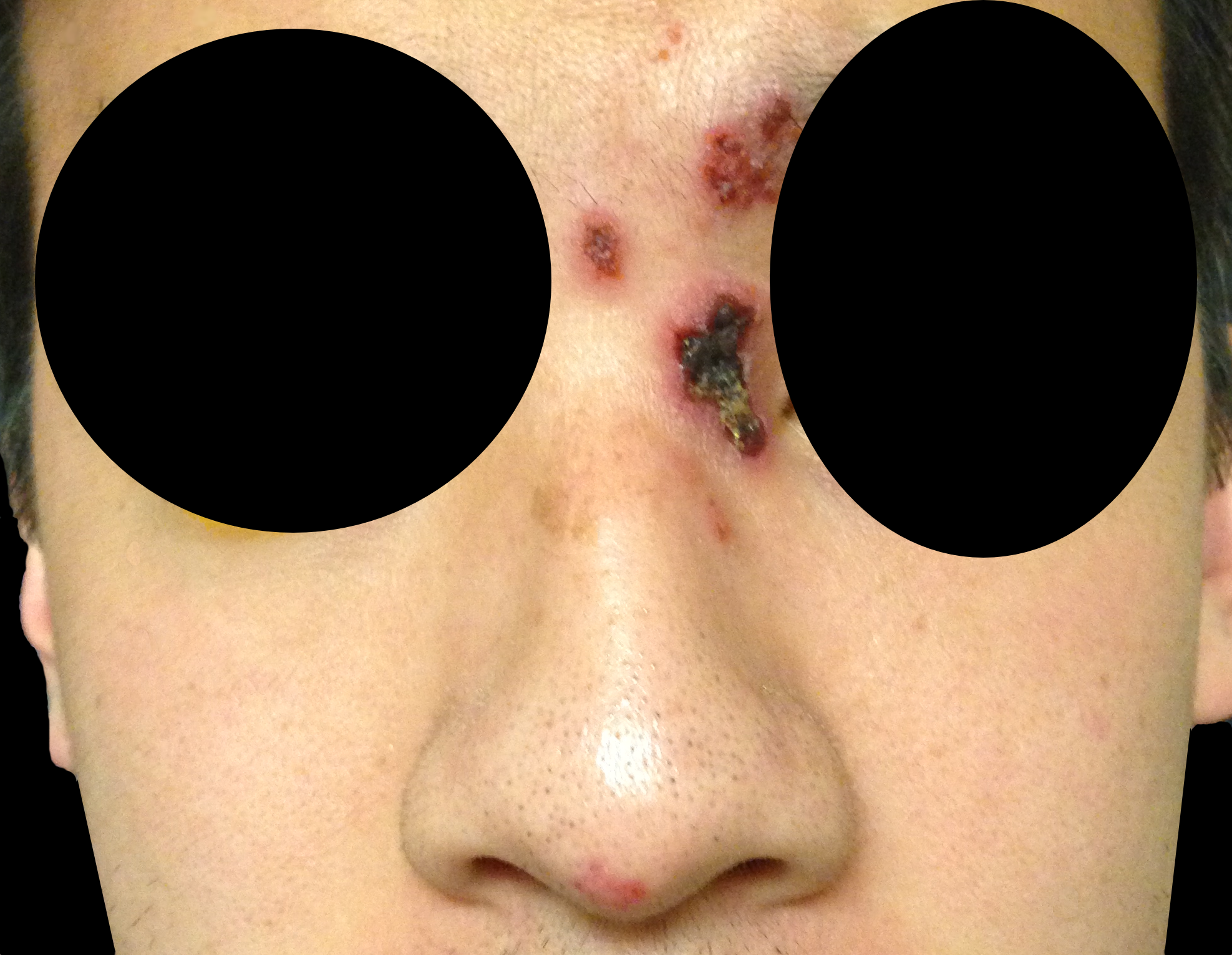
32 year-old male presents with the following. What's the diagnosis?
 

Show Answer
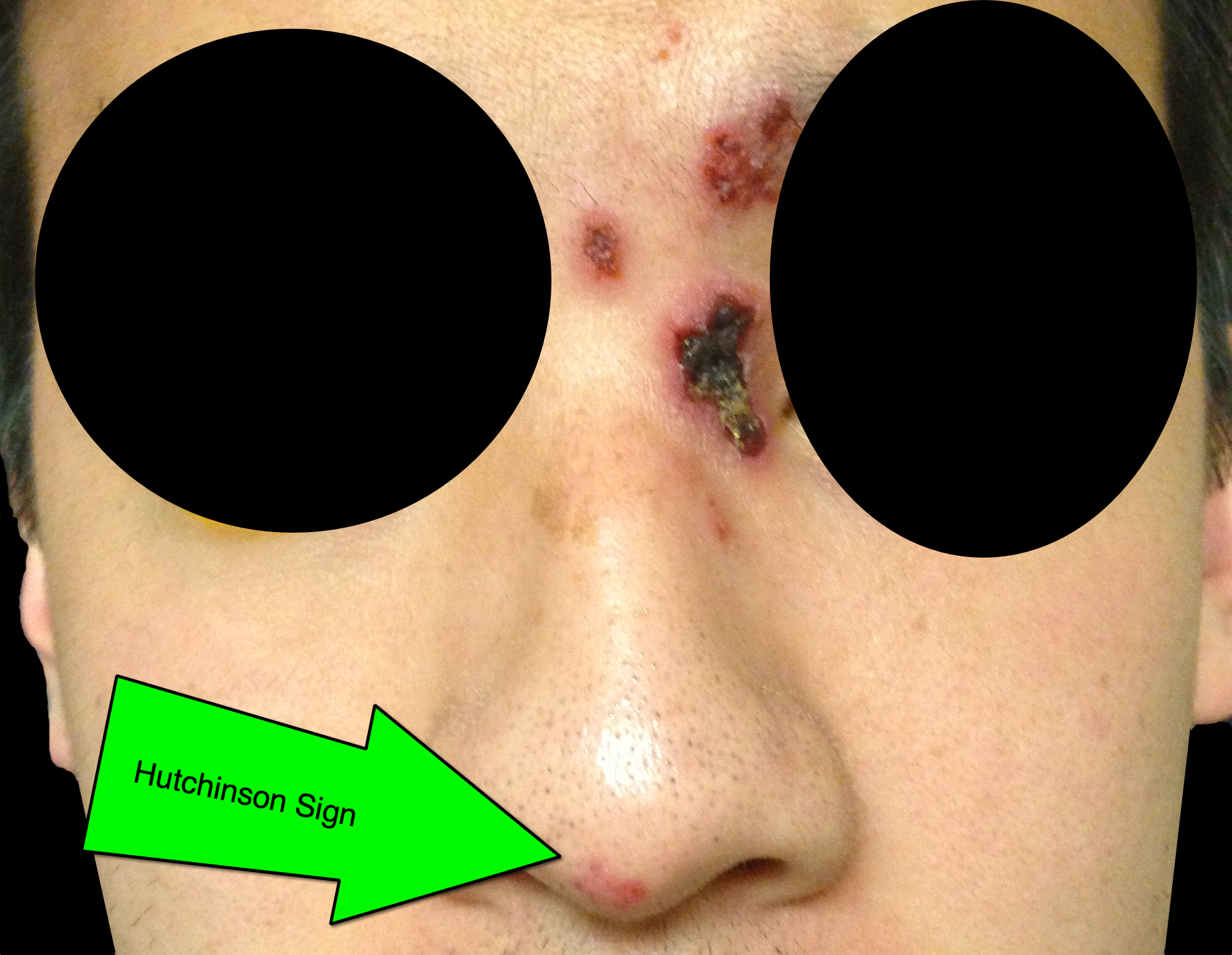
Answer: Hutchinson's Sign

Herpes zoster ophthalmicus (HZO)

  • HZO occurs when varicella-zoster, becomes reactivated in the ophthalmic division (V1) of the trigeminal nerve; any of the V1 branches may be affected (supraorbital, lacrimal, and nasociliary branches); nasocilliary innervates the globe.
  • Most patients develop only a periorbital rash, but a minority of patients have corneal involvement; the risk is increased in immunocompromised patients.
  • Hutchinson’s sign is when the rash occurs at the tip of the nose; patients with the sign have twice the incidence of ocular involvement, but 33% with HZO do not develop the sign.
  • Corneal involvement can result in significant visual complications and vision loss. Slit lamp exam with fluorescein is required when corneal involvement is suspected; a dendritic pattern may be seen.
  • Patients with eye involvement should be referred for urgent ophthalmologic evaluation following initiation of antiviral medications.

Show References

Follow me on Twitter (@criticalcarenow) or Google+ (+criticalcarenow)


Title: What's the Diagnosis? Images by UMEM Alumni Dr. Ari Kestler

Category: Visual Diagnosis

Posted: 2/24/2014 by Haney Mallemat, MD

Question

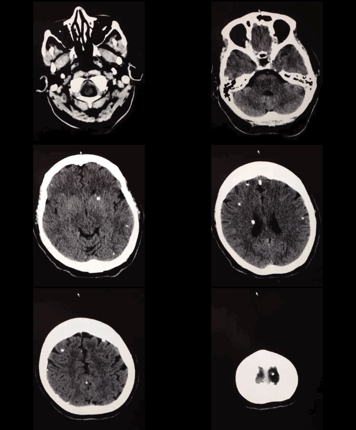
50 year-old with facial weakness and dysarthria. What's the diagnosis?
Show Answer
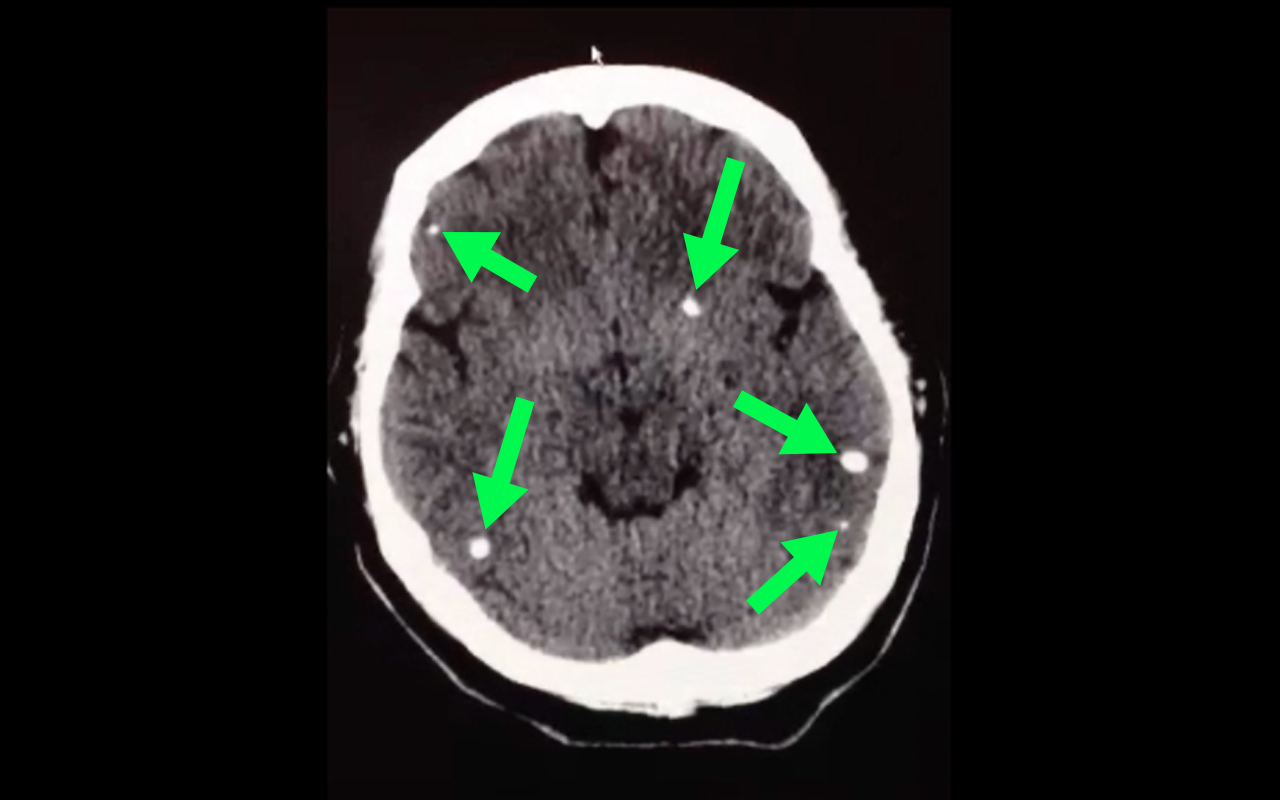
Stroke secondary to neurocysticercosis

Neurocysticercosis

Neurocysticercosis occurs secondary to ingestion of eggs from the Taenia solium tapeworm; ingestion typically occurs from ingestion of contaminated food.

Most patients are asymptomatic but symptoms of neurocysticercosis may include

  • Seizures (most common presentation)
  • Headache
  • Stroke
  • Neuropsychiatric dysfunction

Diagnosis is made via imaging such as CT scan; findings may vary depending on what stage of life the parasite is in (e.g., larvae); lumbar puncture should also be performed if the diagnosis is suspected.

Management should be symptomatic (e.g., seizures receive anticonvulsants) as well as eradication of the active parasites using steroids followed by albendazole (anti-parasitic drug)

Show References

Follow me on Twitter (@criticalcarenow) or Google+ (+criticalcarenow)


Title: Cocaine-Induced Abdominal Pain

Category: Critical Care

Posted: 2/18/2014 by Haney Mallemat, MD

  • The well-known effects of cocaine toxicity include seizures, cardiac ischemia, and rhabdomyolysis. Abdominal pain, however, is a lesser known side-effect and may occur secondary to ischemia, infarction or perforation of the gastrointestinal tract; such cases tend to occur in younger people without known risk factors for ischemia.
  • Ischemia may occur from the direct vasoconstrictive effects of cocaine, but may also occur from its pro-thrombotic effects on the mesenteric vessels; although any segment of the GI tract may be involved, the small bowel is most often affected.
  • Symptoms may vary from mild abdominal pain to bloody diarrhea. Physical exam may reveal peritoneal signs if perforation occurs.
  • CT scan of the abdomen may reveal the diagnosis although angiography may required for diagnosis or to guide revascularization.
  • Management may vary from conservative (i.e., bowel rest and antibiotics) to surgical exploration and bowel resection in selected cases.


Show References

Follow me on Twitter (@criticalcarenow) or Google+ (+criticalcarenow)

Zimmerman, J.Cocaine intoxication. Crit Care Clinics 2012 Oct;28(4):517-26


Title: What's the Diagnosis? Image by Dr.Heather Mezzadra

Category: Visual Diagnosis

Posted: 2/17/2014 by Haney Mallemat, MD

Question

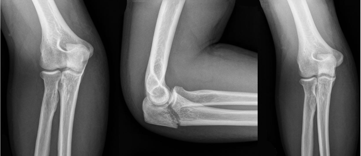
44 year-old construction worker fell off a ladder and presents with elbow pain. What's the diagnosis and what is the most commonly associated nerve injury? 
 

Show Answer
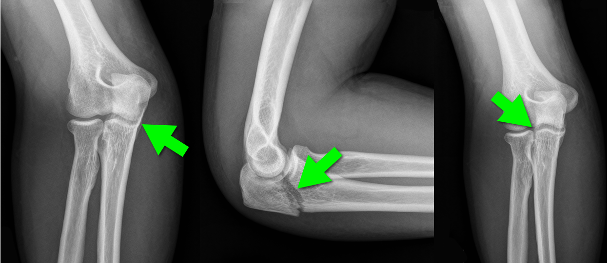
Olecranon fracture; the ulnar nerve is most commonly injured

Pearls for Olecranon Fracture

  • Typically occurs secondary to direct blow or fall on an outstretched hand
  • Patients present with a complaint of pain along the olecranon with an inability to extend the elbow
  • Ulnar nerve injury is common.
  • Non-displaced fractures are typically managed conservatively with placement in a long-arm posterior splint; elbow must be placed in flexion of 70° and forearm is neutral
  • Other types of olecranon injuries require surgical repair 

Show References

Follow me on Twitter (@criticalcarenow) or Google+ (+criticalcarenow)


Title: What's the Diagnosis? Case by Dr. Ryan Spangler (@Emterp2)

Category: Visual Diagnosis

Posted: 2/10/2014 by Haney Mallemat, MD (Updated: 2/10/2014)

Question

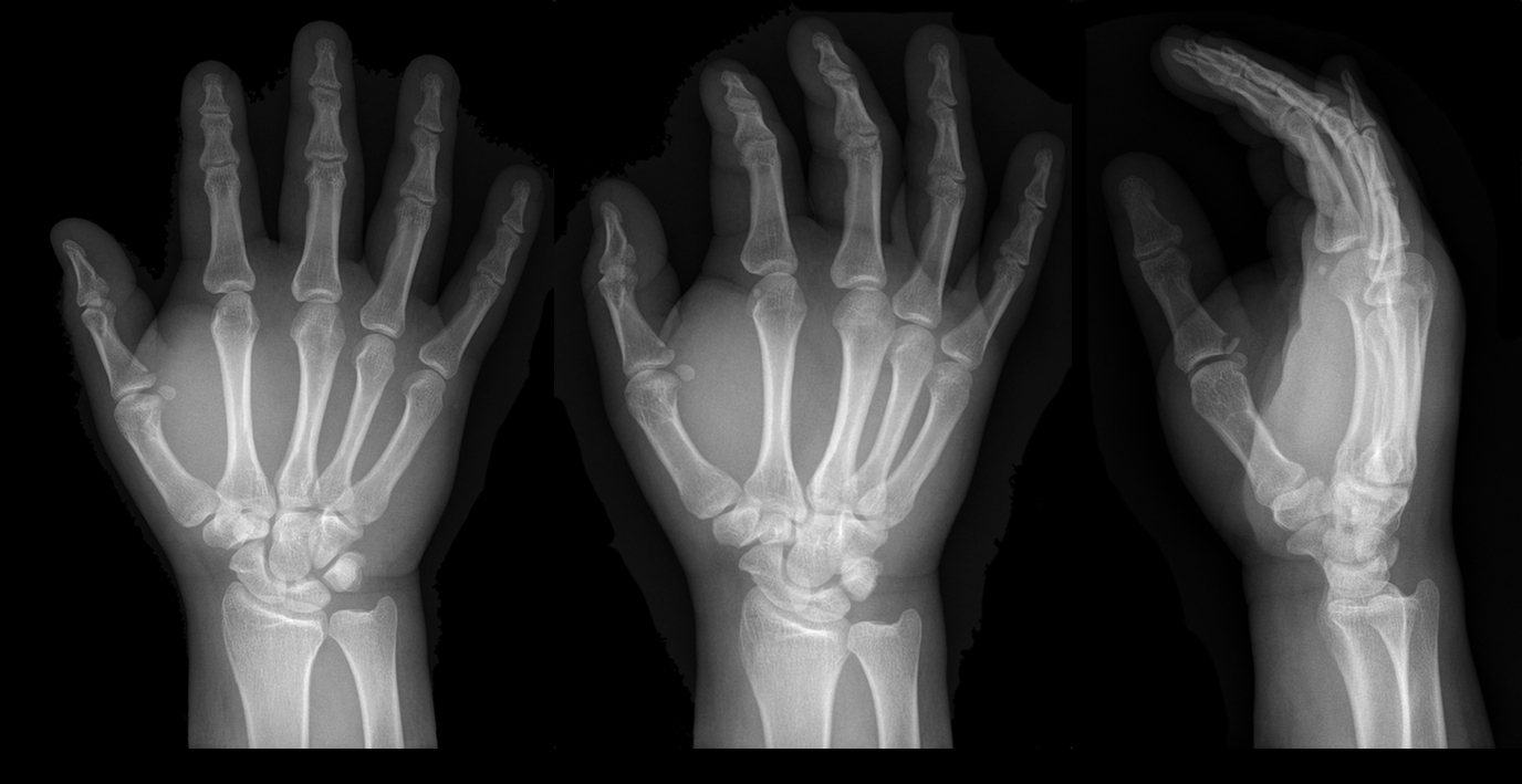
25 year-old male presents after falling off his bicycle. He complains of pain in his right-hand (he is right-hand dominant). What's the diagnosis? 
Show Answer
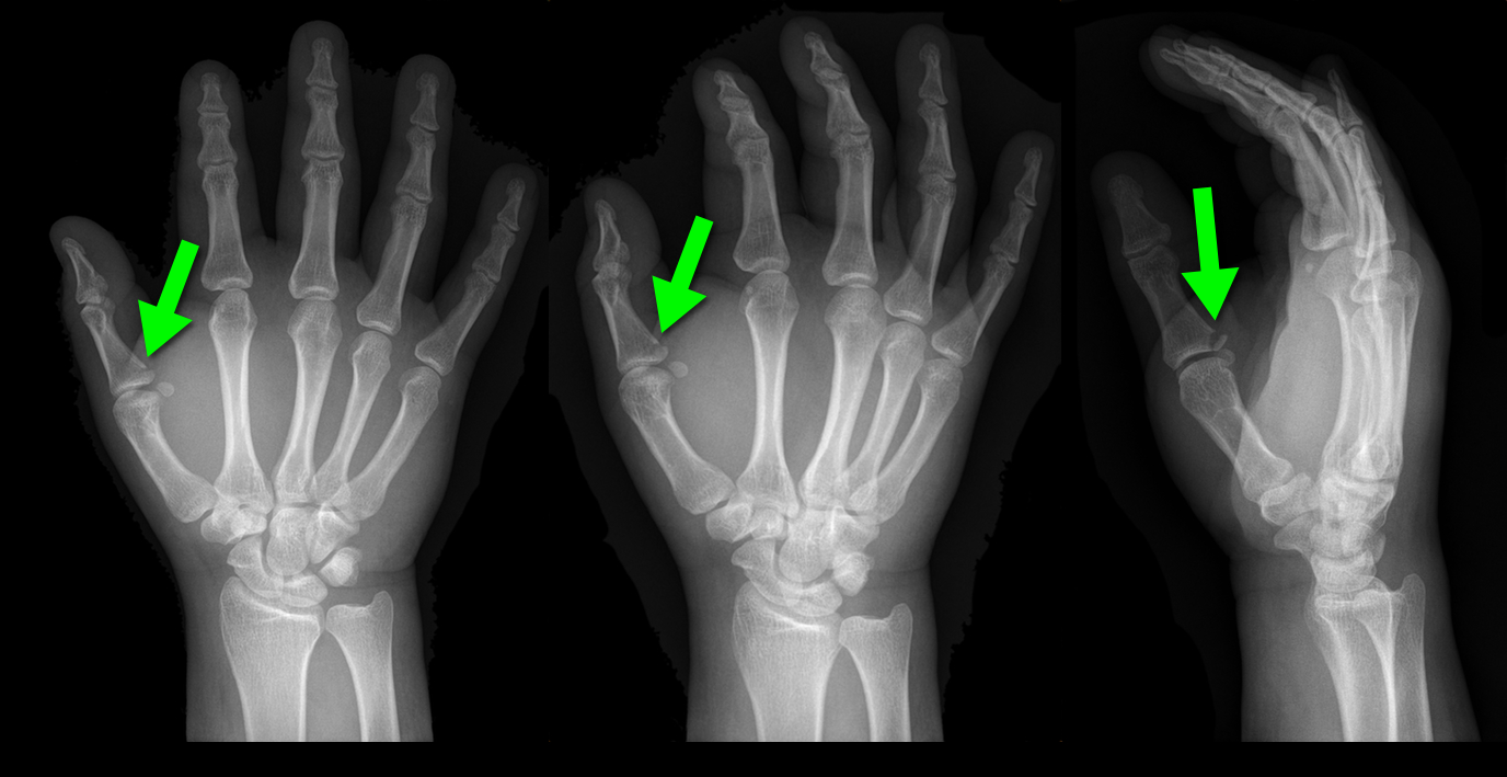
Answer: Gamekeeper's thumb or Skier's thumb

Injury to the ulnar collateral ligament (UCL) secondary

  • repetitive injury to hand
  • acute hyper-abduction injury to the thumb (i.e., during a skiing injury or fall on outstretched hand); most common cause if injury. 

Called “Gamekeeper Thumb” because injury was originally described in gamekeepers who job was to break the necks of captured game (or animals). The act of breaking necks led to a repetitive stress and injury secondary to the valgus forces on the thumb.

UCL can become torn or avulsed from the insertion on the proximal phalanx; the UCL is an important stabilizer of the thumb; improper management can lead to future disability.  

Treatment is with a short-arm thumb-spica cast and follow up with a hand surgeon to determine whether management will be conservative or operative.

Chuter GS, Muwanga CL, Irwin LR (June 2009). "Ulnar collateral ligament injuries of the thumb: 10 years of surgical experience". Injury 40 (6): 652–6 Wheeless' Textbook of Orthopaedics Online: Surgical treatment of acute gamekeeper's thumb

Show References

Follow me on Twitter (@criticalcarenow) or Google+ (+criticalcarenow)


Title: What's the Diagnosis? Images by Dr. Jennifer Guyther

Category: Visual Diagnosis

Posted: 2/3/2014 by Haney Mallemat, MD

Question

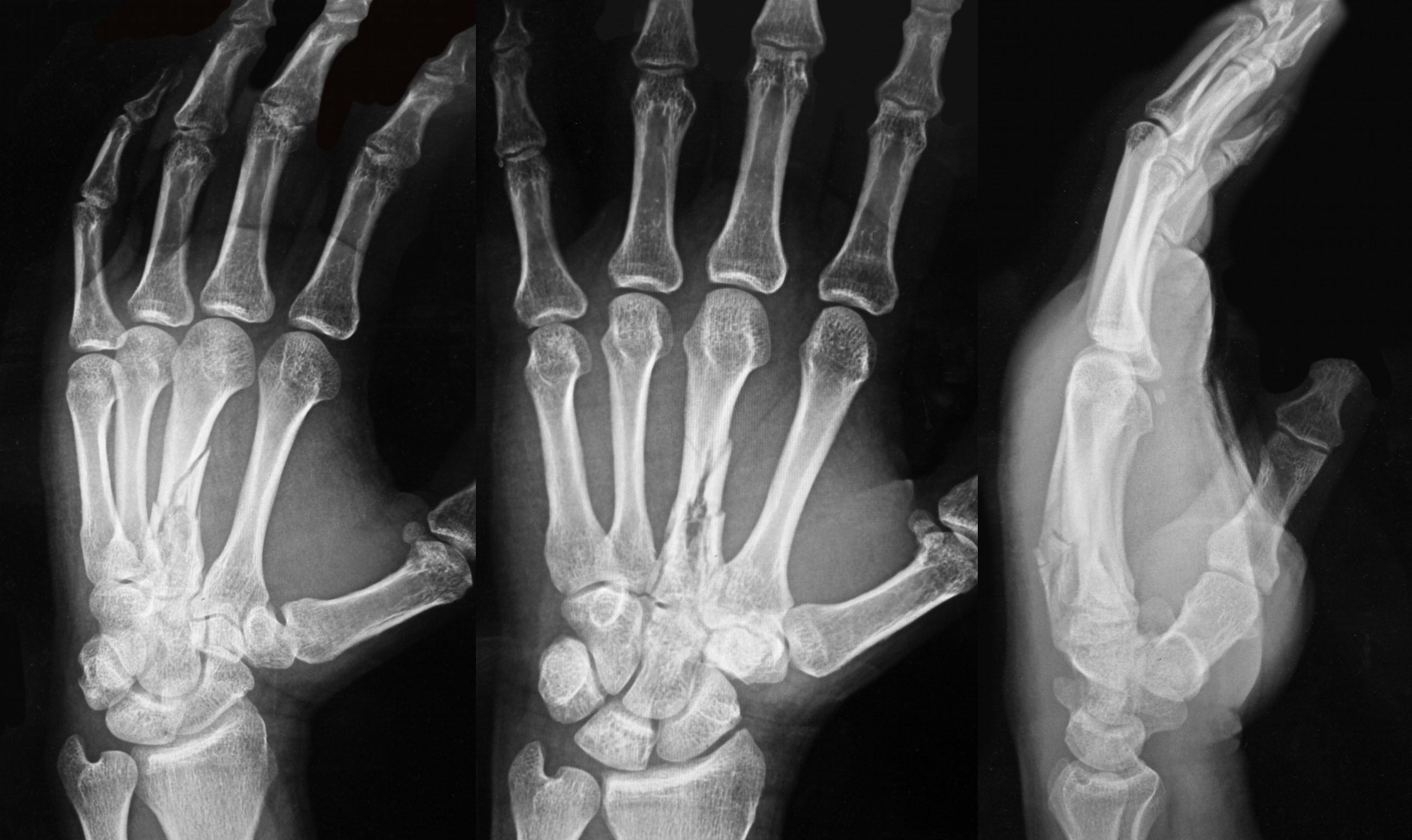
34 year-old left-hand dominant male sustained injury to left hand after his pressurized greasing-gun discharged into the palm of his hand. He has a small lac to the hand but is in extreme pain. On exam his hand is very puffy and he is neurovascularly intact (XR below) What is the next step in management? 
 

Show Answer
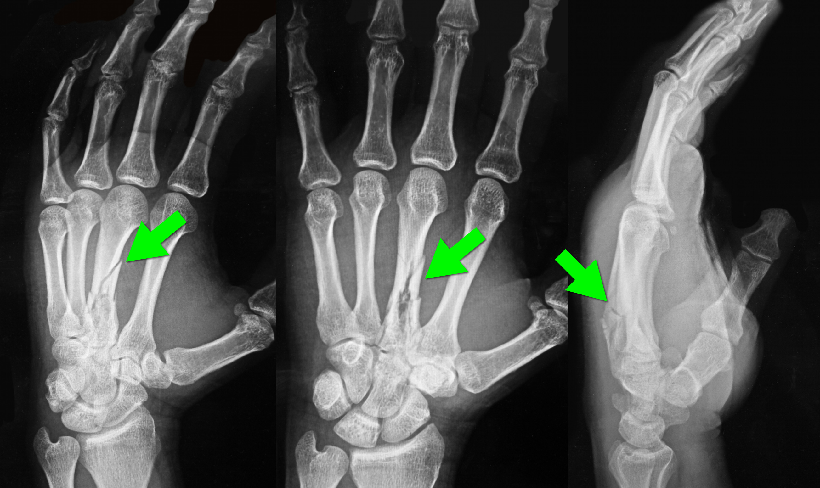
Metacarpal fracture; emergent surgical consult is required

High-Pressure Injection Injuries (HPI) injuries

HPI devices (e.g., grease, paint guns, etc.) may cause injury when the device accidently discharges, typically into the operator’s dominant hand during attempts to clean the nozzle.

Injuries may initially appear benign, however injuries should be considered surgical emergencies because they often require debridement / washout of materials injected into deeper tissues.

Tissue damage is multi-factorial and includes direct injury (e.g., high-pressure tissue damage, vascular compression, etc.) and indirect injury from material injected into tissues (e.g., chemical inflammation / injury, granuloma formation, deep-tissue infections, etc.).

Radiographs may assist surgical planning by identifying subcutaneous air, debris, or unanticipated fractures.

Treatment:

  • High-potential for disability and amputation so prompt aggressive therapy and surgical consult is crucial.
  • Broad-spectrum prophylactic antibiotics and update tetanus
  • Splint the extremity and keep it elevated while awaiting disposition

Show References

Follow me on Twitter (@criticalcarenow) or Google+ (+criticalcarenow)


Title: Necrotizing Skin and Soft Tissue Infections (NSSTIs)

Category: Critical Care

Posted: 1/28/2014 by Haney Mallemat, MD

NSSTIs occur secondary to toxin-secreting bacteria; NSSTIs are surgical emergencies with a high-morbidity / mortality

Risk factors: immunocompromised host (DM, AIDS, etc.), intravenous drug use, malnourishment, peripheral vascular disease

Type I (polymicrobial; most common), Type II (monomicrobial; typically clostridia, streptococci, staph, or bacteroides), Type III (Vibrio vulnificus; seawater exposure)

Signs / Symptoms: pain out of proportion to exam (occasionally no pain at all), skin findings (blistering / bullae, gray-skin discoloration, or “Dishwater-like” discharge), or systemic toxicity (altered mental status, elevated lactate, etc.)

Diagnostic radiology

  • Xray (shows gas); low sensitivity; CT scan (gas / tissue stranding); sensitivity is also low
  • MRI can over-diagnose NSSTI and should not be used routinely
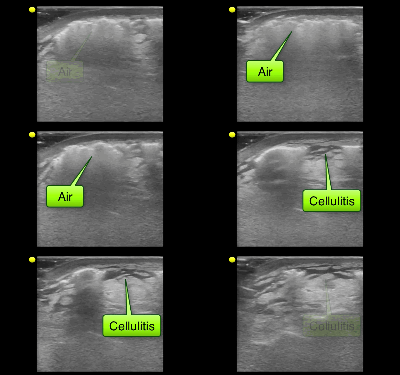
  • Bedside ultrasound may demonstrate fluid or gas collections in deeper tissues (see clip below)

Treatment is emergent surgical debridement with simultaneous hemodynamic resuscitation PLUS broad-spectrum antibiotics; consider clindamycin becuase it has anti-toxin activity

Adjunctive therapies include Intravenous intraglobulin (neutralizes toxins secreted by bacteria) and hyperbaric oxygen
Show References

Follow me on Twitter (@criticalcarenow) or Google+ (+criticalcarenow)


Title: What's the Diagnosis?

Category: Visual Diagnosis

Posted: 1/27/2014 by Haney Mallemat, MD

Question

32 year-old with diabetes presents with fever, erythema, and warmth of his lower extremity; his leg is not particularly painful. He is diagnosed with cellulitis, started on antibiotics, and admitted to the hospital. While boarding in the Emergency Department he becomes rigorous and hypotensive. An ultrasound of his cellulitis is performed and is shown below. What’s the diagnosis?
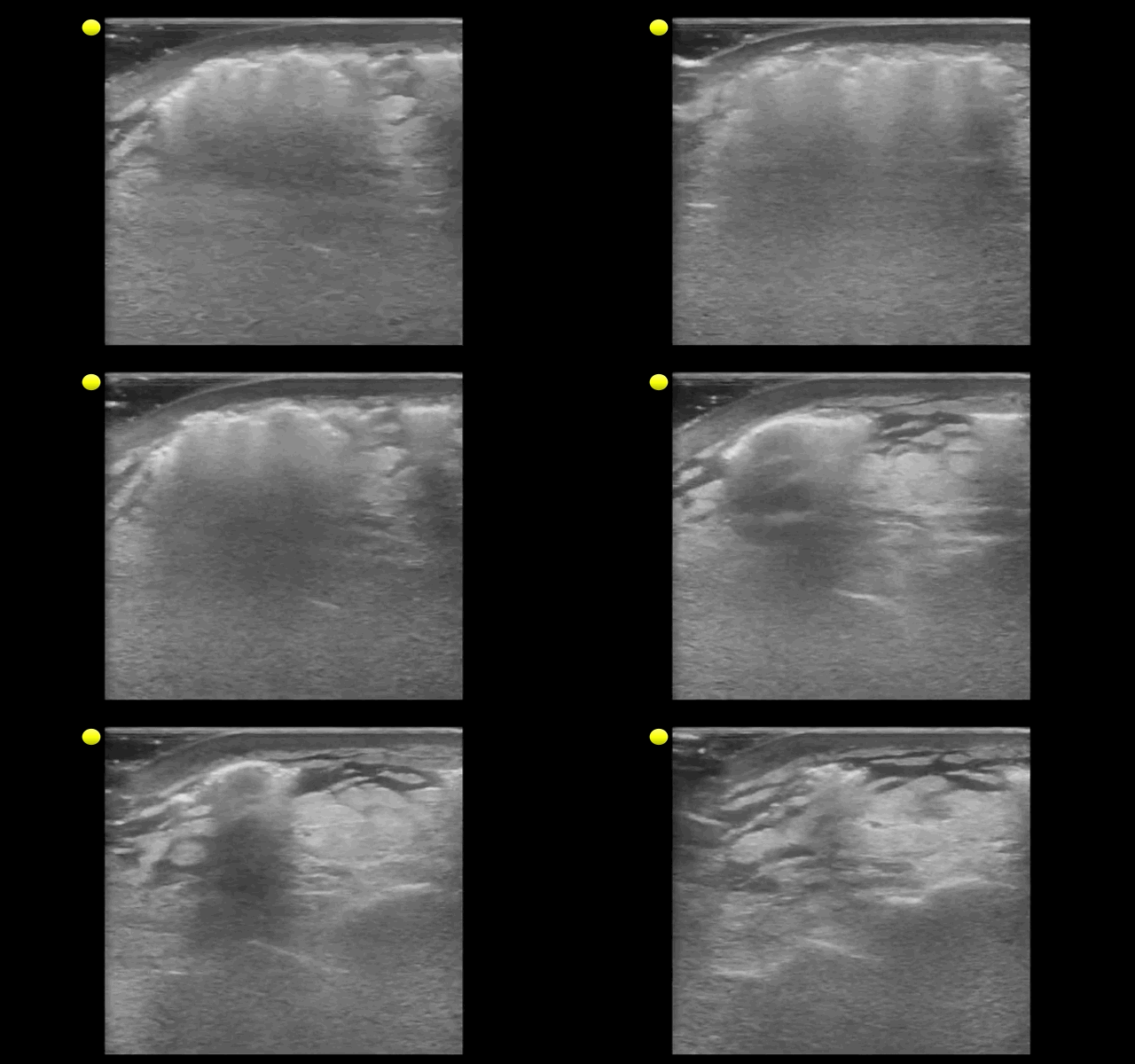
 

Show Answer

"Cobblestoning" of subcutaneous tissue consistent with cellulitis. There are also areas of subcutaneous air raising the concern for necrotizing fasciitis.

Special thanks to Dr. Mike Mallin from the Ultrasound Podcast for the clip. Want to see more amazing ultrasound clips? Check out Sonocloud

Show References

Follow me on Twitter (@criticalcarenow) or Google+ (+criticalcarenow)


Previous  |  1 |  2 |  3 |  4 |  5 |  6 |  7 |  8 |  9 |  10 |  11 |  ... |  17 |  Next
University of Maryland School of Medicine logo
  • Departments
  • Programs
  • Research Centers
  • Institutes
  • Graduate Education
  • CIBR Cores
  • Offices of the Dean
    • University of Maryland, Baltimore
    • University of Maryland Medical Center
    • Medical Alumni Association of the University of Maryland
    • Health Sciences and Human Services Library
University of Maryland School of Medicine
655 W. Baltimore Street
Baltimore, MD 21201
Contact Us

© University of Maryland School of Medicine

    • Webmaster
    • Web Accessibility
    • Site Index
    • UMB Hotline
    • Employment
    • Map