
University of Maryland School of Medicine logo
Emergency Medicine
All Departments
UMSOM Assistant
UMEM Educational Pearls
  • Home
  • Departments
  • Emergency Medicine
  • CME
  • UMEM Educational Pearls
  • CME
  • Courses and Conferences
  • Free Open Access Medical Education
  • UMEM Educational Pearls

Search

81-100 of 321 results by Haney Mallemat

Previous  |  1 |  2 |  3 |  4 |  5 |  6 |  7 |  8 |  9 |  10 |  ... |  17 |  Next

Title: What's the Diagnosis? Image by Dr. Michael Abraham

Category: Visual Diagnosis

Posted: 9/8/2014 by Haney Mallemat, MD

Question

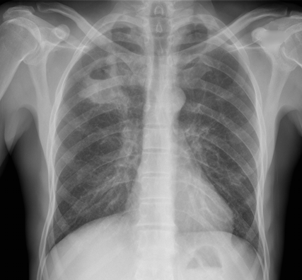
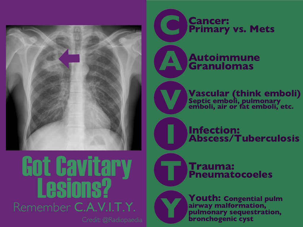
CXR shown below, what's the diagnosis? ...and name 3 differential diagnoses.


Show Answer
Show References

Follow me on Twitter (@criticalcarenow) or Google+ (+criticalcarenow)


Title: Back 2 Basic Series: Under Pressure - Vasopressors Part I

Category: Critical Care

Posted: 9/2/2014 by Haney Mallemat, MD (Updated: 10/1/2014)

Vasopressors are used in shock-states to increase mean arterial pressure (MAP) and improve distal tissue perfusion. Additionally, some agents have effects on the heart to augment cardiac output.

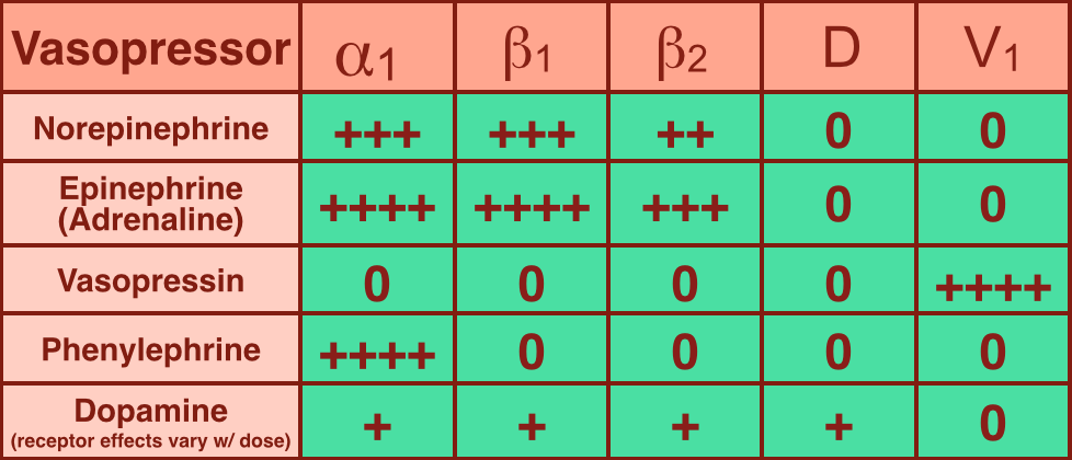
Receptors that vasopressors work on include: 

  • Alpha-1: increase arterial tone (increases MAP) and venous tone to reduce venous pooling and augment cardiac preload 
  • Beta-1: increase inotropy and chronotropy on heart muscle; also increases arterial tone
  • Beta-2 and Dopamine: cause vasodilation but may actually be beneficial because this increases perfusion to cardiac, renal, and GI tissues.
  • V1: arterial vasoconstriction to increase MAP
  • The chart below is a summary; please note that quoted receptor effects vary depending on the source reviewed

Norepinephrine (NE): excellent vasopressor for most types of shock and recommended as a first-line agent in the Surviving Sepsis Guidelines.

  • Works on alpha-1, beta-1, and beta-2 receptors. 
  • Initial dosing 0.05 mcg/kg/min with a maximum dose often cited as 0.5 mcg/kg/min (though there is technically no maximum dose).

Epinephrine (a.k.a. Adrenaline): in several countries the first-line agent for shock (including sepsis).

  • Works similarly to NE on alpha-1, beta-1 and beta-2; it is a more potent inotrope than NE.
  • One downside is the production of lactic acid, which can sometimes lead to confusion when following serial lactates during resuscitation. 
 

Show References

Follow me on Twitter (@criticalcarenow) or Google+ (+criticalcarenow)


Title: What's the Diagnosis?

Category: Visual Diagnosis

Posted: 9/1/2014 by Haney Mallemat, MD

Question

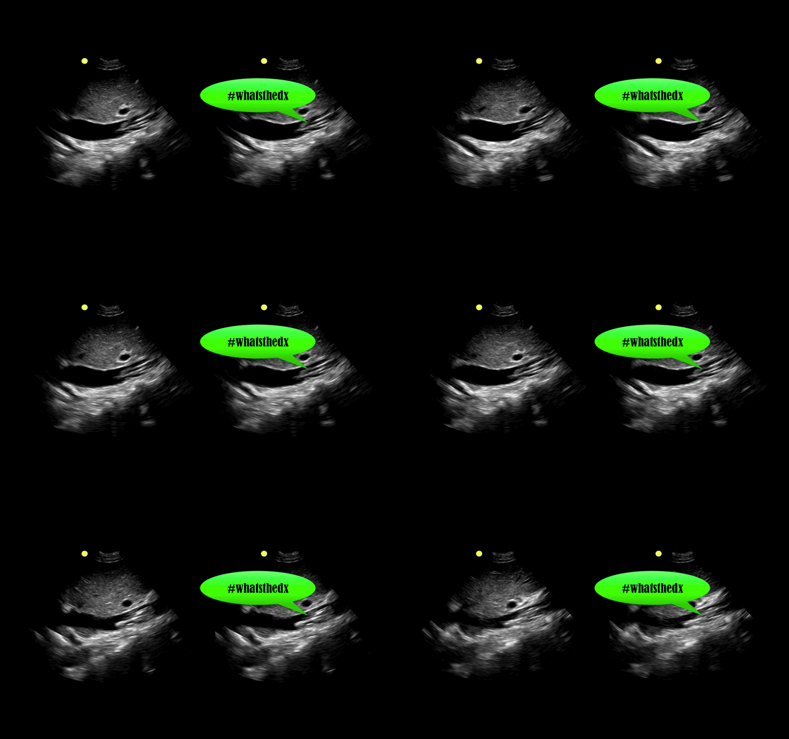
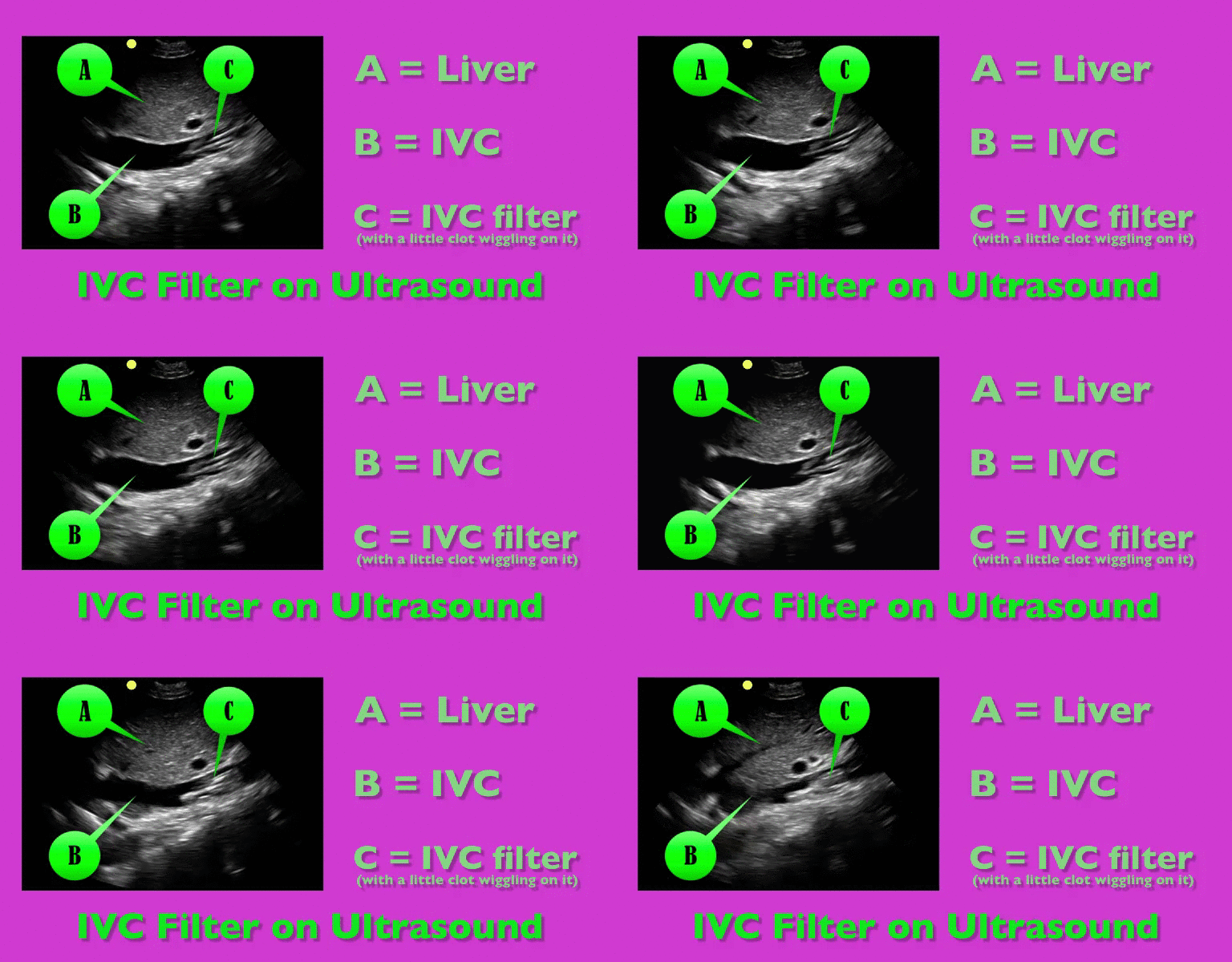
You are scaning the liver with ultrasound and you see this. What's the diagnosis?


Show Answer
Show References

Follow me on Twitter (@criticalcarenow) or Google+ (+criticalcarenow)


Title: What's the Diagnosis?

Category: Visual Diagnosis

Posted: 8/25/2014 by Haney Mallemat, MD

Question

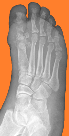
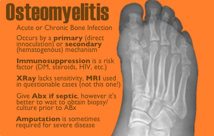
50 year-old female with diabetes complains of pain and discharge from a poorly healing wound. XRay below. What's the diagnosis?
Show Answer
Show References

Follow me on Twitter (@criticalcarenow) or Google+ (+criticalcarenow)


Title: What's the Diagnosis?

Category: Visual Diagnosis

Posted: 8/18/2014 by Haney Mallemat, MD

Question

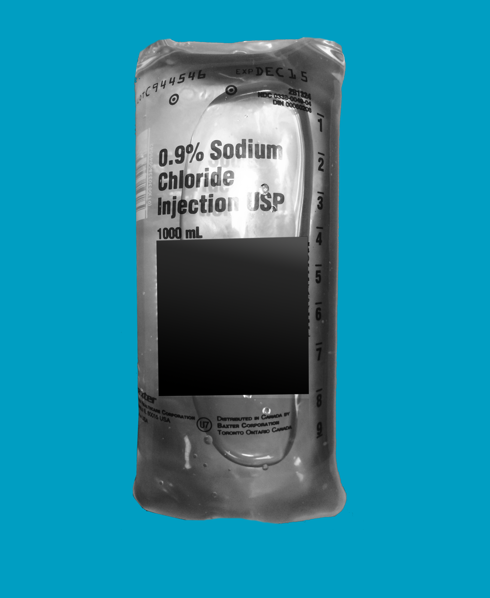
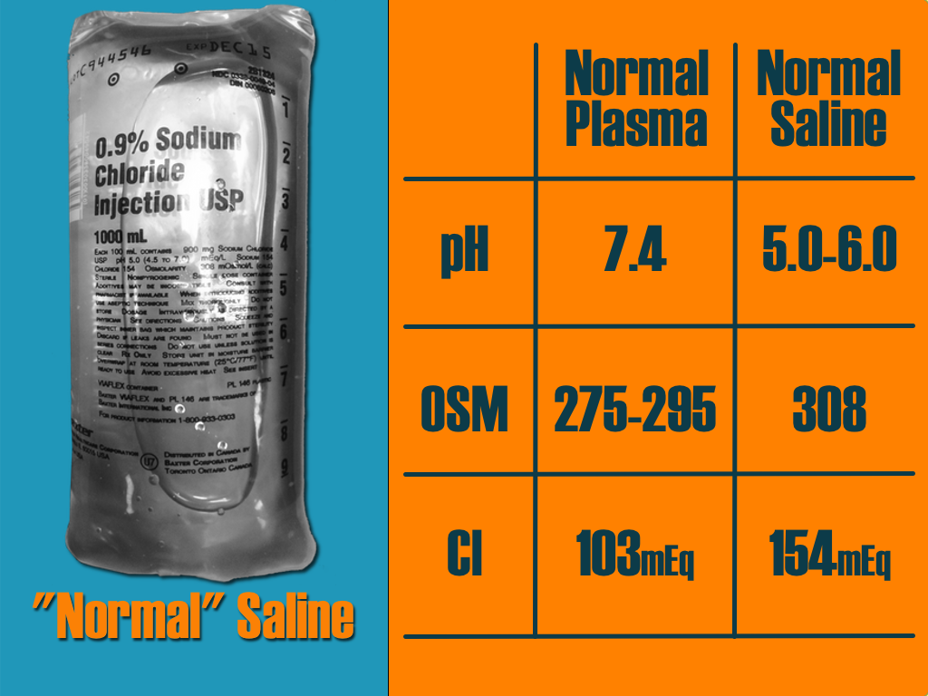
A critically-ill patient requires fluid resuscitation. Someone hands you a bag of this. What’s the pH of this fluid? 
Show Answer
Show References

Follow me on Twitter (@criticalcarenow) or Google+ (+criticalcarenow)


Title: What's the Diagnosis? Image by Dr. Ari Kestler

Category: Visual Diagnosis

Posted: 8/11/2014 by Haney Mallemat, MD (Updated: 8/12/2014)

Question

30 year-old female with complains of night sweats and painful lesions on her fingertips. What’s the diagnosis and list some things to have in the differential diagnosis?

Show Answer

Answer: Osler Nodes

Osler notes are painful, palpable, and erythematous lesions secondary to the deposition of immune-complexes within the pads of digits.

Although classically associated with infective endocarditis, some authors claim that it may be seen in only 10-23% of confirmed cases. Therefore the differential diagnosis should also include:

  • Bacteremia
  • Disseminated gonococcal infection
  • Lupus
  • Vasculitis
  • Drug-induced

Show References

Follow me on Twitter (@criticalcarenow) or Google+ (+criticalcarenow)


Title: Back to Basics Series: Deciphering the Vent

Category: Critical Care

Posted: 8/5/2014 by Haney Mallemat, MD

There are many ventilator modes to choose from, but almost every mode can be distilled down to its basic principles by understanding the “Three T's of Mechanical Ventilation”

Trigger: You must determine whether the vent or patient will trigger a mechanical breath. For example, machine-triggered breaths (a.k.a. control mode of ventilation) are used for paralyzed patients and will deliver a breath after a period of time has elapsed (e.g., if RR is 10/min, then a breath is given every 6 seconds). On the other hand, if a patient’s respiratory drive is intact (a.k.a. assist-mode) than the patient triggers the breath when the vent detects a patient induced change in airflow or airway pressure. These two modes can also be mixed together.

Target: Mechanical breaths must have a specific target, either a target airway pressure or a tidal volume. Because pressure and volume are directly related, pick the variable you want to target and the other parameter will vary depending on the patient’s intrinsic physiology. For example, if you choose to target a specific tidal volume, we may get one plateau pressure in a patient with normal lungs, but a higher plateau pressure in another patient with stiffer lungs.

Terminate: You must decide when the mechanical breath (i.e., inspiration) terminates and expiration begins. Termination occurs: 1) after a set inspiratory time has elapsed in certain pressure-targeted modes, 2) when a predefined target volume has been achieved (i.e., volume-cycled modes), or 3) when airflow has been reduced by a certain percentage (as in pressure-support ventilation; to be discussed separately)

Let’s put this all together by looking at an example: pressure control ventilation (rate = 12/min and target pressure 20cm H20). Trigger: Because this is a “control”, not assist mode, the machine will trigger a breath 12 times per minute or every 5 seconds. Target: Here we chose to have pressure be the target, so when the ventilator triggers a breath it will deliver a constant airway pressure of 20 cmH2O until we tell the vent terminate that breath. Terminate: the constant airway pressure will be turned off after a fixed period of time has elapsed; for this example we will set the inspiratory time as 1 second, then expiration begins. Now, after a few vent breaths we will observe the results of our settings and reassess; if the resulting tidal volume is lower than what we wanted, we will increase the target pressure to increase the tidal volume. If the tidal volume is higher than what we wanted, we will reduce the target pressure to reduce the tidal volume. We can also tweak the inspiratory time to manipulate the tidal volume, but this does so to a lesser degree.

Try to break down your favorite modes of ventilation using the Three T’s and see if this helps you understand vent modes better. 

Show References

Follow me on Twitter (@criticalcarenow) or Google+ (+criticalcarenow)


Title: What's the Diagnosis? Image by Dr. Andrew Crouter

Category: Visual Diagnosis

Posted: 8/4/2014 by Haney Mallemat, MD (Updated: 8/12/2014)

Question

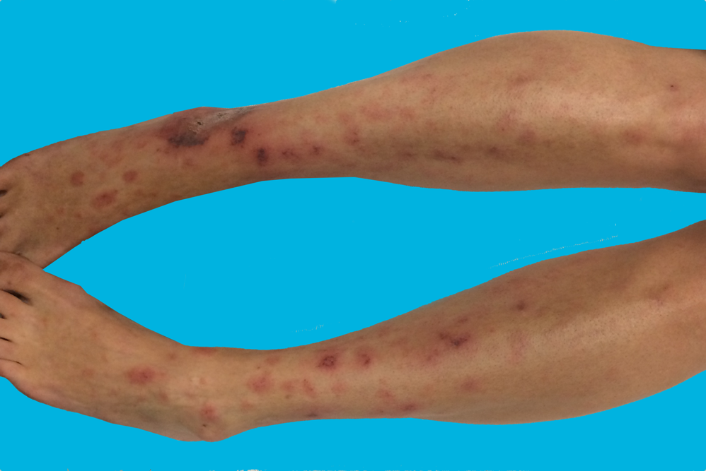
40 year-old female presents with painful lesions and ulcers on lower extremities. She has had this before, but never to this extent. She also has a history of DVT. What’s the diagnosis?
Show Answer

Answer: Livedoid vasculopathy or Livedoid vasculitis (LV)

  • Vascular disease resulting in chronic thrombosis and ulceration of bilateral lower extremities usually in young to middle-aged females. Lesions are painful purpuric macules or papules.
  • A vasoocclusive-type process not an inflammatory one occurring secondary to hyalinization and focal thrombosis of blood vessels. Diagnosis via biopsy.
  • Patients are prothrombotic with an increased risk for VTE’s (altered protein C, increased homocystine, Factor V Leiden mutation, etc.).
  • Treatment goals are to reduce pain, thrombosis (e.g., anticoagulants, antiplatelets, etc.), and superinfected ulcers should be treated with antibiotics covering skin flora. No increased risk of loss of life or limb with the disease.
  • Fantastic differential diagnoses from folks following on Twitter:
    • Polyarteritis nodosum and other vasculitis diseases
    • SLE and anti-phosholipid syndrome
    • Levamisole toxicity
    • Venous stasis ulcers
    • Warfarin-induced skin necrosis

Show References

Follow me on Twitter (@criticalcarenow) or Google+ (+criticalcarenow)


Title: What's the Diagnosis? Case by Dr. Swati Singh

Category: Visual Diagnosis

Posted: 7/28/2014 by Haney Mallemat, MD (Updated: 7/28/2014)

Question

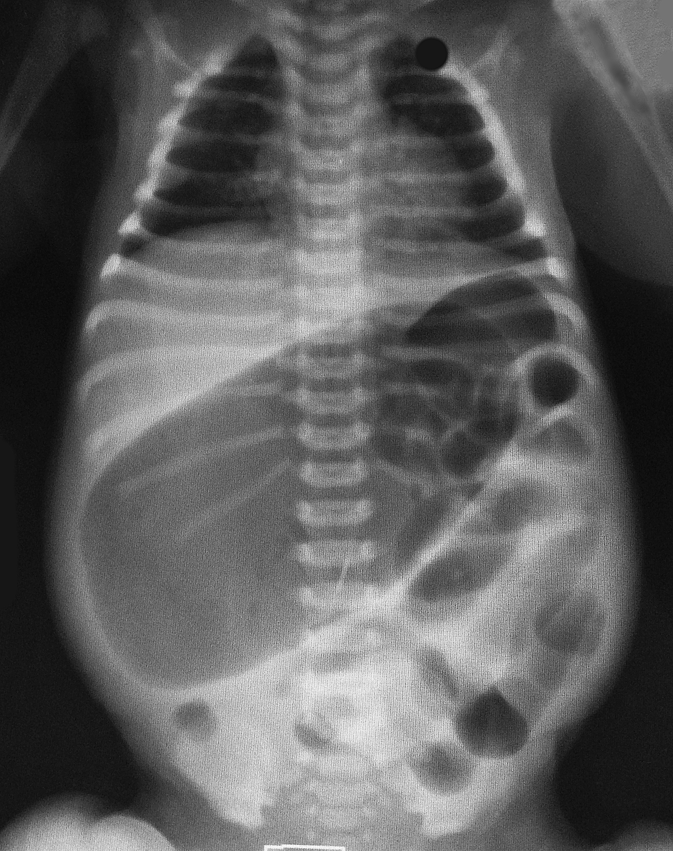
2-day old baby boy presents with forceful vomiting of entire feeds, bloated belly, and has not passed stools since birth. What's the diagnosis?
Show Answer

Colonic atresia

Colonic Atresia

  • Condition in which a part of the colon has not formed correctly; colon is completely blocked or missing altogether. The colon is the rarest site of atresia in the GI tract.
  • Diagnosis may be suggested in prenatal sonograms and is usually revealed in affected newborns shortly after birth.
  • Patients usually present with vomiting, abdominal distention, and failure to pass meconium.
  • Differential includes:
    • Small bowel atresia

    • Meconium plug syndrome

    • Hirschsprung's disease 

  • Treatment is supportive and ultimately surgical correction

Show References

Follow me on Twitter (@criticalcarenow) or Google+ (+criticalcarenow)

http://emedicine.medscape.com/article/934014-overview


Title: What's the Diagnosis? Case by Dr. Bennett Myers

Category: Visual Diagnosis

Posted: 7/21/2014 by Haney Mallemat, MD (Updated: 7/22/2014)

Question

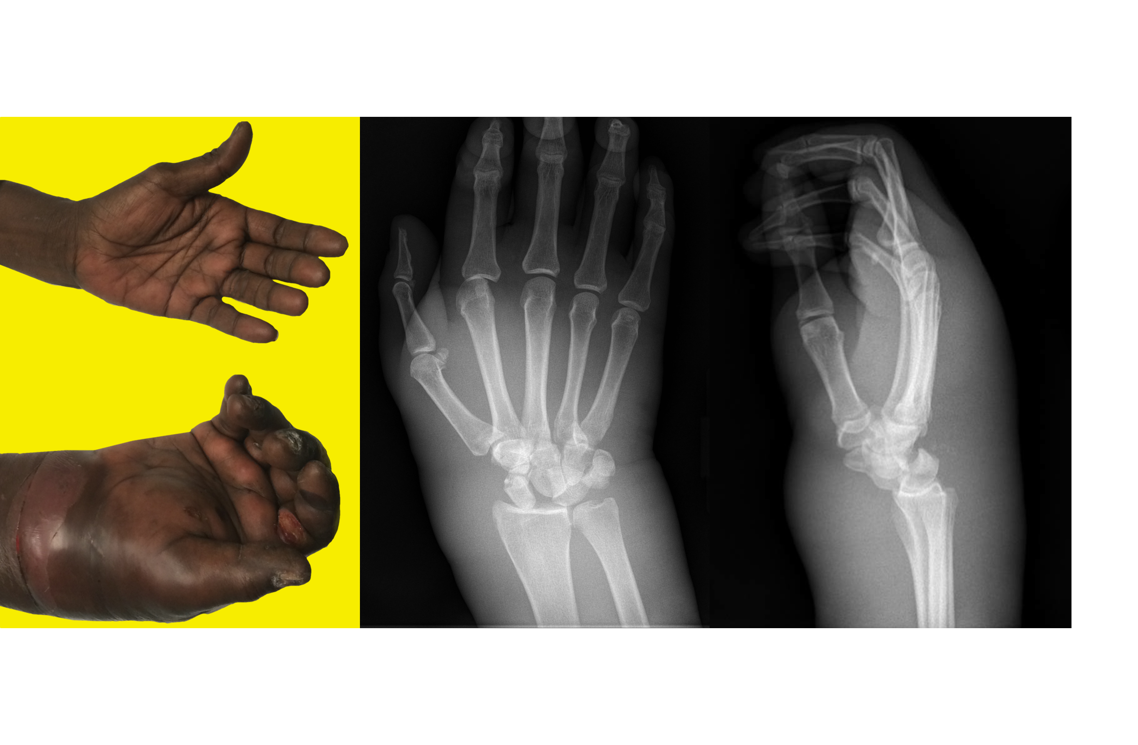
45 year-old right-hand dominant patient presents with right hand pain from a prior injury to hand. Patient has also been injecting subcutaneous heroin into hand for relief. What's the diagnosis?
 

Show Answer
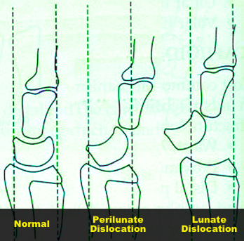
Perilunate dislocation (volar displacement)...and severe hand cellulitis.

Perilunate and Lunate dislocations

  • Lunate dislocation: normal alignment of the distal radius, carpels, and metacarpals; the lunate is displaced 
  • Perilunate dislocation: lunate will demonstrate normal alignment and the remainder of the carpels and metacarpals will typically be displaced in a dorsal direction (occasionally in a volar direction as in this case)
  • These dislocations occur following high-energy trauma, typically with a hyperextended and ulnar deviated hand
  • Plain films are usually enough to make diagnosis, but CT may be used to assess for associated occult fractures (e.g., diagnose co-existing scaphoid fracture


Show References

Follow me on Twitter (@criticalcarenow) or Google+ (+criticalcarenow)


Title: What's the Diagnosis?

Category: Visual Diagnosis

Posted: 7/14/2014 by Haney Mallemat, MD

Question

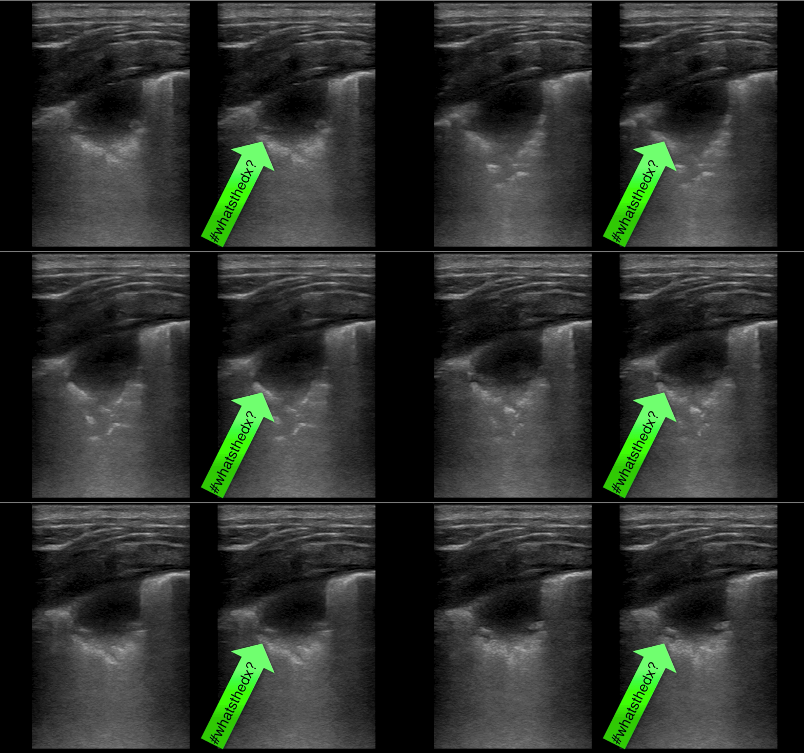
30 year-old presents with cough & fever. CXR shows mild right lower lobe pneumonia. The lung ultrasound of the right lower lobe is shown below. What's the diagnosis? 
Show Answer

Answer: Pulmonary abscess

  • Patient initially denied IV drugs of abuse, but later disclosed use when this was found
  • Chest CT demonstrated several pulmonary abscesses and the patient was admitted for a workup of endocarditis 

Bottom-line: Not only is ultrasound faster than a CXR in diagnosing pneumonia, unexpected diagnoses may be found that change management

Show References

Follow me on Twitter (@criticalcarenow) or Google+ (+criticalcarenow)


Title: Back to Basics Series: Respiratory Failure & Non-Invasive Ventilation

Category: Critical Care

Posted: 7/8/2014 by Haney Mallemat, MD

  • When considering starting a patient on non-invasive ventilation (NIV), ask yourself whether the patient is having a problem of oxygenation (Type I respiratory failure) or a problem of CO2 removal or ventilation (i.e., Type II respiratory failure); don’t forget both types can be present, simultaneously
  • Examples of Type I problems are pneumonia and pulmonary edema; examples of Type II problems are COPD, drug overdose, and neuromuscular disease (e.g., myasthenia gravis). Once the underlying problem is identified, selecting the type of NIV is straight-forward. 
  • There are only two interventions for type I disorders: 1) increase fio2 and/or 2) increase mean airway pressure (positive end-expiratory pressure; a.k.a. PEEP). There are only two interventions for type II disorders: 1) increase tidal volume and/or 2) increase respiratory rate 
  • Continuous positive airway pressure (CPAP) only provides support for type I problems (i.e., can titrate FiO2 and PEEP); CPAP does not provide a tidal volume or a respiratory rate (needed for type II support)
  • Bi-level positive airway pressure (BPAP) provides support for type II problems; tidal volume can be titrated by increasing the pressure support and a respiratory rate can be dialed in.

Editors note: The new Back 2 Basic series will review essential critical care concepts on the first Tuesday of each month. Want a specific topic reviewed? Contact us by email or Twitter.

Show References

Follow me on Twitter (@criticalcarenow) or Google+ (+criticalcarenow)


Title: What's the Diagnosis? Image by UMEM alumni Dr. Adam Friedlander

Category: Visual Diagnosis

Posted: 7/7/2014 by Haney Mallemat, MD (Updated: 7/7/2014)

Question

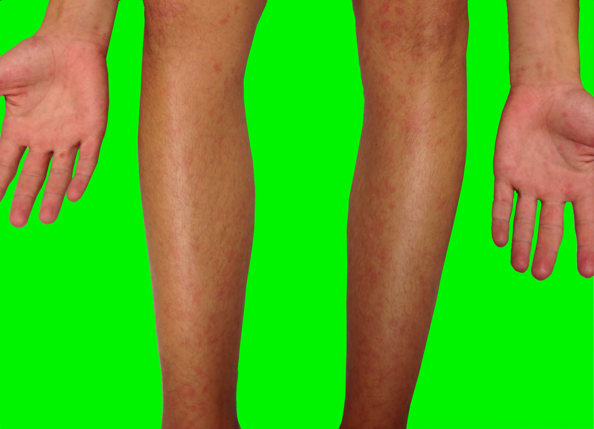
10 year-old male complains of fever and rash (shown below); no other complaints. He went camping 10-days ago. What’s the diagnosis...and what medication(s) should he receive?
Show Answer

Rocky Mountain spotted fever (RMSF)

  • Tick-borne disease caused by the organism Rickettsia rickettsia; endemic in North, Central, and South America; especially prevalent in southeastern and south-central US
  • Consider diagnosis in patients with unexplained fevers, even without history of a tick bite (reported in only 70%)
  • Common symptoms include:
    • Fever
    • Headache
    • Maculopapular rash (see below)
    • GI symptoms (e.g., nausea, vomiting, etc.)
    • Myalgias
    • CNS  (encephalitis, delirium, seizures, etc.)
  • Classic maculopapular rash appears up to 7 days after fever; starts on wrists and ankles then centripetally spreads to involve the trunk and extremities (including the palms and soles); face is typically spared. In children, periorbital edema is a diagnostic clue.
  • Up to 25% mortality if untreated, but is 5% with appropriate antibiotics; doxycycline is the preferred drug for adults and children; chloramphenicol is an alternative agent and used primarily in pregnant women.

Show References

Follow me on Twitter (@criticalcarenow) or Google+ (+criticalcarenow)


Title: What's the Diagnosis? Case by UMEM alumni Dr. Ari Kestler

Category: Visual Diagnosis

Posted: 6/30/2014 by Haney Mallemat, MD

Question

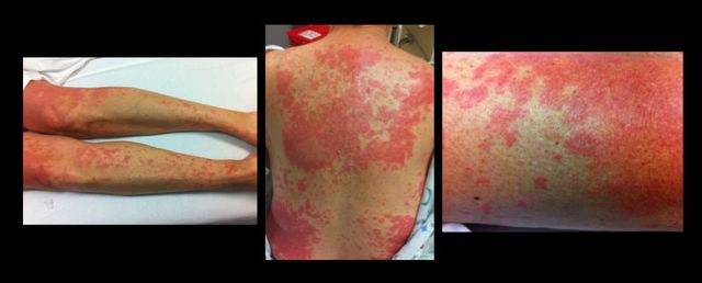
49 year-old female on trimethoprim/sulfamethoxazole presents with a rash & lesions on her oral mucus membranes. What's the diagnosis?
Show Answer

Answer: Steven-Johnson Syndrome (SJS)

  • SJS: involvement of <10% body-surface area (BSA) PLUS mucus membrane involvement (e.g., ocular, oral, genital)
  • Toxic Epidermal Necrolysis (TEN): involvement of >30% BSA PLUS mucus membrane involvement
  • Occurs secondary to immune-complex hypersensitivity reaction from viral, bacterial, or chemical exposure (Sulfonamides > PCN > Cephalosporins)
  • Management:
    • Remove possible trigger(s)
    • Manage like a severe burn (supportive care with hemodynamic support, electrolyte management, analgesia)
    • Strongly consider admission to an ICU
 

Show References

Follow me on Twitter (@criticalcarenow) or Google+ (+criticalcarenow)


Title: What's the Diagnosis?

Category: Visual Diagnosis

Posted: 6/23/2014 by Haney Mallemat, MD (Updated: 6/23/2014)

Question

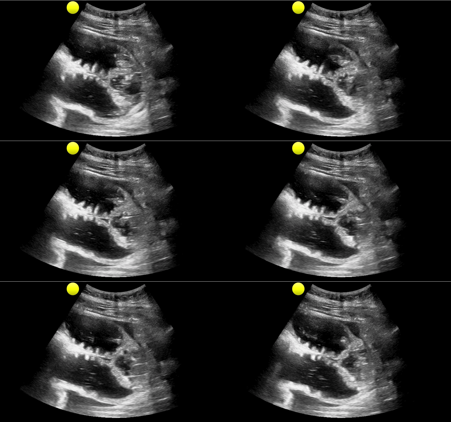
35 year-old female presents with nausea and vomiting 1 week post-op for an abdominal surgery. Abdominal ultrasound is below; what's the diagnosis? 
Show Answer

Small bowel obstruction (SBO)

Ultrasound for Small bowel obstruction (SBO)

  • Wait….ultrasound can be used to detect an SBO? Yes, it can!
  • Historically, abdominal XRay has a 77% and 57% (sensitivity and specificity, respectively) for SBO; ultrasound has 88% and 96% sensitivity and specificity, respectively.
  • Want to learn more about ultrasound for SBO? Click here for a podcast describing the technique by Mike and Matt from the Ultrasound Podcast (@ultrasoundpodcast)
  • Bottom line: Consider using ultrasound as your initial screening test for SBO, rather than an abdominal XR

Show References

Follow me on Twitter (@criticalcarenow) or Google+ (+criticalcarenow)


Title: What's the Diagnosis? Case by Dr. Swati Singh, Amanda Quiller, P.A.

Category: Visual Diagnosis

Posted: 6/16/2014 by Haney Mallemat, MD (Updated: 6/16/2014)

Question

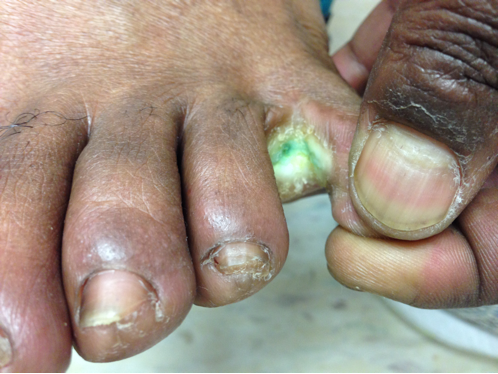
41year-old male without past medical history presents with the image below. What's the diagnosis and what's the most likely causative organism?
Show Answer

Gram-Negative web-space infection; most common etiologic organism is Pseudomonas

Gram Negative Web-space Infections

Gram-negative infections may occur in toe web-spaces of healthy patients, especially in areas with hyperhidrosis (excessive perspiration) and macerated skin.

Common organisms include Pseudomonas; others include Corynebacterium minutissimum (causes erythrasma, chronic superficial infection of the intertriginous areas of the skin), and other gram-negative bacteria

Woods-light examination may be clinically helpful if Pseudomonas is suspected (fluoresce green) and erythrasma will fluoresce coral red. KOH direct examination for fungal elements may show the presence of dermatophytes or Candida. 

Treatment:

  • Moisture reduction of web-spaces by keeping feet dry (e.g., open-toed shoes, drying powders, etc.)
  • Uncomplicated, superficial infections respond well to topical agents active against the causative organisms (e.g., gentamycin ointment)
  • Severe or complicated infections may require oral / parenteral antibiotics (i.e. third-generation cephalosporin or quinolone or aminoglycoside) plus antifungals; tissue removal/surgical debridement may be required.

Show References

Follow me on Twitter (@criticalcarenow) or Google+ (+criticalcarenow)

Wolff, K., & Johnson, R. J. (2009). Fitzpatrick's Color Atlas and Synopsis of Clinical Dermatology. (6th ed., pp. 662-664;698). McGraw-Hill.

http://www.aafp.org/afp/2005/0901/p833.html

http://emedicine.medscape.com/article/1055306-overview


Title: Reversing Warfarin-Associated Bleeding: The EPAHK Study

Category: Critical Care

Posted: 6/10/2014 by Haney Mallemat, MD

  • A feared complication of patients taking vitamin K antagonists (VKA) is life-threatening bleeding (LTB), including intracranial hemorrhage (ICH).
  • Prothrombin complex concentrate (PCC; containing factors 2,7,9,and 10) rapidly reverses VKA-associated bleeding. Despite a rapid reversal of the INR, there is little literature demonstrating a mortality benefit.
  • The EPAHK study was observational-cohort that examined the 7-day mortality of guideline-concordant administration of PCC and vitamin K (GC-PCC-K) for multiple-types of patients with warfarin-associated bleeding.
  • The study demonstrated patients who received GC-PCC-K within 8 hours of presentation had a two-fold decrease in 7-day morality; there was a three-fold reduction when only ICH was considered.


Show References

Follow me on Twitter (@criticalcarenow) or Google+ (+criticalcarenow)

Tazarourte, K. et al. Guideline-concordant administration of prothrombin complex concentrate and vitamin K is associated with decreased mortality in patients with severe bleeding under vitamin K antagonist treatment (EPAHK study). Crit Care. 2014 Apr 24;18(2):R81.


Title: What's the Diagnosis? Case by Dr. Maite Huis in 't Veld

Category: Visual Diagnosis

Posted: 6/10/2014 by Haney Mallemat, MD (Updated: 6/9/2014)

Question

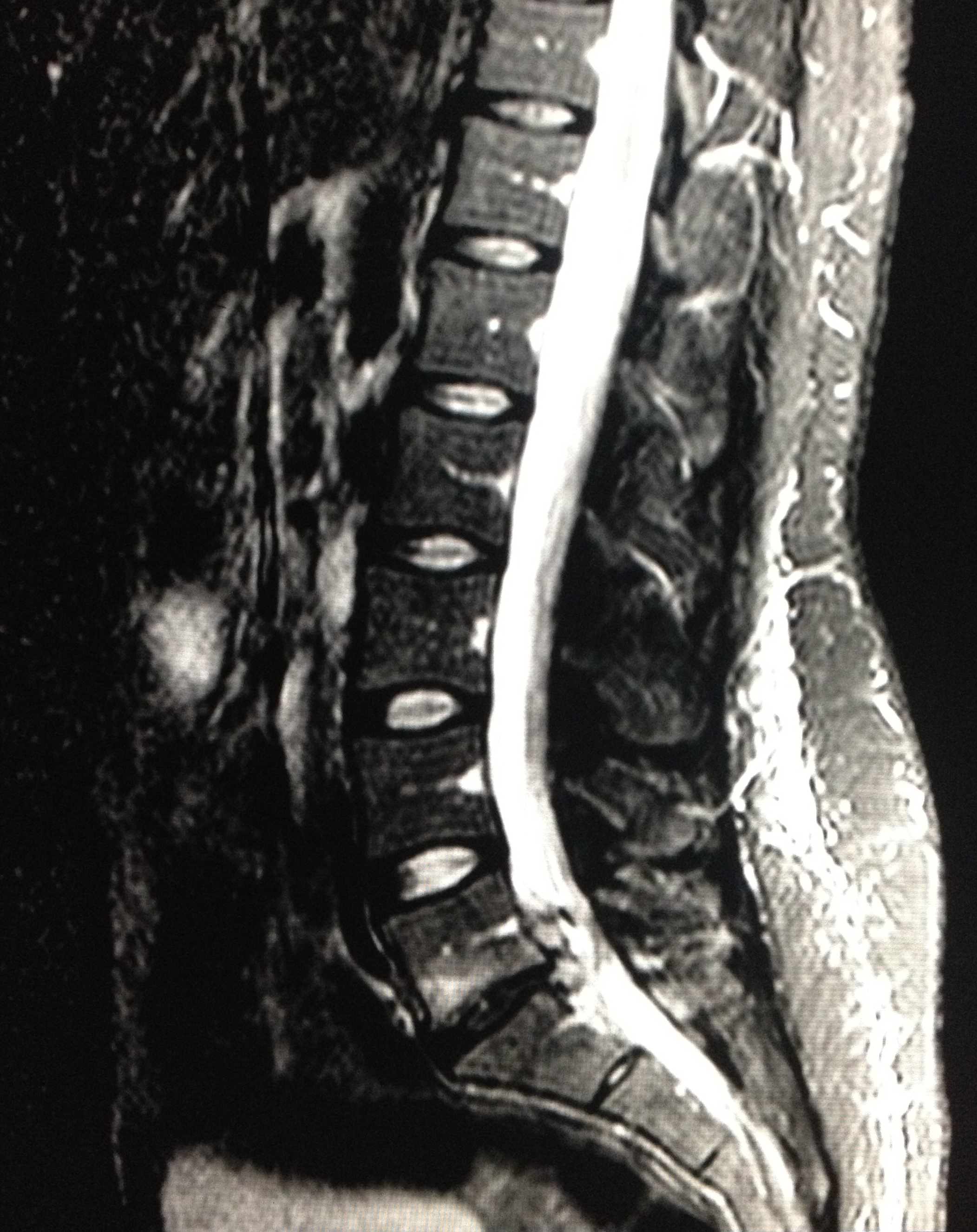
28 year-old female with history of chronic back pain presents with right-foot numbness and inability to move her foot at the ankle joint. What’s the diagnosis and what neurologic finding would you expect to find?
 

Show Answer
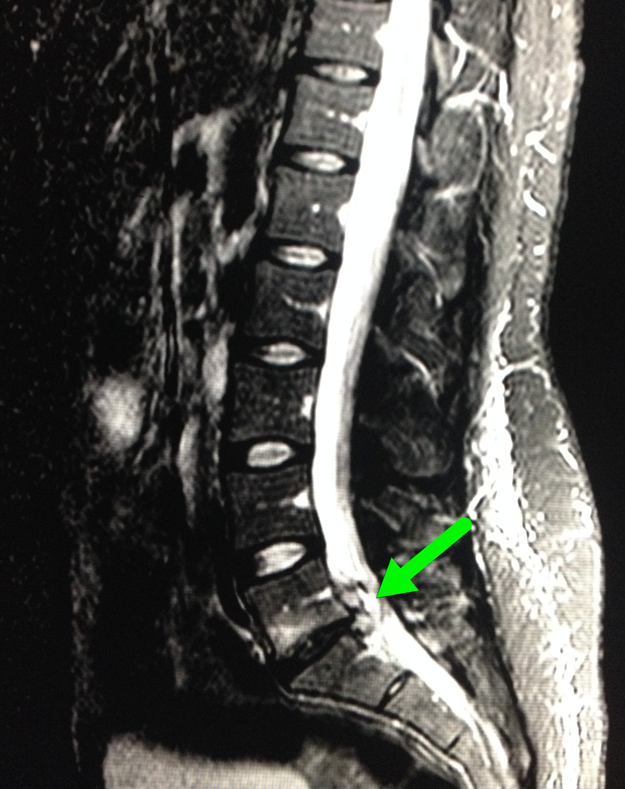
Large disc bulge at L5-S1

  • Patient in the case was found to have reduced:
    • Dorsiflexion (2/5)
    • Plantar flexion (4/5)
    • Reduced sensation in L5-S1 dermatome
  • Neurosurgery emergently took the patient to operating room for an L5-S1 discectomy.
  • Patient discharged the following day with improved motor and sensory findings

Show References

Follow me on Twitter (@criticalcarenow) or Google+ (+criticalcarenow)


Title: What's the Diagnosis? Image by Dr. Brandi Knight

Category: Visual Diagnosis

Posted: 6/2/2014 by Haney Mallemat, MD

Question

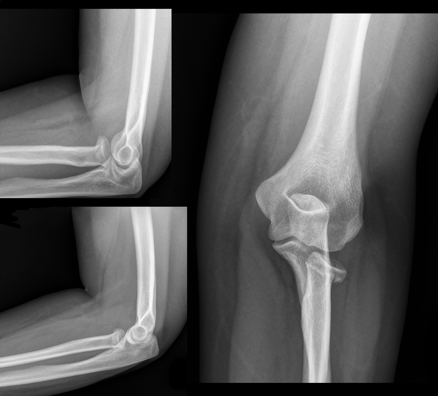
30 year-old female with elbow pain following a fall. What's the diagnosis? (bonus points if you name the fracture with an associated radio-ulnar joint dislocation)
Show Answer
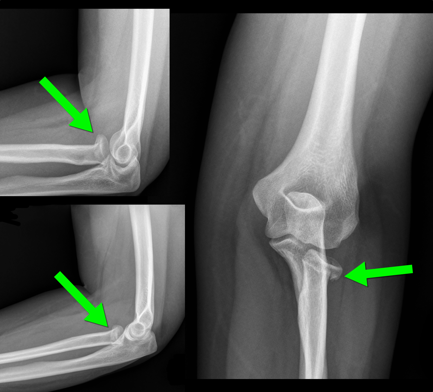
Radial Head Fracture           


  • Typically secondary to fall on an outstretched hand; head of radius collides axially with capitulum of humerus
  • Most common type of adult elbow fracture; radial neck fractures more common in children; 
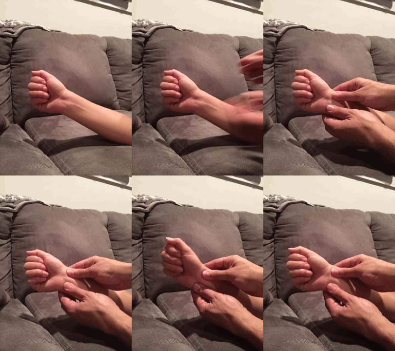
  • Must be on the lookout for the Essex-Lopresti fracture (bonus answer)
    • Radial head fracture + dislocation of the radio-ulnar joint (typically disruption of interosseous membrane
    • Repair early (typically ORIF) otherwise there may be loss of forearm extension, pronation, and supination
    • Look for distal radio-ulnar tenderness / instability by grabbing distal radius and ulnar and moving in opposite directions (see video below)

d


Show References

http://www.wheelessonline.com/ortho/radial_head_frx

Follow me on Twitter (@criticalcarenow) or Google+ (+criticalcarenow)


Title: What's the Diagnosis? Images by Dr. Michael Abraham

Category: Visual Diagnosis

Posted: 5/26/2014 by Haney Mallemat, MD

Question

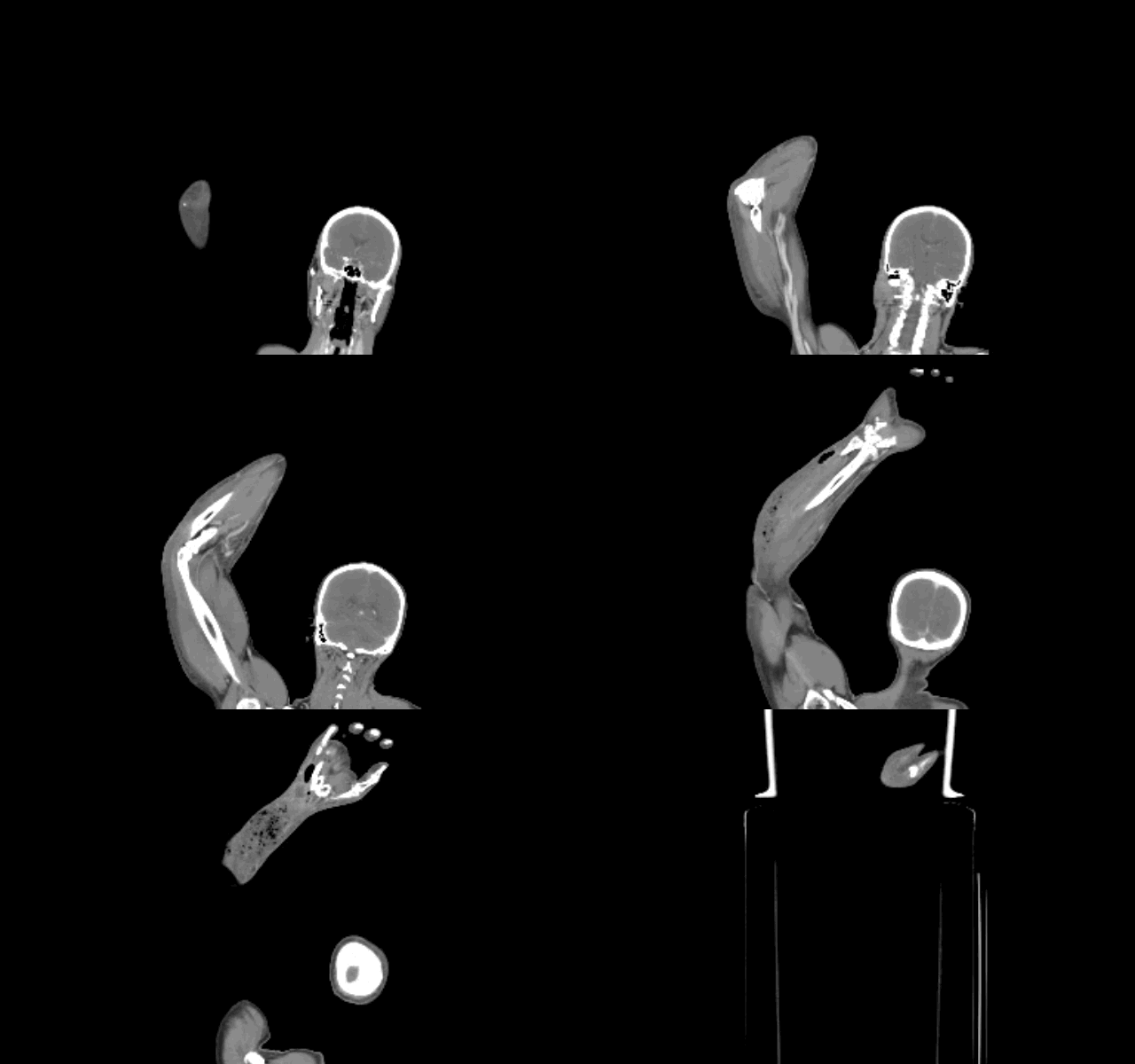
Diabetic patient with active intravenous drug use presents with hypotension, fever, and tenderness of right arm. What's the diagnosis and what antibiotic(s) would you start?
Show Answer

Right arm with necrotizing fasciitis

  • Necrotizing fasciitis (NF) may occur secondary to either mono-microbial (e.g., Strep, Staph, Clostridial species, etc.) or poly-microbial (above in addition to anaerobes and/or gram negative) bacteria; etiology may be unclear from history alone.
  • Urgent surgical debridement is the key to management but because the infection can spread rapidly, antibiotics are essential while awaiting surgery.
  • Empiric antibiotics should provide broad-spectrum coverage as well as cover MRSA if the patient is at increased risk (e.g., recent hospital admission).
  • Clindamycin is often been added for its theoretical anti-toxin effects from certain streptococcal species. Although there is little in vivo data to support this practice, it is still a reasonable approach considering the high-morbidity and mortality from NF
  • Other adjunctive therapies to consider are IVIG and hyperbaric oxygen

Show References

http://lifeinthefastlane.com/education/ccc/necrotising-fasciitis/

Follow me on Twitter (@criticalcarenow) or Google+ (+criticalcarenow)


Previous  |  1 |  2 |  3 |  4 |  5 |  6 |  7 |  8 |  9 |  10 |  ... |  17 |  Next
University of Maryland School of Medicine logo
  • Departments
  • Programs
  • Research Centers
  • Institutes
  • Graduate Education
  • CIBR Cores
  • Offices of the Dean
    • University of Maryland, Baltimore
    • University of Maryland Medical Center
    • Medical Alumni Association of the University of Maryland
    • Health Sciences and Human Services Library
University of Maryland School of Medicine
655 W. Baltimore Street
Baltimore, MD 21201
Contact Us

© University of Maryland School of Medicine

    • Webmaster
    • Web Accessibility
    • Site Index
    • UMB Hotline
    • Employment
    • Map