University of Maryland School of Medicine logo
Emergency Medicine
All Departments
UMSOM Assistant
UMEM Educational Pearls
  • Home
  • Departments
  • Emergency Medicine
  • CME
  • UMEM Educational Pearls
  • CME
  • Courses and Conferences
  • Free Open Access Medical Education
  • UMEM Educational Pearls

Search

61-80 of 321 results by Haney Mallemat

Previous  |  1 |  2 |  3 |  4 |  5 |  6 |  7 |  8 |  9 |  ... |  17 |  Next

Title: What's the Diagnosis?

Category: Visual Diagnosis

Posted: 1/19/2015 by Haney Mallemat, MD

Question

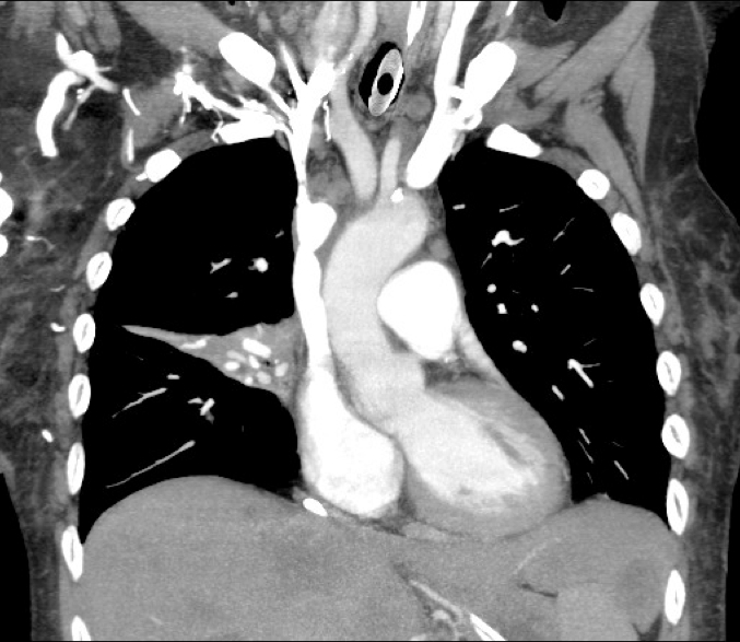
60 year-old male presents with 6 months of weight loss,epistaxis, and increased headache when bending over. What's the diagnosis?
Show Answer
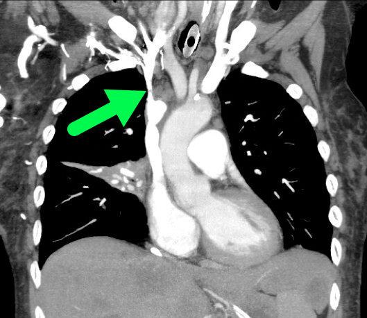
SVC Syndrome

  • SVC syndrome is caused by compression or obstruction of the superior vena cava blocking anterograde flow.
  • Most cases are secondary to extrinsic compression by malignancy. Other causes are secondary to internal obstruction (e.g., thrombosis).
  • Symptoms present sub-acutely, worsen with bending over, and are secondary to increased venous pressure in the head and neck (e.g., epistaxis, headache, tinnitus, conjunctival injection, neck swelling, etc.).
  • Treatment focuses on reversing the underlying cause (e.g., radiation or chemotherapy if due a sensitive tumor) and symptomatic treatment:
    • Steroids, diuretics, and hyperosmolar agents to reduce edema
    • Head of bed elevation to reduce brain edema
    • Anti-emetics to prevent vomiting-associated increases in intracranial pressure

Rice TW, Rodriguez RM, Light RW. The superior vena cava syndrome: clinical characteristics and evolving etiology. Medicine (Baltimore). Jan 2006;85(1):37-42.

Nunnelee JD. Superior vena cava syndrome. J Vasc Nurs. Mar 2007;25(1):2-5

Show References

Follow me on Twitter (@criticalcarenow) or Google+ (+criticalcarenow)


Title: What's the Diagnosis? Case by Dr. Ahmed Alrasheedi @aalrasheedi

Category: Visual Diagnosis

Posted: 1/12/2015 by Haney Mallemat, MD

Question

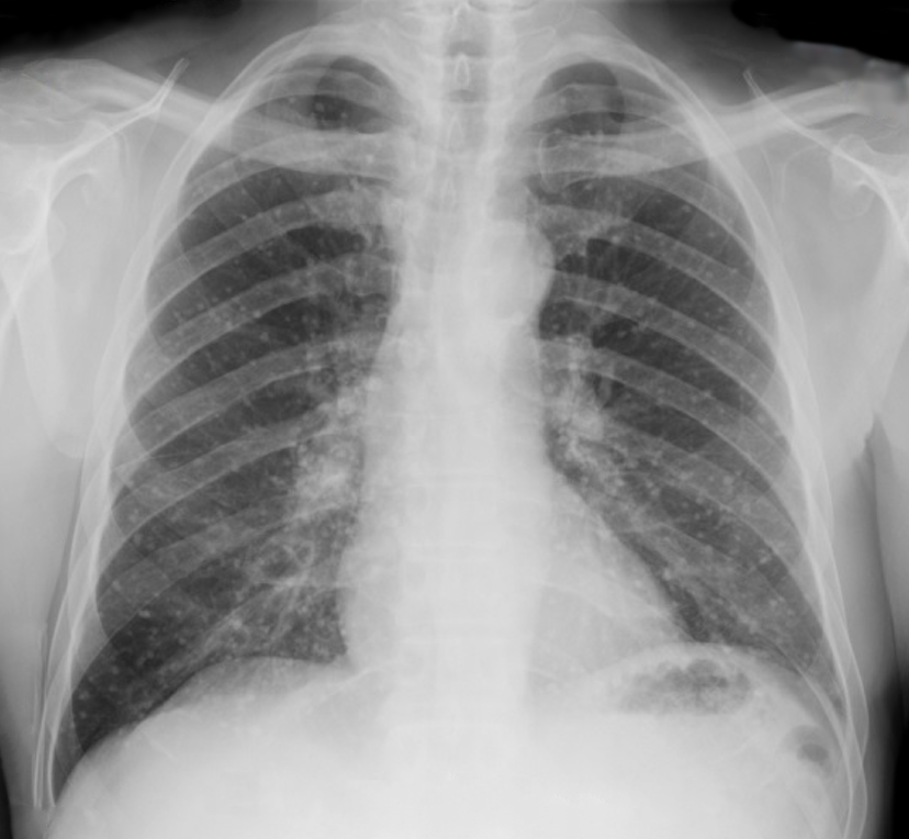
60 year-old male presents with rhinorrhea, body aches, and dry cough for two days. He recently moved here from Kentucky. What’s the diagnosis (please note that there are several differentials based on CXR)?


Show Answer

Answer Histoplasmosis

Histoplasma capsulatum is the fungus that causes histoplamosis, and is endemic to soil in the “Histo belt” of the U.S. (including all of Arkansas, Kentucky, Missouri, Tennessee, and West Virginia and parts Alabama, Illinois, Indiana, Iowa, Kansas, Louisiana, Maryland, Mississippi, Nebraska, Ohio, Oklahoma, Texas, and Virginia); it is found internationally as well.

Outbreaks have occurred following digging up the ground at construction at sites where this yeast lives in the soil, but spores can also become airborne and inoculate people.

Most cases are asymptomatic, but it is associated with a variety of pulmonary syndromes and can have extrapulmonary findings such as splenic calcifications. Disseminated disease typically occurs in immunocompromised patients

Treatment includes itraconazole for mild to moderate disease and amphotericin for disease that is severe or widespread disease.


Differentials:

  • Blastomycosis
  • Septic emboli
  • Sarcoidosis
  • Tuberculosis (milliary)
  • Aspergillosis
  • Carcinoid lung tumor
  • Atypical or viral pneumonias    


Title: What's the Diagnosis? Case by Dr. Joshua McClain and Dr. Yemi Adebayo

Category: Visual Diagnosis

Posted: 1/5/2015 by Haney Mallemat, MD

Question

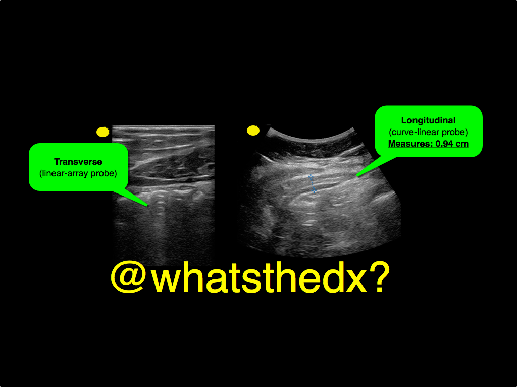
A male patient presents with right lower quadrant pain. The ultrasound is shown at the point of maximal tenderness. The diameter of the structure (image on right) is about 0.94cm. What is this structure and what's the diagnosis?
Show Answer

Appendicitis
  • Ultrasound can be used to diagnose appendicitis; a linear transducer is preferred in thinner / pediatric patients, while a curvilinear probe is may be used larger patients
  • Acute appendicitis causes irritation of the abdominal wall, so when looking for the appendix you need enough depth to visualize the peritoneal wall
  • To perform the scan, start at right upper quadrant in the transverse plane and slide down the abdomen towards the right iliac fossa until bowel gas has disappeared, typically the ileocecal junction. Alternatively, you can start scanning the point of maximal tenderness (i.e. the appendix)
  • Identifying a non-compressible and dilated appendix measuring greater than 6mm in transverse diameter suggesting the diagnosis
  • Color flow can be added to identify hyperemia around the appendix, which increases the specificity of the study.
  • More info on the scan can be found here and two podcasts from the incredible @ultrasoundpodcast guys part I and part II

Show References

Follow me on Twitter (@criticalcarenow) or Google+ (+criticalcarenow)


Title: What's the Diagnosis?

Category: Visual Diagnosis

Posted: 12/30/2014 by Haney Mallemat, MD (Updated: 12/30/2014)

Question

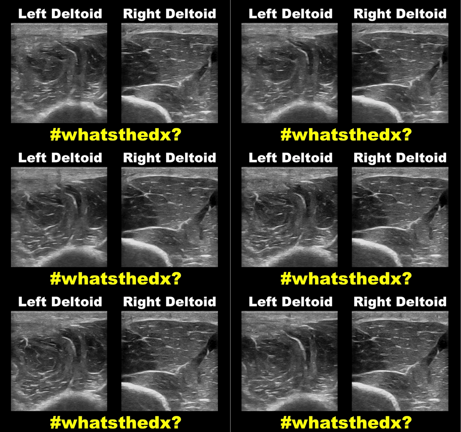
19 year-old male complaining of left arm pain one week after injecting anabolic steroids into his sholder. What's the diagnosis?
Show Answer
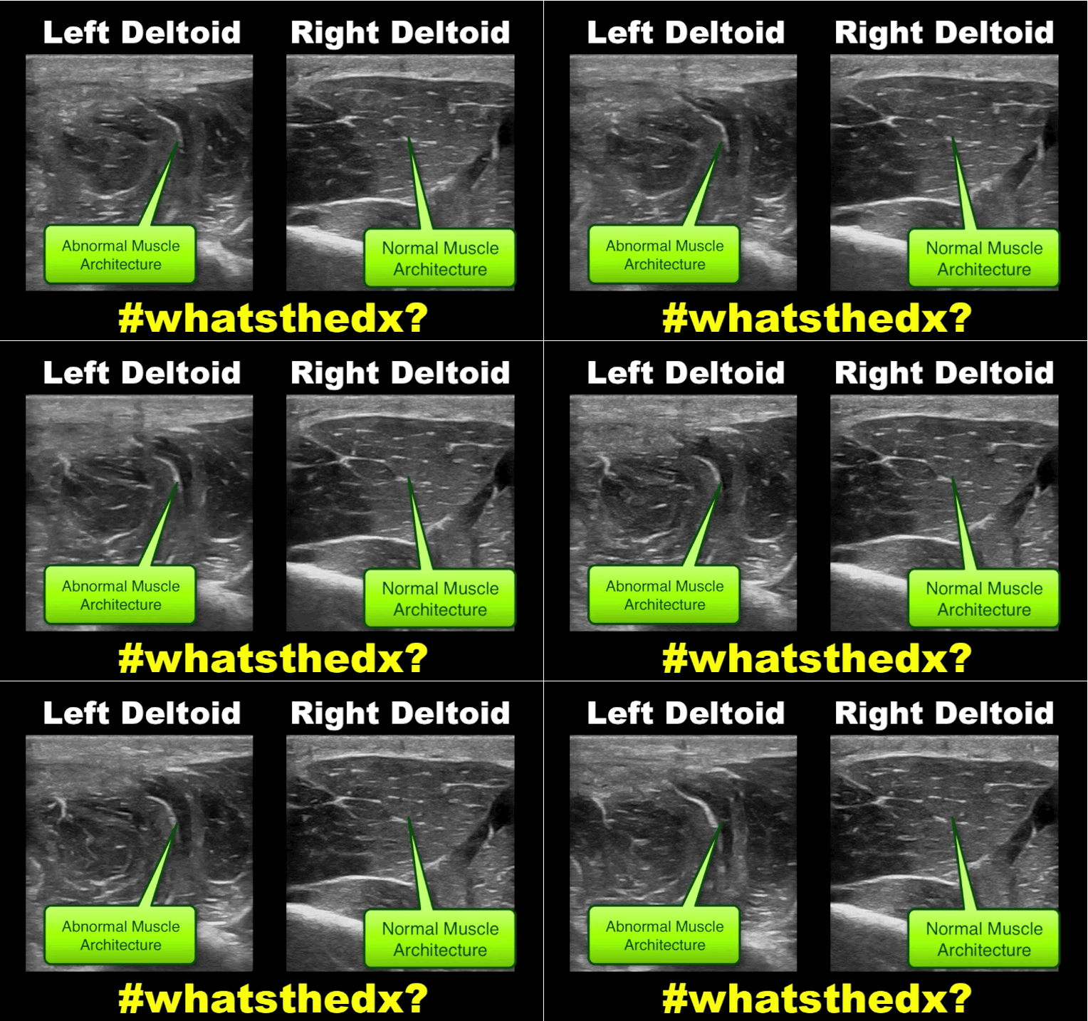
Myositis of the deltoid muscle

Show References

Follow me on Twitter (@criticalcarenow) or Google+ (+criticalcarenow)


Title: Intraarterial therapy: Time for change or time for pause?

Category: Critical Care

Posted: 12/23/2014 by Haney Mallemat, MD

Treating ischemic strokes with interventional therapies (e.g., clot retrievers, stents, intra-arterial tPA, etc.) is nothing new, but there has never been a randomized control trial demonstrating benefit until recently.

The prospective MR CLEAN trial evaluated whether interventional therapies (i.e., either mechanical intervention or intra-arterial tPA) would confer benefit; patients were included if there was an acute occlusion within the proximal intracranial portion of the anterior cerebral circulation.

90% of patients received alteplase prior to randomization; there were 233 patients in the intervention group (alteplase + intraarterial intervention) and 267 patients in the usual care care arm (alteplase only); all patients were treated within 6 hours of symptoms onset

The primary outcome was functional independence at 90 days; an absolute difference of 13.5 percentage points favoring the intervention group was found. There were no significant differences in mortality or symptomatic intracerebral hemorrhage.

Despite these exciting results, we must pause and ask why this was this the first randomized trial demonstrating benefit when previous trials could not? Here are three blogs posts that deep dive this question and raise even more questions:

  • EM Nerd
  • Emergency Medicine Literature of Note
  • St. Emlyn's

Show References

Follow me on Twitter (@criticalcarenow) or Google+ (+criticalcarenow)

Berhemer, et al. A Randomized Trial of Intraarterial Treatment for Acute Ischemic Stroke. N Engl J Med. 2014 Dec 17.


Title: What's the Diagnosis?

Category: Visual Diagnosis

Posted: 12/23/2014 by Haney Mallemat, MD (Updated: 12/23/2014)

Question

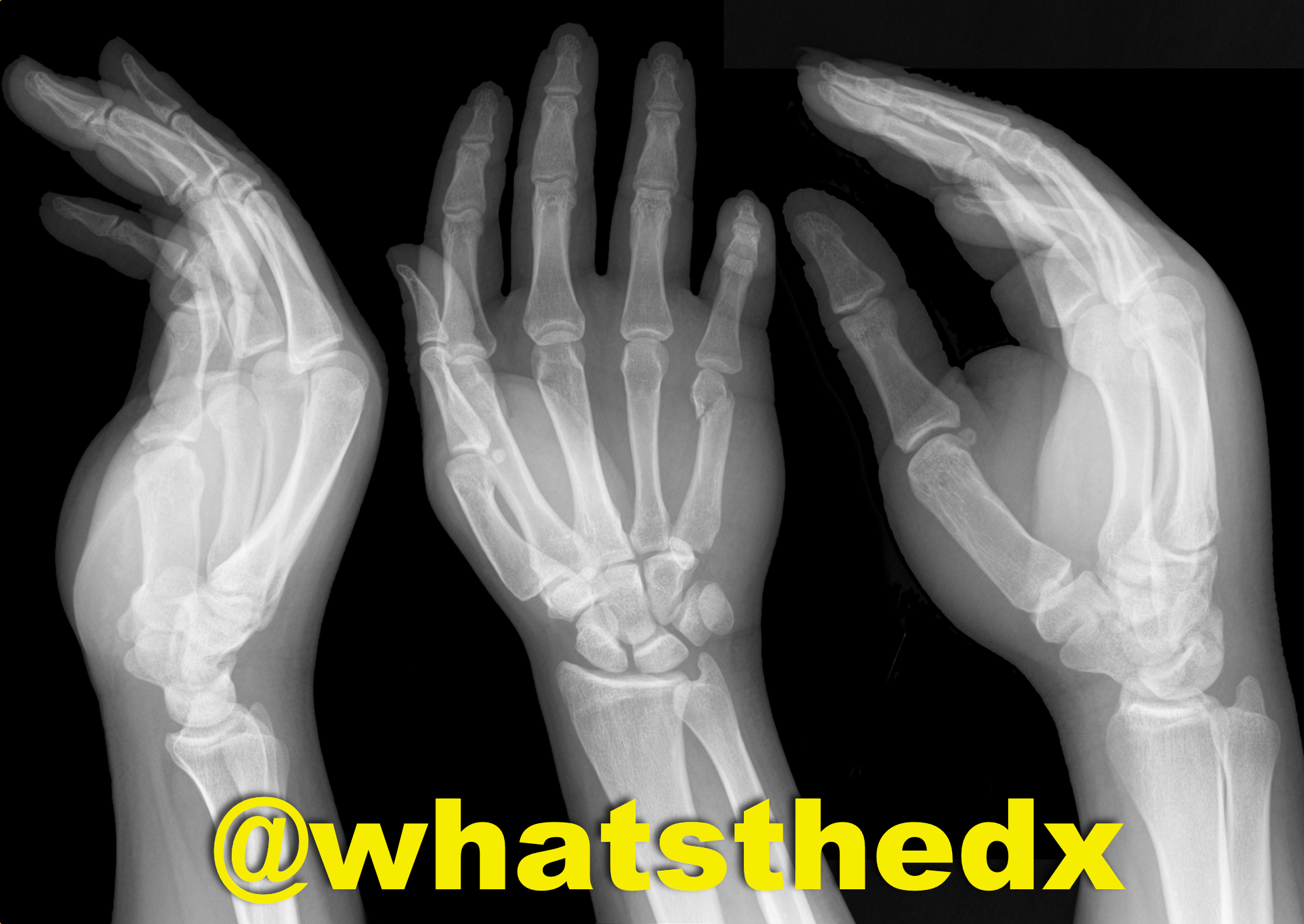
Hand pain following fist versus face. What's the diagnosis and what nerve block would you use?
Show Answer
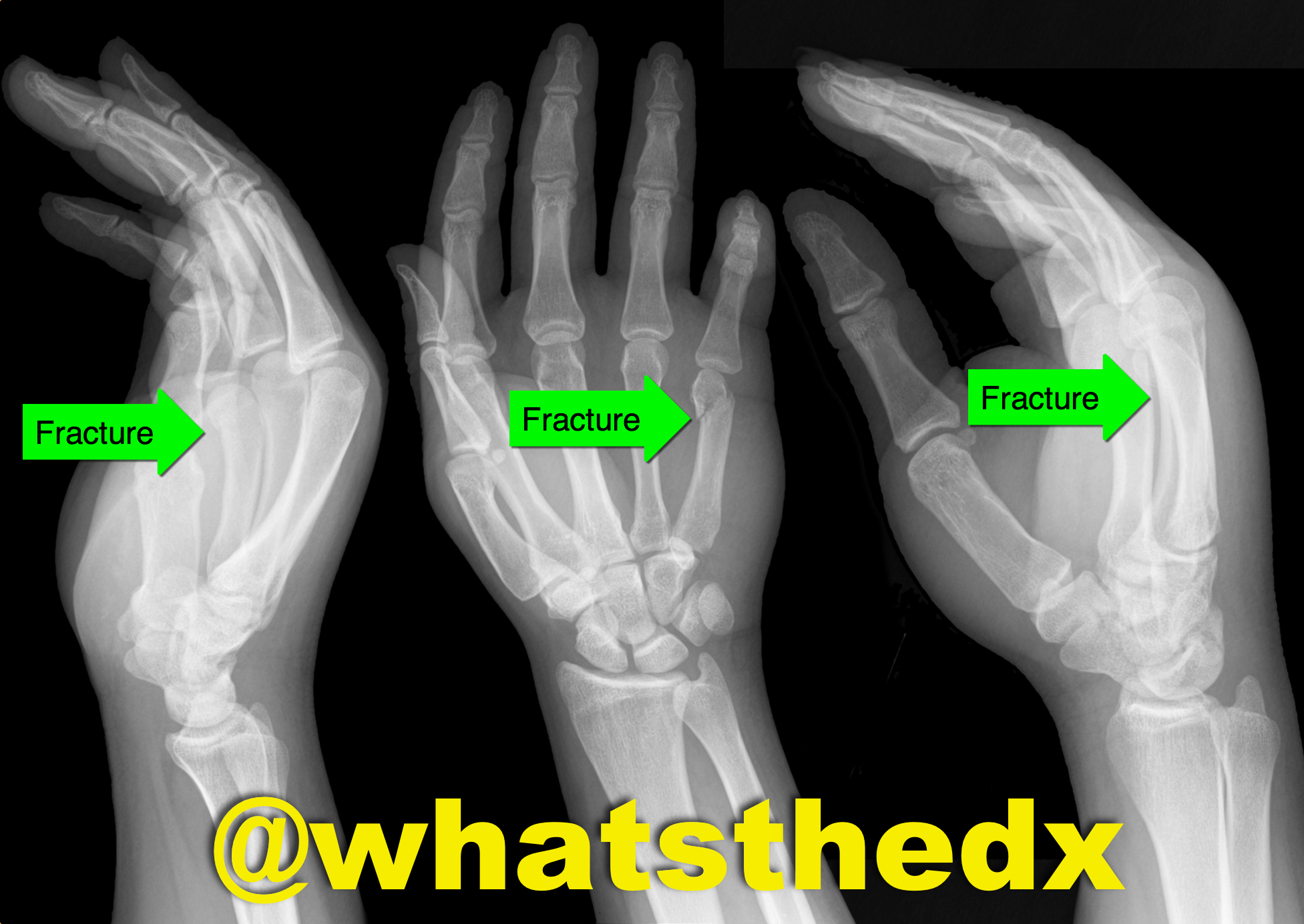
Boxer's fracture. Read more here.

Pain control can be achieved with an ulnar nerve block (e.g., reducation if if angulated). A video for the technique using ultrasound can be found here.

Show References

Follow me on Twitter (@criticalcarenow) or Google+ (+criticalcarenow)


Title: What's the Diagnosis? Dr. Ahmed Alrasheedi

Category: Visual Diagnosis

Posted: 12/15/2014 by Haney Mallemat, MD

Question

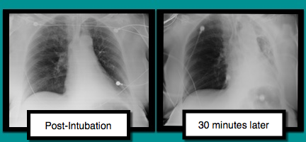
A patient is intubated for respiratory failure and the post-intubation CXR is shown on the left. 30 minutes later the patient desaturates and another CXR is obtained (the one on the right). What’s the diagnosis and what should you do?
Show Answer

Mucus plug

There are several conditions to consider when patients are having difficultly being ventilated. The approach to such a patient can be remembered with a mnemonic found here.

In this case, conditions to consider are:

  • right main-stem intubation (can be seen with ultrasound; here)
  • mucus plug (shift is towards problem)
  • tension pneumothorax (shift is away from problem); use ultrasound

Treating mucus plugs can usually be treated as follows:

  • Deep tracheal suctioning
  • Disconnecting patient from the vent and bagging
  • Chest physiotherapy (i.e., percussing the chest) on the affected area
  • Bronchoscopy, if above does not result in resolution

Show References

Follow me on Twitter (@criticalcarenow) or Google+ (+criticalcarenow)


Title: What's the Diagnosis? Case by Adeleke Oni, MD

Category: Visual Diagnosis

Posted: 12/8/2014 by Haney Mallemat, MD

Question

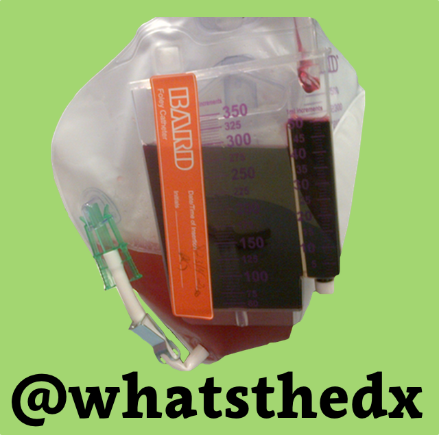
Patient was found in a house fire and was given a certain medication in the Emergency Room. The patient’s urine turned this color (red), what’s the diagnosis?
Show Answer

Hydroxocobalamin (i.e., Cyanokit)

Cyanide toxicity

  • Occurs secondary to combustion of natural and synthetic products producing cyanide
  • Patients recovered from fires should have a lactate level drawn; levels greater than 10 are concerning for cyanide toxicity.  
  • The Cyanokit contains hydroxocobalamin, a form of vitamin B12 that is used as the antidote.
  • Hydroxocobalmin has many side-effects including self-limited hypertension, flushing of the skin, and red urine for up to 14 days.  
  • See a previous UMEM post by Dr. Bryan Hayes for more information 

Show References

Follow me on Twitter (@criticalcarenow) or Google+ (+criticalcarenow)


Title: What's the Diagnosis? Case by Dr. R. Gentry Wilkerson

Category: Visual Diagnosis

Posted: 12/1/2014 by Haney Mallemat, MD

Question

Patient presents with dyspnea. What's the diagnosis and name three potential causes (can be specific to the case or in general)?
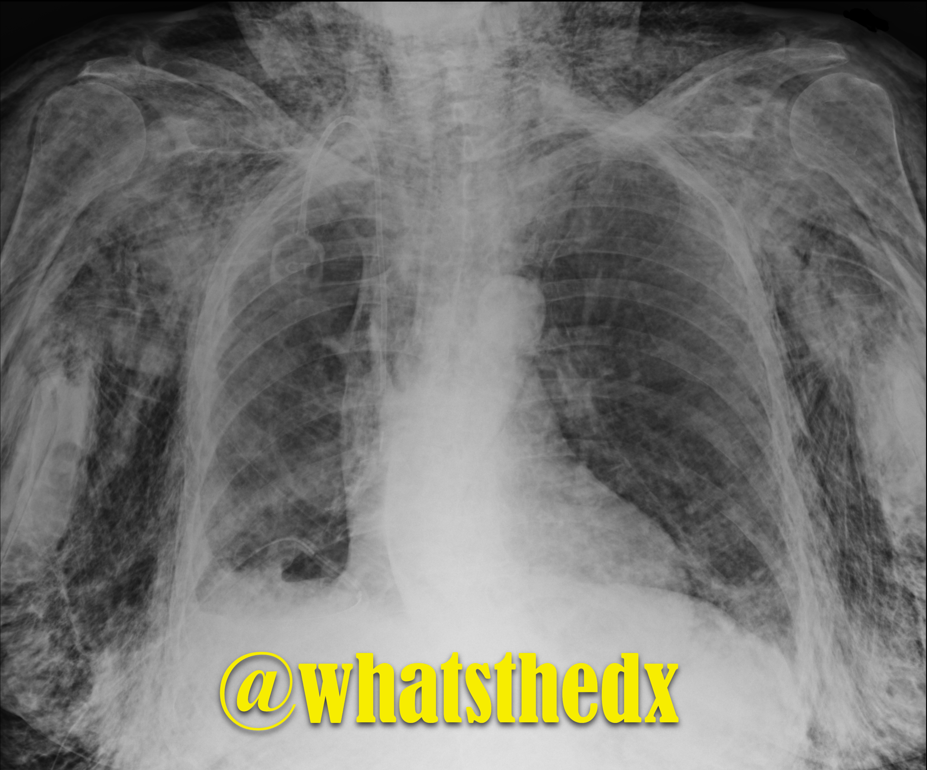
Show Answer
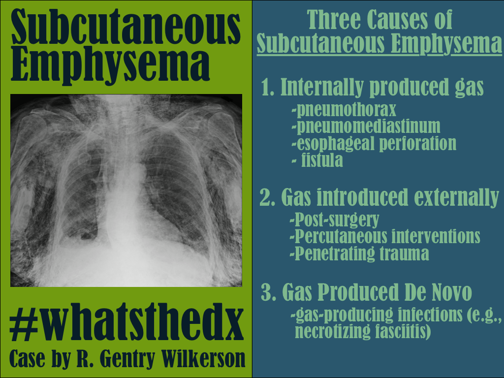
Show References

Follow me on Twitter (@criticalcarenow) or Google+ (+criticalcarenow)


Title: Tips For The Resuscitationist #1*: AEIOU

Category: Critical Care

Posted: 11/25/2014 by Haney Mallemat, MD

Running a successful resuscitation not only means doing everything right, but also remembering all the things that can go wrong. A.E.I.O.U. is a simple mnemonic that can help you remember the simple things that are sometimes forgotten during a medical resuscitation.

A – Advanced airway equipment to bedside, as well as checking the correct placement of the Airway if a patient is intubated in the field. Also consider adding another A, for Arterial line; early placement can help with pulse checks and an accurate assessment of blood pressure should there be return of spontaneous circulation (ROSC); the femoral site is fast and accurate.

E – End-tidal CO2 (ETCO2) helps detect ROSC. Ask for the ETCO2 monitor to be set up right after you receive notification of an arrest in transit; ETCO2 requires time to set-up / calibrate

I – Intraosseous line(s); compared to peripheral or central venous access, IO’s are faster, safer, and any medication can be administered through it, including vasopressors / inotropes.

O – Order (i.e., “who’s who in the Resus room?); You may be the team leader or you may be assisting, but it is important that you, and everyone else in the room, know their role prior patient arrival. If you are leading the resus, be sure everyone knows who you are, and assign everyone in the room a specific task (e.g., chest compressions, IO placement, etc.). If you are assisting and have not been assigned a task, ask the resus leader what you can do to help. If there is nothing immediate for you to do then take the initiative to de-clutter the room and step outside; be nearby and ready to help, if needed.

U – Ultrasound; can help prognosticate and detect reversible causes (e.g., pericardial tamponade). Have the ultrasound machine in the room prior to patient arrival. It should be powered on, with the proper probe connected, and in the proper mode. The most experienced ultrasonographer should scan the patient during a pulse check; experience is vital because hands-off time should be minimized.


*Tips for the Resuscitationist (#TFTR) is a new series to help you to better manage your critically ill patients. Do you have an idea for a topic or do you have a tip you would like to share? Send it to us via twitter @criticalcarenow (use (#TFTR)). You can also email us here. 

Show References

Follow me on Twitter (@criticalcarenow) or Google+ (+criticalcarenow)


Title: What's the Diagnosis? Image by Dr. Yitschok Applebaum, Dr. Jonathan Hoover, and Dr. Semhar Tewelde

Category: Visual Diagnosis

Posted: 11/24/2014 by Haney Mallemat, MD (Updated: 11/25/2014)

Question

Patient with syncope and then falls down the stairs. What's the diagnosis? (hint: be very,very careful)
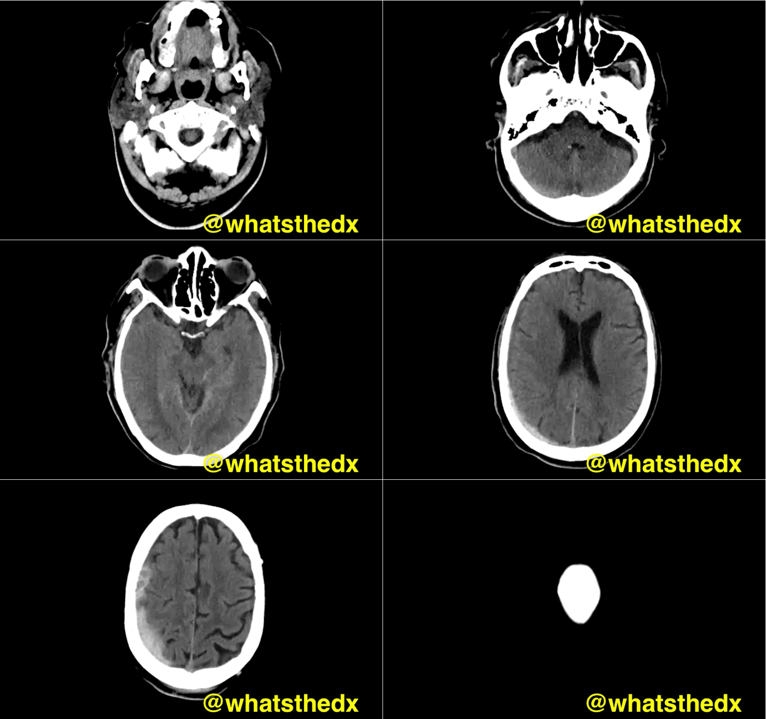
Show Answer
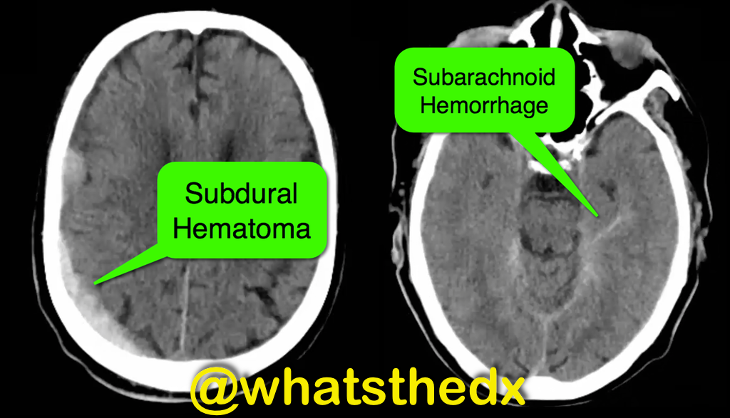
Right subdural hematoma and left subarachnoid hemorrhage


Show References

Follow me on Twitter (@criticalcarenow) or Google+ (+criticalcarenow)


Title: What's the Diagnosis? Images by Dr. Denise Fraga and Dr. Nicole Cimino-Fiallos

Category: Visual Diagnosis

Posted: 11/17/2014 by Haney Mallemat, MD

Question

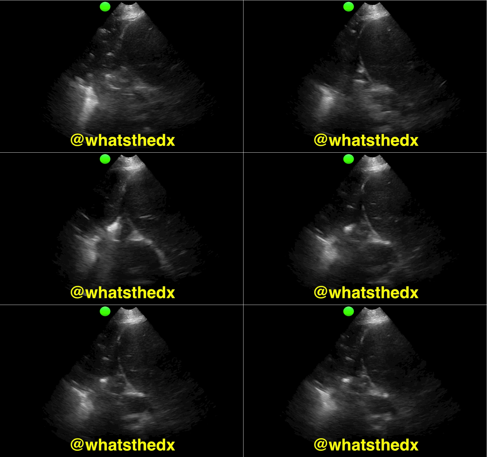
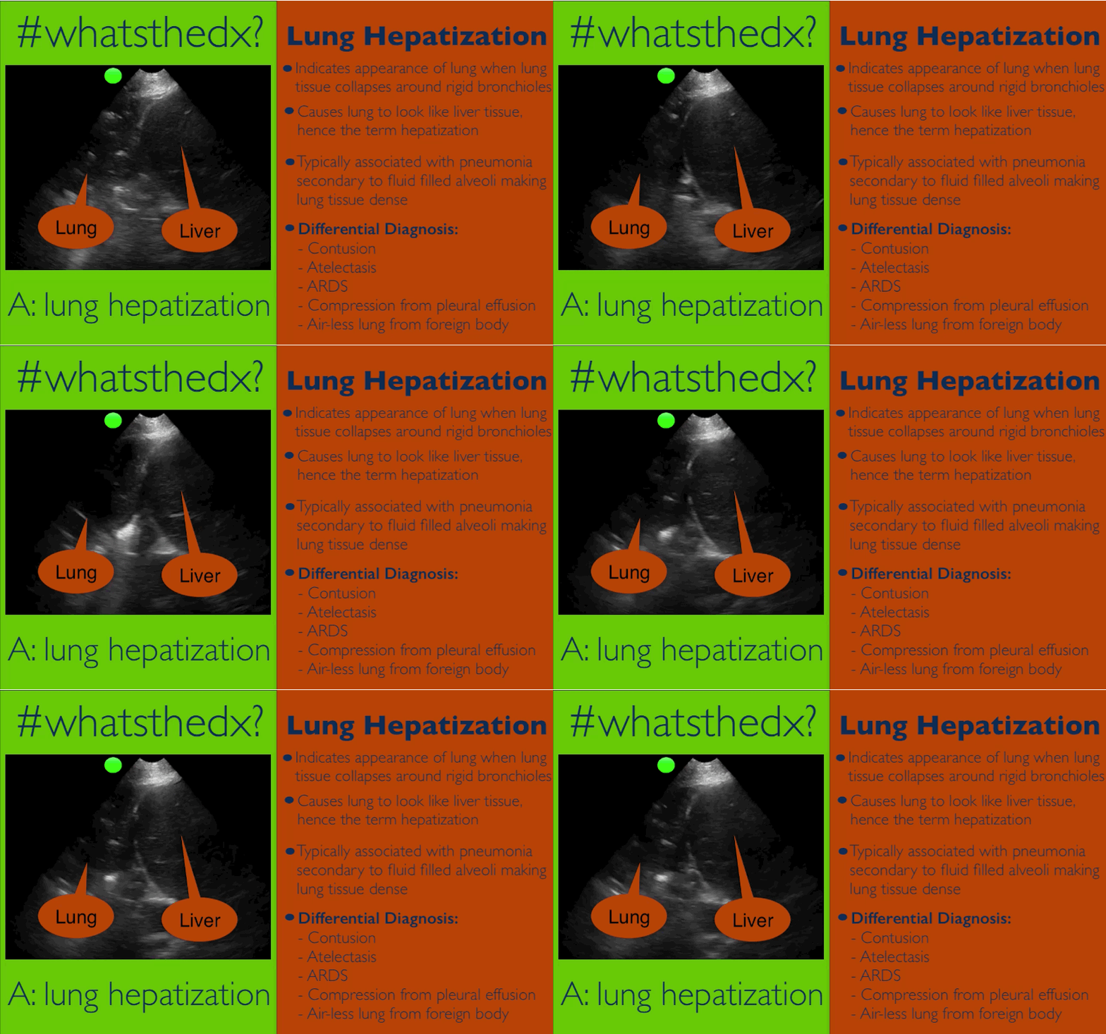
Patient presents with dyspnea & hypoxemia (pulse oximeter is 80%). The "stat" CXR is delayed, but ultrasound is not. What's the diagnosis and what are some differential diagnoses?
Show Answer
Show References

Follow me on Twitter (@criticalcarenow) or Google+ (+criticalcarenow)


Title: What's the Diagnosis?

Category: Visual Diagnosis

Posted: 11/11/2014 by Haney Mallemat, MD

Question

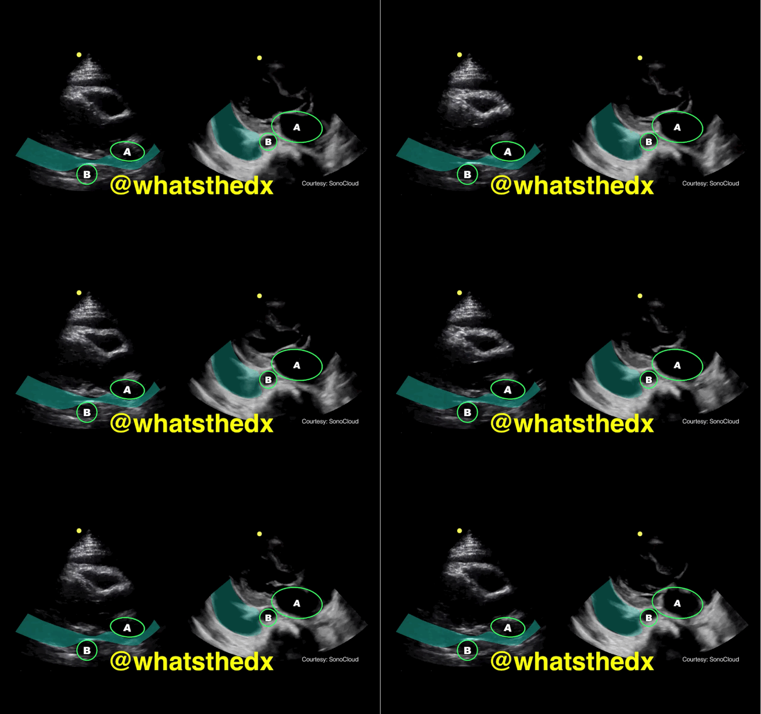
Parasternal long-axis of two different patients. What is the:

  1. structure labeled “A”
  2. structure labeled “B”
  3. diagnosis on the left?
  4. diagnosis on the right?

Show Answer

Answer: 

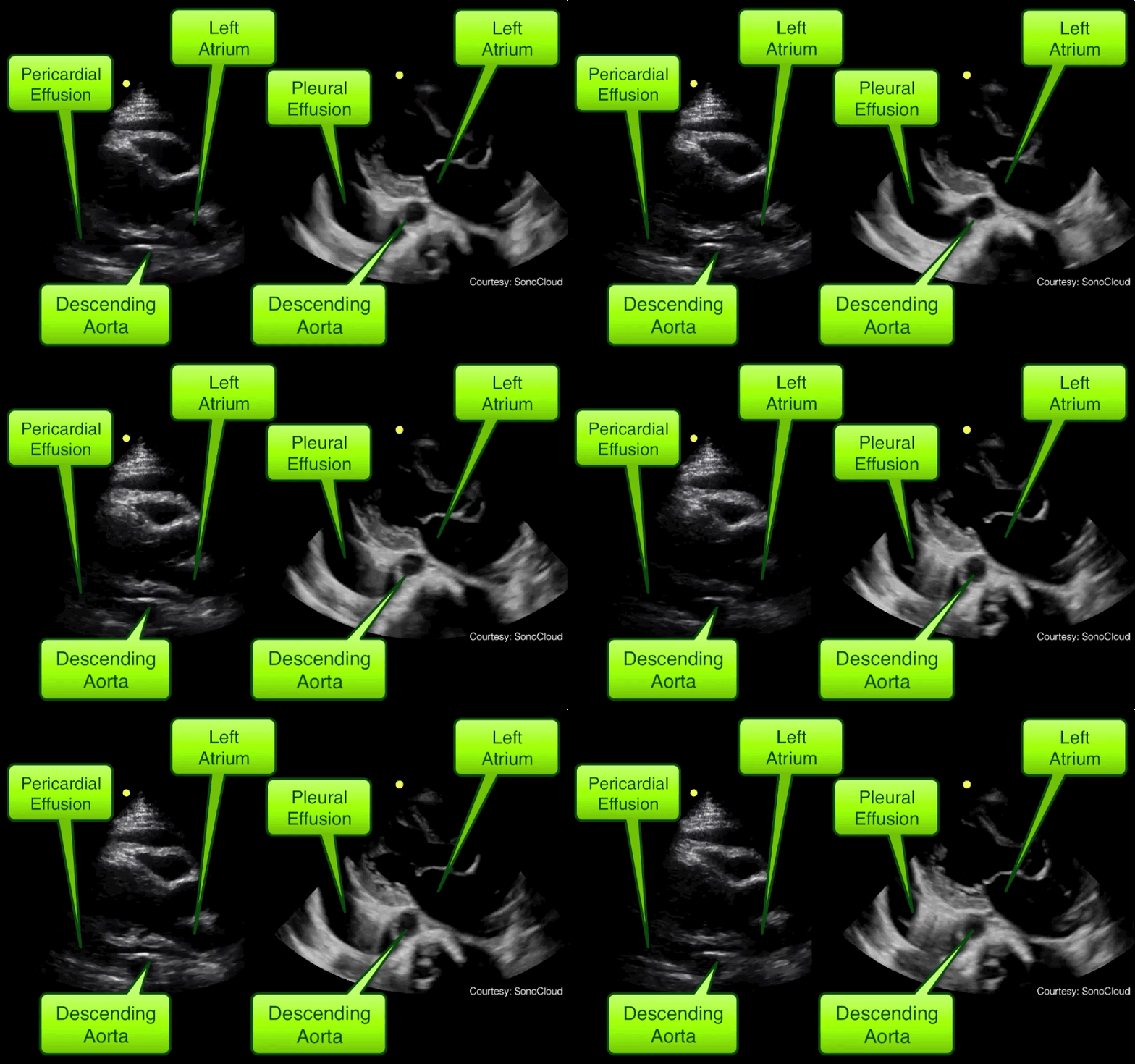
  1. A = Left atrium
  2. B = Descending aorta
  3. Left clip = Pericardial effusion
  4. Right clip = Pleural effusion

Take home pearl: when there is fluid behind the heart, the parasternal long-axis view of the heart is helpful to distinguish between a pleural effusion and a pericardial effusion.

  • Step 1: Find the descending aorta and left atrium
  • Step 2: Is there fluid that crosses in-between the two?
    • If there is, then there is a pericardial effusion
    • If there is no fluid between, then it is a left pleural effusion

Show References

Follow me on Twitter (@criticalcarenow) or Google+ (+criticalcarenow)


Title: What's the Diagnosis? Clip by Dr. Mike Stone (@bedsidesono)

Category: Visual Diagnosis

Posted: 11/4/2014 by Haney Mallemat, MD (Updated: 11/5/2014)

Question

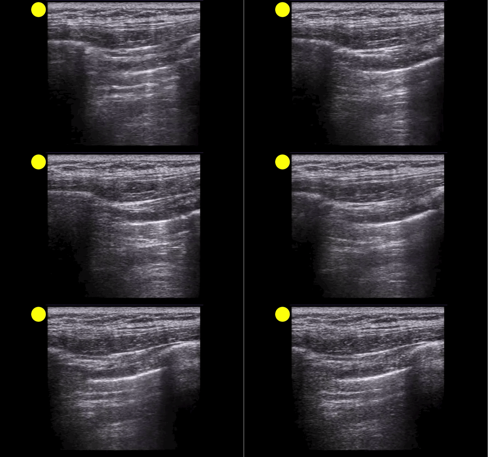
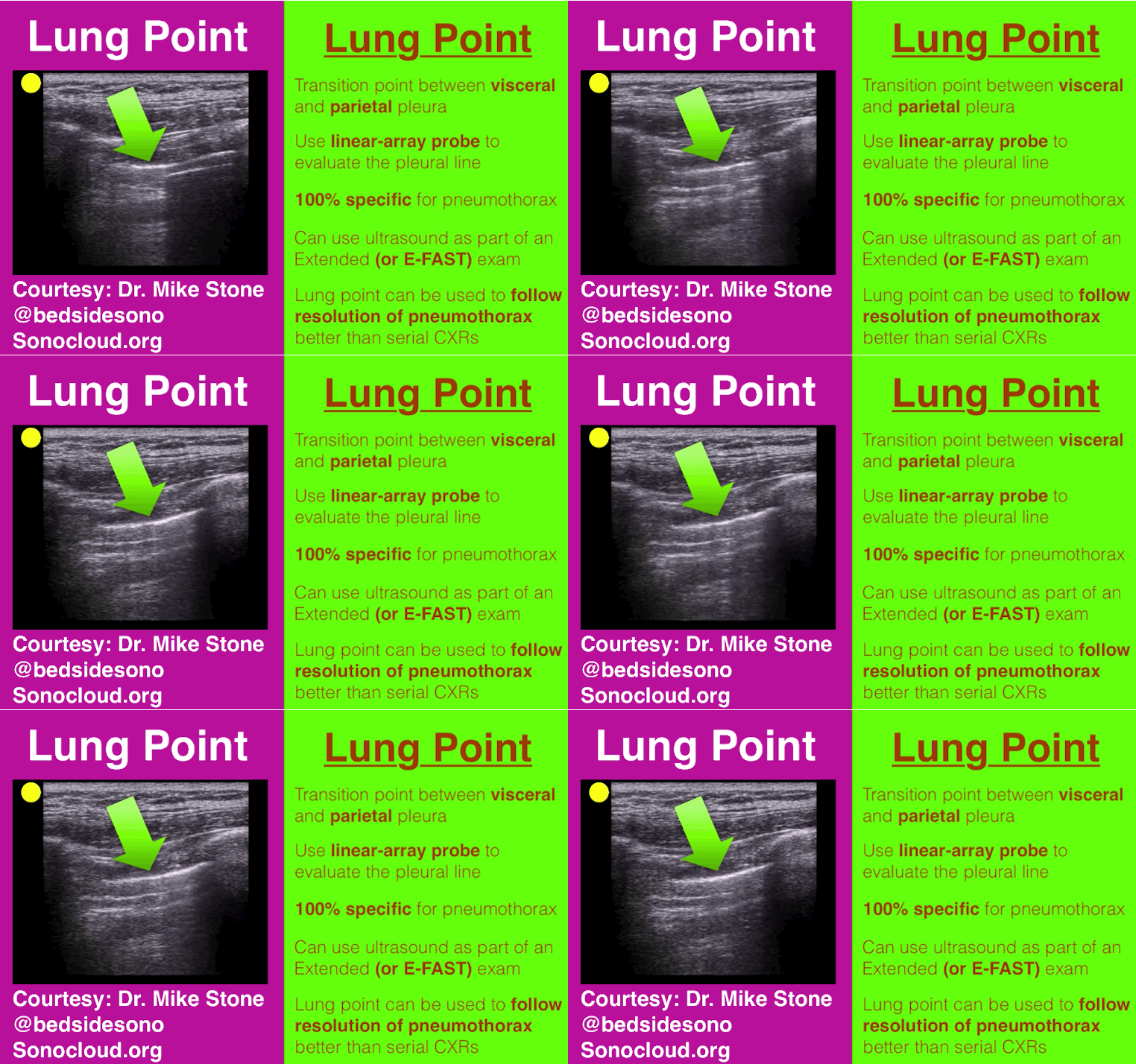
A patient presents post-motor vehicle crash with chest pain and dyspnea. The lung ultrasound is shown below. What's the diagnosis?
Show Answer
Show References

Follow me on Twitter (@criticalcarenow) or Google+ (+criticalcarenow)


Title: What's the Diagnosis?

Category: Visual Diagnosis

Posted: 10/20/2014 by Haney Mallemat, MD (Updated: 11/4/2014)

Question

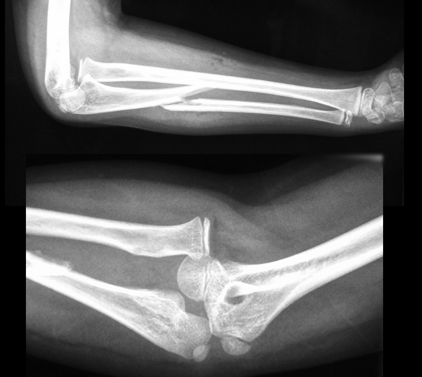
13 year-old right-hand dominant following assault with blunt object. What’s the diagnosis?
Show Answer

Monteggia Fracture

  • Ulnar fracture with dislocation of proximal radioulnar joint (often subtle); do not confuse with Galeazzi fracture (radial-shaft fracture with distal radioulnar dislocation)
  • Interosseous membrane (between radius and ulna) transmits forces to radioulnar joints and causes associated dislocations.
  • Secondary to fall on outstretched hand with arm in hyper-pronation or with direct trauma to forearm as in defensive wounds (e.g., nightstick injury).Suspected forearm fractures should include Xray of the wrist, forearm, and elbow
  • Children may be treated with closed reduction and immobilization; adults usually require open reduction and internal fixation. Radial head dislocations should be reduced within 6-8 hours because of associated articular damage and nerve injury.
  • Radial, ulnar, and median nerve neuropraxias (motor / sensory deficits) may complicate injury.

Show References

Follow me on Twitter (@criticalcarenow) or Google+ (+criticalcarenow)


Title: What's the Diagnosis?

Category: Visual Diagnosis

Posted: 10/13/2014 by Haney Mallemat, MD

Question

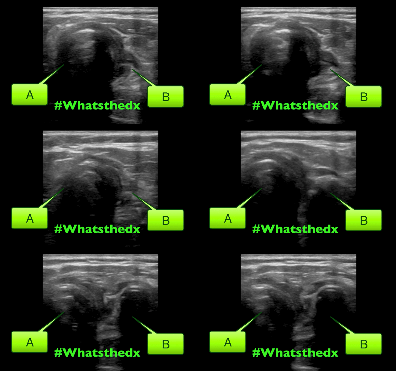
A neck ultrasound is performed during endotracheal intubation. What is labeled "A", what is labeled "B" and what's the diagnosis?
Show Answer
Answer:

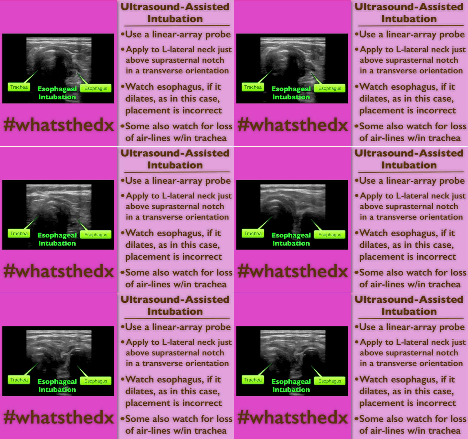
A: Trachea

B: Esophagus

This is an esophageal intubation! Take out the tube and try again. Check out this podcast for more on the technique from my good friends at the ultrasound podcast

Here's a summary:

Show References

Follow me on Twitter (@criticalcarenow) or Google+ (+criticalcarenow)


Title: What's the Diagnosis? By Dr. Ari Kestler and Dr. Ashley Strobel

Category: Visual Diagnosis

Posted: 10/6/2014 by Haney Mallemat, MD

Question

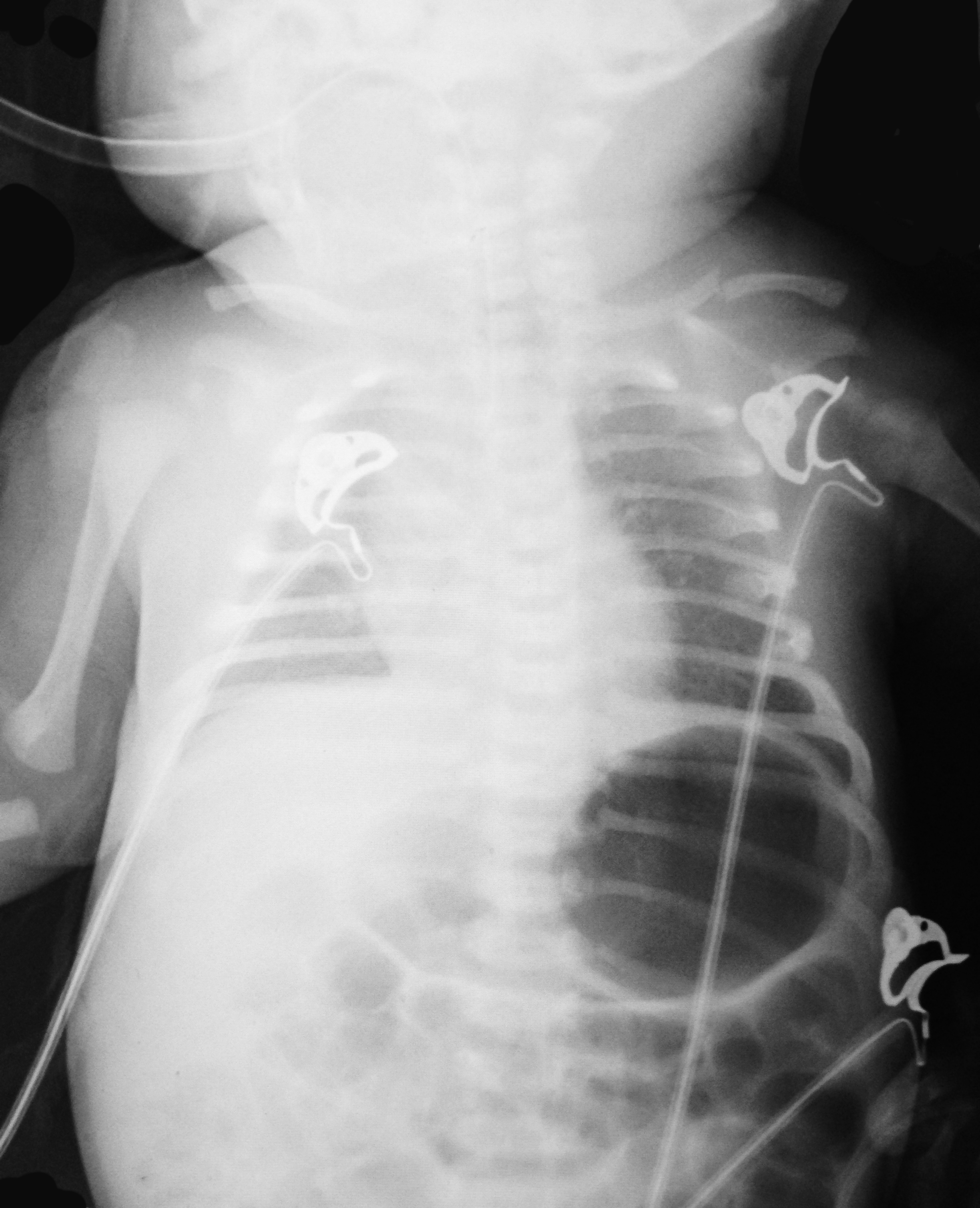
7d old child presents with difficulty feeding,vomiting one time, and now with intermittent apneic episodes. What's the diagnosis? (Careful....this one is tricky!)
Show Answer
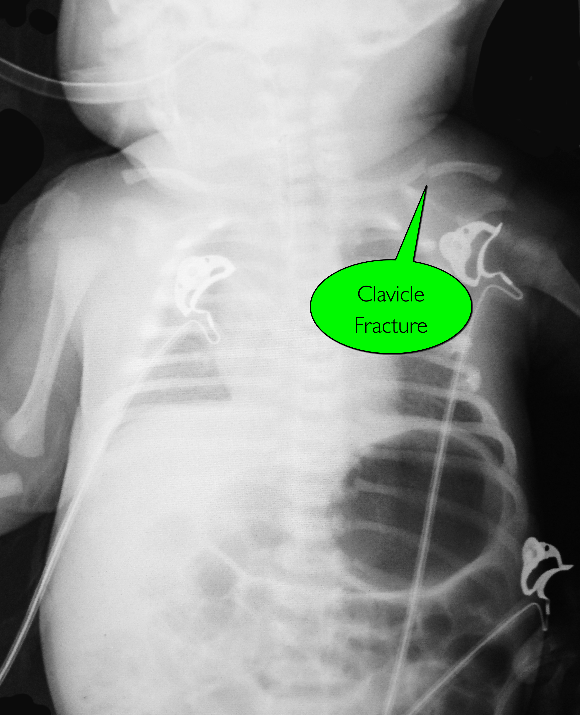
Answer: Workup should include an ALTE and sepsis evaluation PLUS evaluation for non-accidental trauma

Non-accidental trauma (NAT)

NAT is most prevalent in children 0-3 months of age.

Classic metaphyseal lesions, rib fractures, and fractures in various stages of healing are most commonly described in children.

How do we know this is not just birth trauma from a shoulder dystocia, large for gestational age, or difficult vaginal delivery?

  • Key is dating the fracture (see referenced article for more info)
  • Callus not present prior to 8 days of life and callus thickness decreases inversely with fracture age.
  • Subperiosteal new bone formation is highly unlikely in fractures less than 7 days old

Subperiosteal new bone formation appears as a:

  • hazy cortical margin or a thin layer of bone separated from the original cortex by a discrete lucent interval
  • linear hyperdensity parallel to the cortex of the bone
  • see referenced article for images

NAT Work-up:

  1. CT head without contrast if 2 yo
  2. Skeletal survey if 2 yo
  3. AST, ALT, amylase, lipase, CBC with manual diff, BMP, urinalysis, urine toxicology
  4. Consults (Ophthalmology, Social Work, Child Protection, etc.)

Show References

Follow me on Twitter (@criticalcarenow) or Google+ (+criticalcarenow)

Walters MM, Forbes PW, Buonomo C, and Kleinman PK. Healing Patterns of Clavicular Birth Injuries as a guide to fracture dating in cases of possible infant abuse. Pediatric Radiology. October 2014; 44: 1224-1229


Title: Back 2 Basics Series: Need More Squeeze - Part II Inotropes

Category: Critical Care

Posted: 9/30/2014 by Haney Mallemat, MD (Updated: 10/1/2014)

The last Back to the Basics post discussed the use of vasopressors to improve hemodynamics by increasing arterial (and venous) tone. This time we’ll discuss the use of agents to increase inotropy for patients with severe systolic dysfunction / failure.

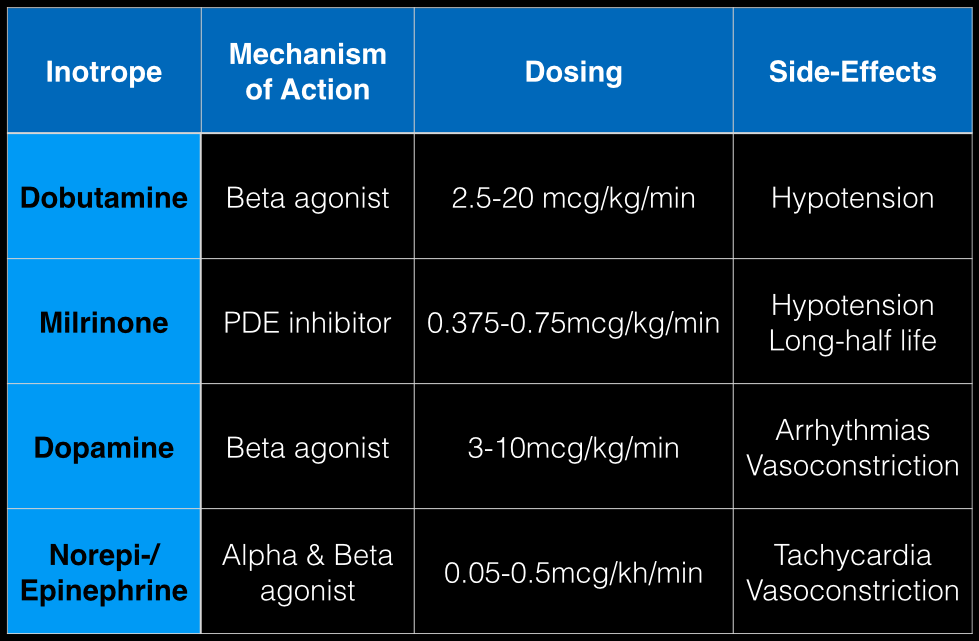
Dobutamine: a direct b1 and b2-receptors agonist. It has no peripheral vasoconstrictor properties, so if blood pressure increases it occurs secondary to increased cardiac output. Unfortunately, blood pressure may be decreased in some patients due to its peripheral vasodilatory effects; in these cases it may need to be used with a vasopressor.

Milrinone: augments contractility by increasing intracellular Ca levels via cellular phosphodiesterase inhibition. Because it does not work on beta-receptors, it might be preferred for patients taking beta-blockers requiring inotropic support. It may cause peripheral vasodilation and hypotension, but this may be a benefit if pulmonary artery pressure is elevated as reductions in pulmonary artery pressure lead to improvements in right ventricular function. It has a long-half life and should be avoided in patients with renal impairment.

Dopamine: chemical precursor to norepinephrine and technically a vasopressor. At moderate doses (3-10 mcg/kg/min) it works on beta-receptors to increase myocyte contractility. At higher doses works primarily as a vasopressor, which may reduce cardiac output due to higher afterload.

Norepinephrine/epinephrine: has alpha and beta properties that lead to increased peripheral vasoconstriction, but also increases inotropy and chronotropy (faster heart rate)
Show References

Follow me on Twitter (@criticalcarenow) or Google+ (+criticalcarenow)a


Title: What's the Diagnosis? Case by Dr. Maite Huis in 't Veld

Category: Visual Diagnosis

Posted: 9/29/2014 by Haney Mallemat, MD (Updated: 10/1/2014)

Question

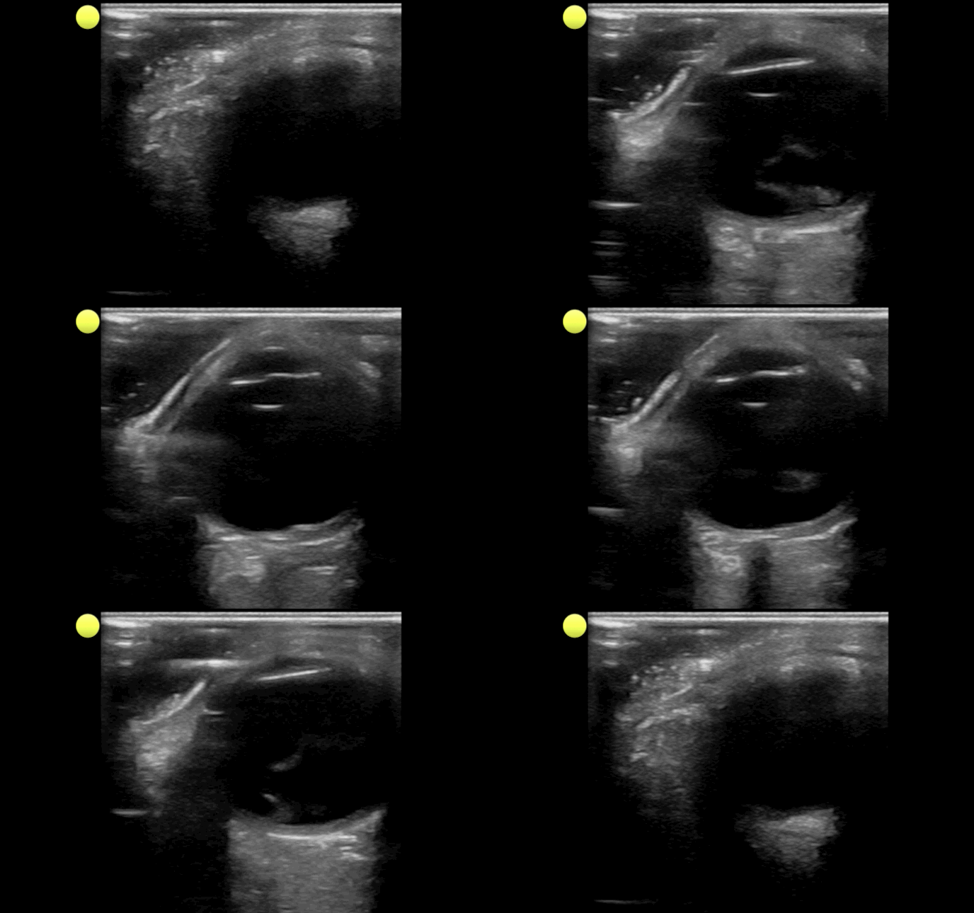
Person presents with painless vision loss after seeing flashes of light. Ultrasound is below. What's the diagnosis?
Show Answer
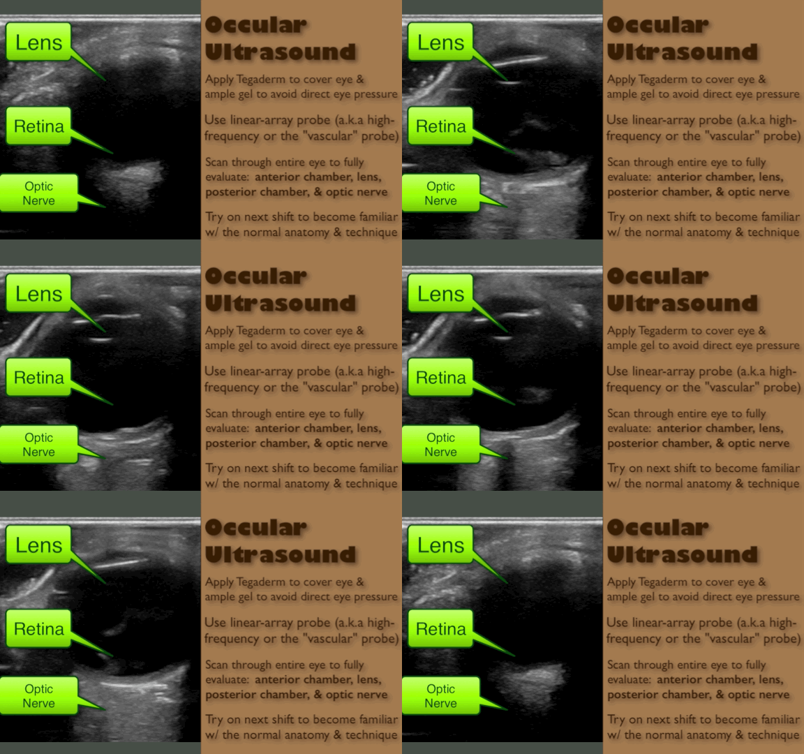
Answer: Vitreous hemorrhage with retinal detachment

Show References

Follow me on Twitter (@criticalcarenow) or Google+ (+criticalcarenow)


Title: What's the Diagnosis? Case by Dr. Leen Alblaihed

Category: Visual Diagnosis

Posted: 9/15/2014 by Haney Mallemat, MD (Updated: 10/1/2014)

Question

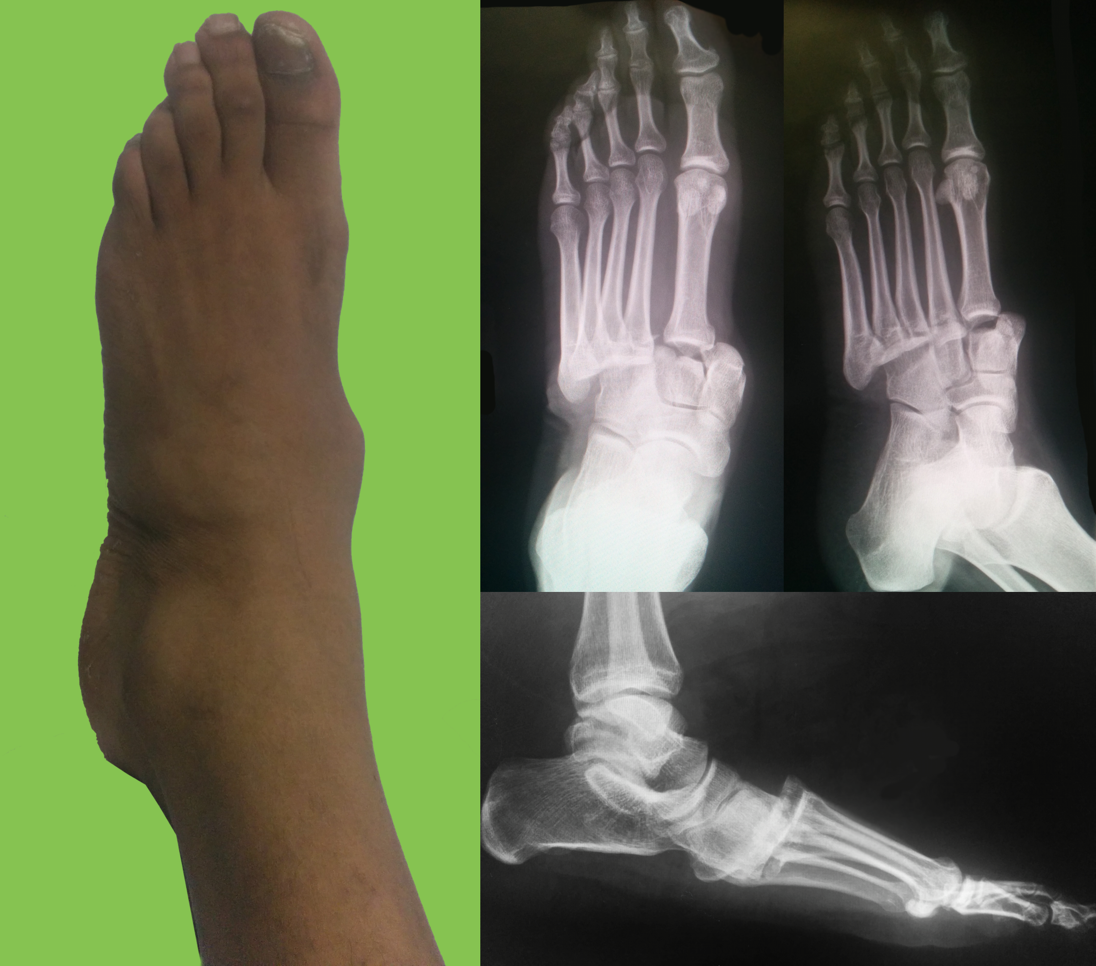
Football player complains of sudden foot pain after begin tackled. What’s the diagnosis? 
Show Answer

Lisfranc fracture-dislocation

  • Lisfranc joint consists of the tarsometatarsal joint (i.e., mid-foot) complex
  • Injuries occur when bones are fractured/avulsed or there is ligamentous disruption, secondary to a direct force or sudden rotation on a downward pointing forefoot; classic injury was fall from a horse while foot is stuck in the stirrup
  • Examine the Lisfranc joint:
    • Hold heel and twist mid-foot; assess for tenderness
    • Piano-key test: move toes up or down (increases stress in mid-foot)
    • Ask patient to stand on one foot with tip-toes; if not painful, then Lisfranc joint is intact
  • X-ray:
    • Fleck sign: fleck of bone between the first and second metatarsal bases, representing avulsion fracture of the Lisfranc ligament
    • >2 mm displacement between medial cuniform and base of 2nd metatarsal
    • If suspicion remains with normal x-ray, obtain weight bearing x-rays
    • No pain but Lisfranc on x-ray, consider Charcot’s joint
  • Treatment is usually surgical. If fracture is stable and non-displaced (<2 mm) use a short leg cast and non-weight bearing for 6 weeks.

Show References

Follow me on Twitter (@criticalcarenow) or Google+ (+criticalcarenow)


Previous  |  1 |  2 |  3 |  4 |  5 |  6 |  7 |  8 |  9 |  ... |  17 |  Next
University of Maryland School of Medicine logo
  • Departments
  • Programs
  • Research Centers
  • Institutes
  • Graduate Education
  • CIBR Cores
  • Offices of the Dean
    • University of Maryland, Baltimore
    • University of Maryland Medical Center
    • Medical Alumni Association of the University of Maryland
    • Health Sciences and Human Services Library
University of Maryland School of Medicine
655 W. Baltimore Street
Baltimore, MD 21201
Contact Us

© University of Maryland School of Medicine

    • Webmaster
    • Web Accessibility
    • Site Index
    • UMB Hotline
    • Employment
    • Map