Search
Subscribe to daily UMEM Pearls (FREE!) via:
1-20 of 76 results by Kami Windsor
Background: The 2025 AHA guidelines changed their recommended initial IV atropine dose for unstable bradycardia from 0.5 mg to 1.0 mg, while the 2025 ERC guidelines continue to recommend an initial dose of 0.5 mg. A 2025 review found limited comparative studies and noted that using 0.5 mg as an initial dose seems “clinically reasonable" [1].
Now: One tertiary care center performed a retrospective propensity-score matched study [2] comparing the 2 groups of patients with unstable bradycardia who had received an initial dose of 0.5 mg versus 1 mg in their ED between 2020 and 2025.
- Primary outcome = First-Dose Success (reaching a heart rate >50 bpm within 3 minutes without additional therapy).
- Notably excluded were patients with noncardiac secondary bradycardia (hyperkalemia, drowning, trauma, etc.) and those with pacemaker failure, amongst others.
Results in the propensity score-matched cohort:
- First-dose success was significantly higher in the 1.0 mg vs the 0.5 mg group (45 vs 25%; p=0.007)
- NNT of 6 (95% CI, 3-18)
- The 1.0 mg group had lower rates of:
- Additional atropine requirement (26 vs 48%, p=0.003)
- Other rescue therapy requirement (51% vs 74%, p=0.002)
- ICU admission (53% vs 74%, p=0.005).
- No difference between groups in 30-day mortality or subsequent permanent pacemaker placement
Bottom Line: Acknowledging the small size & retrospective nature of the study, it appears that an initial dose of 1mg IV atropine helps us work smarter--not harder--in the stabilization of patients with unstable bradycardia.
Show References
Not all patients with an acute PE will be crashing and critically ill, but it seemed worthwhile to remind everyone that there are new guidelines and recommendations from AHA/ACC/ACCP/ACEP/CHEST/SCAI/SHM/SIR/SVM/SVN/XYZLMNOP about the management of patients with acute pulmonary embolism in the 2026 AHA/ACC Joint Committee statement. A few key takeaways, with highlights for the sicker PE patients:
- New Classifications A through E for acute PE (see images below)
- Get familiar with the clinical scores! Hestia, PE Severity Index (PESI), simplified PESI (sPESI)
- ED discharge recommended for Category A and supported for Category B
- LMWH recommended over unfractionated heparin when parenteral AC is needed, unless contraindicated
- DOACs recommended over warfarin unless contraindicated
Highlights for the sicker PE patients, i.e. Categories C+:
- Get a look at the RV! (POCUS, CT, formal echo)
- Further stratify Category C patients/identify Category D earlier
- Find out how close to decompensation the patient might be
- Inform your management if the patient decompensates
- For PE patients with e/o RV strain (C2+ per this document; for me, particularly those C3+ with respiratory complaints as a marker of poor pulmonary perfusion, or Category D+), consider use of inhaled vasodilators
- Be careful with any sedation even if normotensive – decreasing preload / blunting the body's compensatory adrenergic response can be disastrous, have hemodynamic support available
- If you have to intubate, choose induction meds wisely and have hemodynamic support ready
- For patients with Category D-E acute PE:
- Norepinephrine = initial vasopressor of choice for hypotension due to modest inotropic effects; max at 15mcg/min due to effects on pulmonary vascular resistance at higher doses, if second vasopressor needed, reach for vasopressin
- Dobutamine as additional inotropic support OR for normotensive shock
- Avoid fluid boluses unless patient is also hypovolemic, and then give small boluses (250mL) only
- Consider advanced therapies for Category D and particularly E
- PE Response Team (PERT) Consultation recommended – and depending on where you practice, can help get the patient transferred if advanced therapies are an option
For a great breakdown and further discussion of the new guidelines, I recommend checking out the Life in the Fast Lane blogpost here.


Show References
Click the link for below to read the bulleted, abridged version of the Executive Summary of the Updated SSC Guidelines for Adults with Sepsis and Septic Shock 2026…
- Strength of guidelines provided as conditional “suggestions” or strong “recommendations"
- Amount of certainty given existing evidence (very low [VL], low, moderate)
- Note “very low” certainty may simply indicate there isn't a study or any reliable data
- Please refer to the article (linked in References) for given rationales from the SSC
Show Additional Information
Show References
A crucial part of cardiac arrest management is identification of the underlying rhythm, with key aspects of management diverging depending whether shockable (pulseless ventricular tachycardia/pVT or ventricular fibrillation/VF) or unshockable (pulseless electrical activity/PEA or asystole).
A recent study prospectively evaluated adult atraumatic out-of-hospital-cardiac-arrests (OHCAs) presenting to the ED, to determine what percentage of cases had “Occult VF” – VF found point-of-care echocardiogram but not by ECG. The researchers only included cases with simultaneous ECG and echo assessments for the initial 3 pulse checks. Echo and ECG determinations for the study were adjudicated by research team members.
They found that:
- 5.3% of patients had occult VF (43/811), with ECG reading PEA in 81.4% (35/43) and asystole in 18.6% (8/43)
- Of the 202/811 patients with VF on ECG, only 23.3% had echo VF
- Interestingly, patients with ECG VF but no VF on echo had a lower rate of ROSC and successful defibrillation than those with Occult VF
- There was no statistically significant difference in survival to hospital discharge between the two
- A higher percentage of patients evaluated by TEE compared to TTE were found to have occult VF (12.5% vs 4.9%)
Major limitations:
- Only have the cases with simultaneous ECG + echo for the first 3 pauses
- Only have the recorded echo clips, which are not the same as in-the-moment viewing during the arrests
- No comparison to how research team interpreted ECGs to how the treating team managed
Bottom Line: Point-of-care echocardiogram continues to have value in the management of cardiac arrest, potentially changing management and affecting post-ROSC decisions. Ensuring high-quality CPR, with appropriate defibrillation and anti-arrhythmic strategies, remains paramount in management of shockable OHCA.
Show References
We have all been there – an ED patient with circulatory shock requiring vasoactive medications and, therefore, an arterial line for accurate and close monitoring of the MAP and appropriate titration of the infusions. But does it save lives?
The recently published NEJM article by Muller et al. takes a look at noninvasive BP monitoring (NIBP) by cuff versus early arterial catheterization in patients with hypotension and evidence of tissue hypoperfusion:
- Open-label, pragmatic, parallel-group, noninferiority, multicenter RCT across 9 ICUs in France
- Adult patients enrolled within 24h of ICU admission, randomized to NIBP (n=506) or arterial line placement within 4h of enrollment (n=504)
- 15% of NIBP group received art line during study period as deemed necessary by predefined safety criteria (unable to get NIBP or SpO2, for ex)
- 50% septic shock, >90% medical patients, 90% on pressors at randomization
- Notable exclusions: BMI >40, high-dose vasopressors (total norepi tartrate* + epi infusion rate >2.5 mcg/kg/min)
- Findings:
- No difference in primary outcome of 28-day mortality (34.3% NIBP vs. 36.9% art line)
- No difference in 90 day mortality, 28-day ventilator, vasopressor, or RRT-free days
- More arterial puncture attempts in the NIBP group (742 vs. 269 per 1000 ICU days)
- No increase in arterial line-associated infections or ischemia
- More (8 vs 1%) hematoma or hemorrhage at art line site in arterial line group
- More patients in NIBP group reported serious pain/discomfort related to device (13 vs 9%)
Bottom Line: This trial indicates that in appropriately-selected patients with shock, such as those not on high doses of vasopressors, with BMI < 40 and an ability to consistently obtain NIBP measurements, early arterial line placement in the ED for vasopressor titration is unlikely to improve outcomes. It is important to note other potential indications for arterial line placement (severe hypoxia, inability to obtain reliable SpO2 with need for ABG monitoring, cardiac arrest, pain related to NIBP cuff monitoring, intracranial hemorrhage, etcetera) may still make arterial line placement in the ED prudent and better for overall patient care.
*France refers to norepi by the tartrate formulation dose, US refers to the base norepi dose (ratio is 2:1 tartrate: base).
Show References
Q: Can you use high flow nasal cannula (HFNC) to manage acute hypercapnic respiratory failure?
A: It probably depends.
Background: While we now frequently utilize HFNC as an initial therapy for most acute hypoxic respiratory failure, its appropriateness in managing acute respiratory failure with hypercarbia has historically been opposed. With more recent data indicating that HFNC may be as good as noninvasive ventilation (NIV) for management of hypercapnia as well, this seemed like a good time to point out a few things:
- Most of the existing studies are small, with a notable amount of heterogeneity
- These studies look at mild to moderate hypercapnia, not severe
- There are various amounts of crossover from HFNC to NIV as rescue
- Most acute hypercapnic studies involve COPD, not other etiologies such as obesity hypoventilation, etc.
The RENOVATE trial was a larger multicenter randomized noninferiority trial looking at HFNC vs NIV in all-comer acute respiratory failure, summarizing that HFNC was noninferior in the primary composite outcome of death + intubation at 7 days.
BUT this conclusion is not clearly supported in the smaller COPD (or acute cardiogenic pulmonary edema) subgroup:
- The median pH / PaCO2 for HFNC was 7.32 / 55 mmHg and for NIV was 7.3 / 64
- 13% of the HFNC group were on NIV prior to randomization
- 23% crossed over to NIV use
- Posthoc analysis indicated possible harm with HFNC in the COPD group
What does seem to be clear across studies that HFNC has the capacity to clear some CO2 and is by and large better tolerated than facemask NIV.
Bottom Line: For mild-moderate acute COPD exacerbations with patient intolerance or exclusion criteria for NIV therapy, trialing HFNC is a reasonable option. For patients with severe acute or acute on chronic hypercapnia, as indicated by a [pseudo-arbitrary] pH < 7.25 and PaCO2 >70-80, noninvasive ventilation should be your go-to… or be ready to promptly intubate if/when the high flow fails.
Show References
Whether you agree or disagree that “roc rocks and succ sucks,” evidence shows that approximately 3-4% of intubated patients experience awareness while paralyzed [1,2], and more of these patients are in the rocuronium subgroup [2,3,4]. Rocuronium acts in a dose-dependent fashion; the relatively standard 1-1.2 mg/kg in emergency department rapid sequence intubation (RSI) can result in a duration of paralysis can of up to 60-90 minutes. Commonly used sedatives in RSI, however, such as etomidate and ketamine, wear off quickly, before before rocuronium's paralytic effects have abated.
A recent single-center study showed that the majority of patients (60%) receiving rocuronium for paralysis during rapid sequence intubation (RSI) received no additional sedation until more than 15 minutes after induction, whether in the ED or ICU [5].
Patients experiencing awareness during paralysis with post-traumatic stress disorder [1,2] including distress from being restrained, feeling procedures, and feeling of impending death.
Bottom line: Start appropriate dose sedation promptly after RSI, especially with rocuronium, to avoid short- and long-term distress to patients.
Show References
A recent retrospective cohort study out of China investigated an escalating energy (200 > 300 > 360J) versus fixed energy (200 > 200 > 200 J) defibrillation strategy in OHCA with ventricular fibrillation requiring repeated defibrillations.
Notes:
- 342 adult patients with OHCA receiving prehospital defibrillation from 2017-2023
- *Cheskes et al.'s DOSE-VF for refractory VT/VF published in November 2022
- Defibrillation energy strategy dependent on which biphasic AED was used prehospital; ultimately 64% escalating, 36% fixed low-energy.
- Total 782 defibrillations, mean age 58 years, 80% male
Results:
- Equivalent outcomes after 1st shock in both groups (which makes sense as both groups started with 200J defibrillations)
- More patients in the escalating energy group with VF termination (93% vs 75%, p<0.001) and change to an organized rhythm (64% vs 47%, p<0.001)
- In the refractory VF population (required >2 shocks), more organized rhythms after 360J than the 3rd 200J defibrillation (35% s 18%, p=0.003).
Caveats:
- Retrospective
- No assessment of possible shock-related myocardial injury differences between groups
- No commentary on other OHCA management (like anti-arrhythmics)
Bottom Line: For patients with OHCA VF, if the first shock does not succeed, try try again – at a higher dose.
Show References
A 2023 retrospective cohort study comparing amiodarone to lidocaine for in-hospital cardiac arrests (IHCA) with refractory VT/VF found that use of lidocaine was associated with increased chance of ROSC, 24 hour survival, survival to discharge, and favorable neurologic outcome at hospital discharge.[1]
Now, a recent study comparing amiodarone to lidocaine in the pre-hospital setting for OHCA has found similar results. [2] Another retrospective cohort study using propensity score matching, they evaluated 23,263 adult patients with OHCA and defibrillation refractory VT/VF managed by 1700 EMS agencies.
Use of lidocaine was associated with greater odds of prehospital ROSC, fewer post-drug administration defibrillations, and greater odds of survival to discharge.
In comparison to earlier trials, these studies are some of the first demonstrating benefits to lidocaine use over amiodarone that reach statistical significance, but of course have all the limitations that come with retrospective studies and are not further analyzed in the context of etiologies for cardiac arrest or application of post-ROSC care.
Bottom Line: If you happen to be someone who reaches for amiodarone as your go-to, it may be time to start considering lidocaine.
- Initial dose: 1 to 1.5 mg/kg IV/IO.
- For refractory VF may give additional 0.5 to 0.75 mg/kg IV push, repeat in 5 to 10 minutes; maximum 3 doses or total of 3mg/kg.
Show References
For those of us living in a world where ED boarding is a reality and ICU beds are in short supply, a re-up on the basic tenets of post-arrest care to optimize survival and neurologic outcomes in patients with sustained ROSC after OHCA:
- Actively prevent fever in comatose patients. (“Comatose” = lack of meaningful response to verbal commands.) There may be a subset of patients comatose after ROSC who benefit from actual therapeutic hypothermia, but fever is definitely harmful. Tylenol is not going to cut it; be ready to start active cooling methods to avoid fever, and give yourself a cushion. Starting cooling efforts at 37.9 is probably not going to work to avoid reaching 38.0 deg C.
- Avoid hypotension and maintain a MAP > 65mmHg; in patients with signs of increased ICP or chronic uncontrolled hypertension, consider a MAP goal > 80mmHg. The literature is still not quite clear that higher MAP targets improve outcomes, but MAPs <65 are associated with poorer neurologic recovery.
- Target normoxia with an oxygen saturation between 92-98%. Hypoxia and hyperoxia are associated with poorer neurologic function. An O2 sat of 100% doesn’t tell you whether your PaO2 is 100 or 300, so aim for a lower value.
- Target normocarbia to mild hypercarbia (PCO2 35-55). Arterial PCO2 affects cerebrovascular tone, but the data indicates no difference in outcomes between normocarbia and mild hypercarbia up to 55mmHg.
- Monitor for seizures with EEG as soon as possible in comatose patients. Treating seizures with Keppra is appropriate and burst suppression with propofol is reasonable. “Prophylactic” antiepileptics are not beneficial and are discouraged.
- Early coronary angiography is only clearly indicated for ST elevations on EKG post-ROSC. Studies have not found a benefit in short or longer term survival for early catheterization in patients without ST elevations, although it may still be beneficial depending on the patient’s clinical scenario.
- Utilize bedside (or formal) echocardiography to help guide management in patients with hypotension after cardiac arrest. Whether fluids, vasopressors, or inotropes are needed, bedside echo can inform what you do.
- Early neuroprognostic determination acutely in the ED is largely impossible. Except in cases with clear goals of care refusing life-support, life sustaining measures should not be removed based on comatose state, prolonged downtime, presence of cerebral edema without herniation, etcetera.
Show References
Two recent studies (see “Additional Information” for more study details) published in the New England Journal of Medicine evaluated the outcomes of OHCA, comparing drug administration via intraosseous devices versus intravenous access, neither demonstrating benefit to one strategy over the other in terms of sustained ROSC or 30-day survival. [1,2] While there were a few limitations, these results are generally in line with existing literature. Although it is worth noting that some studies signal improved outcomes with IV access, the time to intervention seems to be the more important metric related to outcome. [3-5]
Bottom Line: Intraosseous devices remain rapid and easy to place devices that can provide access for drug administration when IV access is unable to be obtained. In patients with difficult access, use an IO to administer meds, fluids, or blood products as indicated while you and your team work on more definitive IV access and focus on high-quality CPR.
Show Additional Information
Show References
Question
The incidence of opioid-overdose-related deaths has clearly increased in the past decade, with recent estimates of up to 17% of OHCA being opioid-related in 2023. [1,2] The use of naloxone for opiate reversal in overdose is well-established, with reasonable inference but no formal proof that its use could help in opioid-associated out of hospital cardiac arrest (OA-OHCA). [3] The August publication of two trials [4,5] retrospectively examining naloxone administration in OHCA offers some perspectives…
- Patients receiving naloxone for OHCA are:
- More often be younger, with fewer comorbidities, but more often unwitnessed than their non-naloxoned counterparts
- More likely to have opioid OD as a presumed etiology
and
- Naloxone administration is associated with:
- Increased rates/odds of ROSC and survival to hospital discharge, whether OD is suspected or not
- And “early” naloxone (given prior to EMS IV/IO access) is associated with increased rates of DC with good neuro outcome in PEA compared to receipt after IV/IO access or none at all
[View “Visual Diagnosis” for slightly more detail on the referenced studies.]
Bottom Line: While prospective trials are absolutely needed to offer more definitive evidence regarding the use of empiric naloxone in nontraumatic OHCA, the rising incidence of OA-OHCA in the U.S. and current findings are convincing enough to encourage early naloxone administration, especially in populations with higher incidence of opioid use.
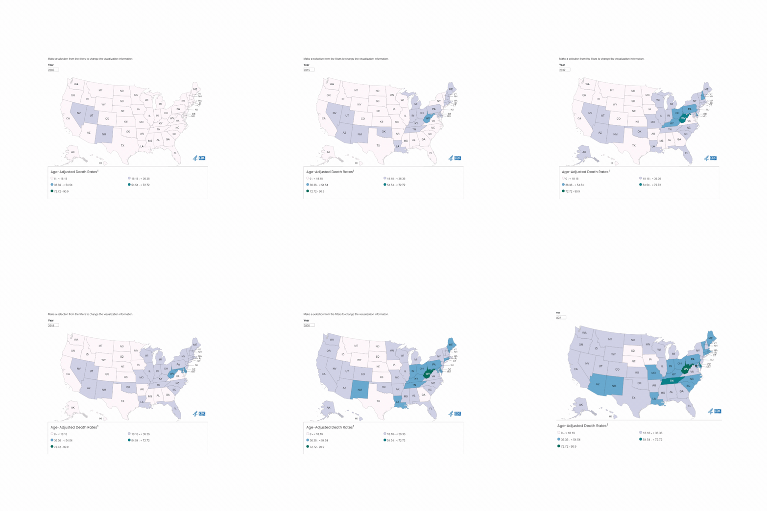
U.S. Mortality due to Opioid Overdose (CDC data)
Show Answer
Show References
Attachments
This May, the Society of Critical Care Medicine (SCCM) published new recommendations [1] for the use of corticosteroids in critical illness (separate from patients with known adrenal insufficiency or on chronic steroids), namely:
- “Suggesting” for all septic shock with continued vasopressor requirement not just “refractory” (requiring 2+ pressors)
- Matches the 2021 Surviving Sepsis Campaign Guidelines suggestion [2]
- “Suggesting” for ARDS (acute onset, bilateral infiltrates not due to cardiac dysfunction or volume overload, PaO2: FiO2 </= 300)
- Matches the 2024 American Thoracic Society Clinical Practice Guidelines suggestion [3]
- Does not explicitly exclude influenza+ ARDS, in which steroids have previously been associated with worsened outcomes [4]
- “Recommending” for patients with bacterial community acquired pneumonia and new O2 requirement
- New guidelines from ATS/IDSA not yet updated from 2019; support primarily from 2023 CAPE COD trial [5]
Bottom Line:
For severe bacterial pneumonia and septic shock, ED physicians should feel comfortable administering a dose of hydrocortisone 50mg IV as hydrocortisone 200mg/day is an accepted regimen for these disease processes.
For patients with ARDS who remain boarding in the ED, EM docs should discuss initiation of steroids with their intensivists, whether the institutional preference is for dexamethasone 20mg IV (per DEXA-ARDS) [6] or methylprednisolone 1mg/kg/day (per Meduri)[7].
Show References
Background: Sepsis remains a common entity associated with a relatively high rate of inpatient mortality, with timely recognition and treatment being key to improving patient outcomes. Various screening and warning scores have been created to attempt to identify sepsis and those patients at high risk of mortality earlier, but have limited performance because of suboptimal sensitivity and specificity.
A prospective observational study compared the performance of a variety of these scores (SIRS, qSOFA, SOFA, MEWS) as well as a machine learning model (MLM) against ED physician gestalt in diagnosing sepsis within the first 15 minutes of ED arrival.
- 2550 patients deemed by EMS or triage nurse as potentially critically-ill
- Excluded trauma, cardiac arrest, acute MI, stroke activation, patients in labor
- Seen by ED attendings (94%) / senior residents (6%) at a single urban academic center
- Visual analog scale assessment, 0-100% likelihood that patient has sepsis
- VAS >50% treated as ED physician gestalt in favor of sepsis
- 275 patients ultimately with discharge diagnosis of sepsis present on arrival to hospital
- Initial VAS outperformed all scores (AUC 0.90; 95% CI 0.88 to 0.92) both at 15 minutes and 1 hour
Although not without its limitations, this study highlights the importance and relative accuracy of physician gestalt in recognizing sepsis, with implications for how to develop future screening tools and limit unnecessary exposure to unnecessary fluids and empiric broad spectrum antibiotics.
Bottom Line: In the era of machine learning models and AI, ED physicians are not obsolete. Even at 15 minutes, without lab results and diagnostics, our assessments lead to appropriate diagnoses and care. In this new normal of prolonged wait times and ED boarding, ED triage and evaluation models that optimize early physician assessment are of the utmost importance.
Show References
In cardiac arrest, avoidance of excessive ventilation is key to achieving HQ-CPR and minimizing decreases in venous return to the heart. The controversy regarding BVM vs definitive airway and OHCA outcomes continues, but data indicates that mechanical ventilation during CPR carries no more variability in airway peak pressures and tidal volume delivery than BVM ventilation [1], with the AHA suggestion to keep in-hospital cardiac arrest patients with COVID-19 on the ventilator during the pandemic [2].
So, can we automate this part of CPR?
Two recent studies looked at mechanical ventilation (MV) compared to bagged ventilation (BV) in intubated patients with out-of-hospital-cardiac arrest (OHCA).
Shin et al.'s pilot RCT evaluated 60 intubated patients, randomizing half to MV and half to BV, finding no difference in the primary outcome of ROSC or sustained ROSC, or ABG values, despite significantly lower tidal volumes and minute ventilation in the MV group [3].
Malinverni et al. retrospectively compared MV and BV OHCA patients from the Belgian Cardiac Arrest Registry, finding that MV was associated with increased ROSC although not with improved neurologic outcomes. Of note, patients across the airway spectrum were included (mask, supraglottic, intubated), and the mechanical ventilation was a bilevel pressure mode called Cardiopulmonary Ventilation (CPV) specific to their ventilators, specifically for use during cardiac arrest [4].
Bottom Line: Larger randomized trials will be necessary to get a definitive answer as to how mechanical ventilation affects outcomes in OHCA, but in instances where the cause of arrest is not primarily pulmonary (severe asthma, pneumothorax) and the ED is short-staffed or prolonged resuscitations are likely (such as in accidental hypothermic arrests), it is probably reasonable to keep patients on the ventilator:
- in a control mode
- with a target tidal volume of 6ml/kg,
- a PEEP of 5-8cmH2O (depending on habitus)
- and an FiO2 of 100% while still in arrest.
- Set the trigger to “off” to avoid additional breaths triggered by chest compressions
- Pressure alarms may need adjustment to allow asynchronous breath delivery during chest compressions
Show References
Post-arrest shock is a common entity after ROSC. There is support for the use of continuous norepinephrine infusion over epinephrine to treat shock after ROSC, due to concerns about increased myocardial oxygen demand and associations with higher rates of rearrest [1,2] and mortality [2,3] with the use of epinephrine compared to norepinephrine, and increased refractory shock with use of epinephrine infusion after acute MI [4].
An article in this month’s AJEM compared norepinephrine and epinephrine infusions to treat shock in the first 6 hours post-ROSC in OHCA [5]. With a study population of 221 patients, they found no difference in the primary outcome of incidence of tachyarrhythmias, but did find that in-hospital mortality and rearrest rates were higher in the epinephrine group.
Bottom Line: Absent definitive evidence, norepinephrine should probably be the first pressor you reach for to manage post-arrest shock, especially if there is strong suspicion for acute myocardial infarction.
Show References
Background: Acutely intoxicated / poisoned patients are commonly encountered in the ED, with the classic teaching that a GCS < 9 is an indication to intubate for airway protection. But we’ve probably all had a patient who was borderline, or who we thought was still protecting their airway pretty well despite a lower GCS. Are we risking our patient’s health and our careers by holding off on intubation? Maybe not.
The NICO trial, a multicenter, randomized controlled trial, looked at patients presenting by EMS with GCS <9 due to suspected poisoning, without immediate indication for intubation (defined by signs of respiratory distress with hypoxia, clinical suspicion of any brain injury, seizure, or shock with systolic BP <90 mmHg). They found that withholding intubation with close monitoring, compared to the standard practice of intubating at the EMS or ED physician’s discretion, resulted in:
- No deaths in either group
- Fewer intubations (18.1% vs 59.6%; AR difference 41.5%, 95% CI -54.1 to -30.9)
- Fewer intubation-associated adverse events (6% vs. 14.7%; 95% CI -16.6 to -0.7)
- Decreased incidence of pneumonia (6.9% vs 14.7%; 95% CI -15.9 to 0.3)
- Fewer ICU admissions (39.7% vs. 66.1%) and decreased hospital and ICU LOS
Comparing the patients who were intubated in each group, there was no significant difference between groups in:
- Rate of intubation-associated adverse events or first-pass failure
- Median ICU or hospital length of stay
Notes:
- French study – EMS setup there is different from ours in the US
- Median GCS = 6, study population skewed young and male (mean age 33yo, 62% male)
- Mostly alcohol or benzodiazepine intoxication
- Unblinded study
Bottom Line: Without clear indication for intubation such as respiratory distress or accompanying head bleed, etcetera, intubation for mental status alone shouldn't be dogma in acute intoxication. Close monitoring will identify need for intubation, without apparent worsened outcomes due to a watchful waiting approach.
Show References
Background: For better or worse, the combination of “vanc-and-zosyn” has long been a go-to empiric regimen for the treatment of septic shock. Piperacillin-tazobactam is known to cause decreased creatinine secretion into the urine leading to an increased serum creatinine without any actual physiologic harm to the kidney, but the results of previous studies have led researchers to posit an increase in actual AKI with the vanc and zosyn combo. This concern has led to some physicians choosing cefepime for anti-pseudomonal gram-negative coverage instead, despite its known potential for neurotoxicity and cefepime-associated encephalopathy.
The ACORN trial: The recently published ACORN trial compared cefepime to piperacillin-tazobactam in adult patients presenting to the ED or medical ICU with sepsis or suspected serious infection. The primary outcome was a composite of highest stage of AKI or death at 14 days.
- Single-center, unblinded, pragmatic, randomized control trial
- 2500 patients, approx. 20% with chronic kidney disease
- Approximately 77% received vancomycin as well
- ~20% antibiotic crossover in each group
Results:
- No difference between groups in the primary outcome, or in major adverse kidney events, even in subgroup that also received vancomycin
- No difference in hospital length of stay, vasopressor days, ventilator days
- Slightly higher incidence of delirium or coma in the cefepime group
Bottom Line: Good antibiotic stewardship would probably decrease the frequency of vanc-and-zosyn administration, but concern for renal dysfunction alone shouldn’t guide the choice between cefepime or piperacillin-tazobactam, even in those with CKD, and even in those patients also receiving vancomycin.
Show References
The BRASH syndrome (Bradycardia, Renal failure, AV nodal blockade, Shock, Hyperkalemia) has been increasingly described in the literature in the past 3-5 years.
The inciting factor is generally considered to be something that prompts acute kidney injury, often hypovolemia of some sort. Rather than AV nodal blocker overdose or severe hyperkalemia causing conduction problems, the combination of AV nodal blocker use (most often beta-blockers, but can be any type) and hyperkalemia (often only moderate) has a synergistic effect on cardiac conduction with ensuing bradycardia that can devolve into a cycle of worsening renal perfusion and shock.
Treatment is supportive, but most effective when the syndrome is recognized and all parts simultaneously managed. ED physicians should be familiar with its existence for targeted whole-syndrome stabilization and to avoid diagnostic delay.
- Shock – If hypovolemic, IV fluid resuscitation. Concomitantly or if still hypotensive, epinephrine infusion is recommended as it provides both chronotropy and inotropy, and also assists with hyperkalemia.
- Hyperkalemia – usually mild/moderate; IV calcium for any ECG abnormalities, intracellular shifting medications, and kaliuresis (may require high-dose loop diuretics, with IV fluids if needed to maintain volume)
- Bradycardia – will usually respond to IV calcium and chronotropy (epinephrine, isoproterenol); pacing rarely but sometimes needed
- Renal failure – IVF and perfusion support as noted above, but patients may require dialysis if renal failure is severe and hyperkalemia is unable to be medically managed
Show References
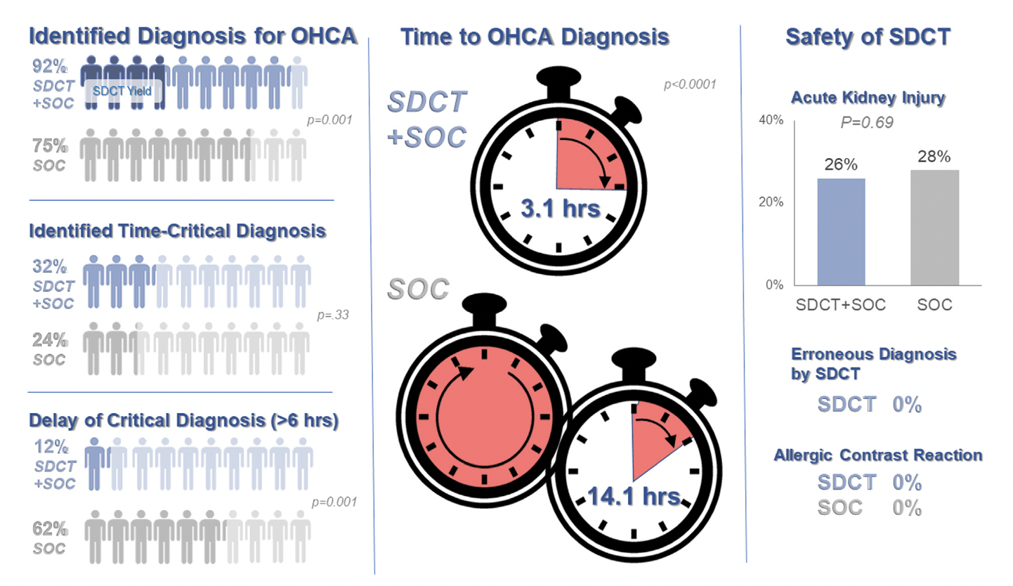
Background: Prior evidence1,2 has suggested that early “pan-scan” after ROSC provides clinically-relevant information that assists in the care of the patient in question, when the cause of OHCA is unclear.
The recent CT FIRST trial looked at patients pre- and post- implementation of a protocol for head-to-pelvis CT within 6 hours of ROSC for adult patients without known cause or evidence of possible cardiac etiology, stable enough for scan. *Patients with GFR <30 were excluded from assignment to CT, although were included in the post/CT cohort if their treating doctors ordered CT scans based on perceived clinical need. To balance this, a similar number of patients with GFR <30 were included in the pre/“standard of care” cohort.
- Pre/SOC cohort (143 pts) vs. Post/SOC+CT cohort (104 pts)
- CT protocol: Dry head CT, CTA chest, venous phase CT abd/pelvis
- In pre/SOC group, CTs ordered by treating docs in 52% (one or mix of the above CTs)
Outcomes After Protocol (Pre- vs. Post-):
- Increased identification of OHCA diagnosis (75% vs. 92%, p = 0.001)
- In SOC + CT group, diagnosis only found by CT in 13%
- In SOC group, diagnosis only found by CT in 17%
- Faster OHCA diagnosis (14.1h vs. 3.1h, p= 0.0001)
- Fewer delays in time-critical diagnoses* (62% vs. 12%, p= 0.001) *both OHCA dx and resuscitation-related injury
- No difference in ultimate diagnosis of time-critical diagnoses, rates of AKI, or survival to hospital discharge, allergic contrast reactions (0), scan complications (0), inappropriate treatments based on CT findings (0)
Bottom Line: Early pan-CT allows for earlier definitive diagnosis and stabilization without increase in adverse events. While this earlier diagnosis does not seem to yield better survival, earlier stabilization may provide some benefits in terms of resource allocation and disposition, a notable benefit during our current crisis of staffing shortages and ED boarding.