
Elevated intracranial pressure (ICP), defined as >20mmHg, is frequently encountered in patients with severe traumatic brain injury (TBI). A step-wise approach would include:
1. Analgesia and sedation: frequently forgotten.
2. Hyperosmolar agents: both hypertonic saline and mannitol can be used. Neither is superior.
3. Induced arterial blood hypocarbia using hyperventilation (must monitor for cerebral ischemia)
4. Barbiturates (last resort due to side effects)
5. Surgical:
a. CSF drain
b. Decompressive craniectomy: benefits challenged by the DECRA study
Stocchetti N, Maas AIR. Traumatic Intracranial Hypertension. N Engl J Med 2014; 370:2121-30.
Background
- Artificial nutrition is a staple of critical care
- Patients who are unable to eat, require enteral nutrition (preferred over parental nutrition)
- There are some formulas that are called "immunonutrition" which try to alter the inflammatory response seen in critical illness
- They may contain omega-3 fatty acids and essential amino acids such as arginine or glutamine, and anti-oxidants.
Data
- A recent trial (MetaPlus) was designed to see if immunonutrition could decrease the development of infections in the critically ill
- Compared to regular high protein formulas, there was no difference in mortality, duration of ventilation, or hospital length of stay
What to do
- Immuno-nutrition formulas cannot be routinely recommended
- Use regular high protein formulas
- Start within 48 hours of identifying a need
Show References
Question
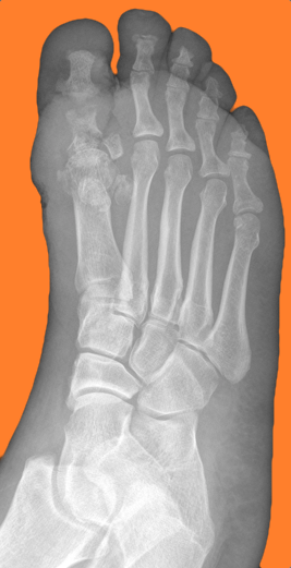
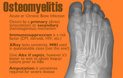
50 year-old female with diabetes complains of pain and discharge from a poorly healing wound. XRay below. What's the diagnosis?
Show Answer
Show References
GRACE Score
- The Global Registry of Acute Coronary Events (GRACE) is an international database tracking outcomes of patients presenting with acute coronary syndromes (ACS).
- GRACE score is calculated based on 8 variables: Age, HR, systolic BP, creatinine, killip class, ST-segment deviation on EKG, cardiac biomarkers, and cardiac arrest on admission.
- Several reports have shown that the GRACE score is a better predictor of clinical outcome (risk of death or the combined risk of death or myocardial infarction at 6 months) than the TIMI score.
- A recent study evaluated the relationship between GRACE score & severity of coronary artery disease (CAD) angiographically evaluated by Gensini score in patients with NSTE-MI.
- Results showed that the GRACE score has significant relation with the extent & severity of CAD as assessed by angiographic Gensini score.
- GRACE score was shown to be important both for determining the severity of the CAD and predicting death within 6 months of hospital discharge from NSTE-MI.
Show References
Patellar tendonitis aka jumpers knee
Activity related knee pain due to degenerative, micro injury rather than an inflammatory process
Up to 20% in jumping athletes
Anterior knee pain during or after activity
Bassett Sign:
a) Tenderness to palpation with knee in full extension (patellar tendon relaxed)
b) No tenderness with knee in flexion (patellar tendon tight)
E-cigarette popularity has increased and with that another possible source of toxicity. The most recent MMWR shows how e-cigarette use has increased over the past 5 years. The general toxicity involves nicotine toxicity with nausea, vomiting, eye irritation as the major sources of toxicity. Only one reported death where the nicotine reservoir was accessed and then injected IV in a suicide attempt.
There are some reports of asthma exacerbations but is more likely due to the vapor flavor and not the nicotine.
Attachments
General Background:
Heat transfer is important to understand, especially when evaluating and treating someone who is hyperthermic or hypothermic. Are they really hot (or cold) from exposure, or is there an underlying metabolic or toxicological cause?
Mechanisms of Heat Transfer:
There are 4 main methods of heat transfer:
- Radiation
- Transfer of thermal energy through space by electromagnetic waves
- Dependent on exposed surface area
- Normally about 60% of heat loss
- Conduction
- Transfer of thermal energy through direct physical contact
- Depends on conductivity of the surface (stone floors > water > air)
- Can cause significant hypothermia for someone who is down for an extended period on a stone floor
- Evaporation
- Transfer of thermal energy through converting liquid to a gas
- Occurs through perspiration and respiration
- Can lead to dehydration
- Convection
- Transfer of thermal energy through movement of air or liquid across an object
- Rate of heat loss depends on temperature gradient, density and velocity of moving substance
- Can lead to extreme heat loss, especially when combined with evaporation (wet clothes on a cold, windy day)
Bottom line:
When evaluation someone for hyper- or hypothermia from a potential environmental exposure, be sure to obtain the history about where they were found and the circumstances in which they were found. This can help you develop your diagnostic differential.
Show References
Sepsis Pearls from the Recent Literature
- Sepsis remains one of the most common critical illnesses managed by emergency physicians and intensivists.
- Recent publications and meta-analyses (i.e., ProCESS, ALBIOS, SEPSISPAM) have further refined the management of these complex patients.
- A few pearls from the recent literature:
- Early broad-spectrum antibiotics remains the most important factor in reducing morbidity and mortality.
- Appropriate fluid resuscitation with a balanced crystalloid solution targeting 30 ml/kg. Use a dynamic measure of volume responsiveness to determine if additional fluid needed (i.e., PLR with a minimally invasive or noninvasive cardiac output monitor)
- Maintain adequate tissue perfusion with IVFs and vasopressors (norepinephrine) targeting a MAP > 65 mm Hg. Patients with chronic HTN may benefit from a higher MAP goal. If the diastolic BP is < 40 mm Hg upon presentation, start vasopressors concurrent with IVF resuscitation.
Show References
Question
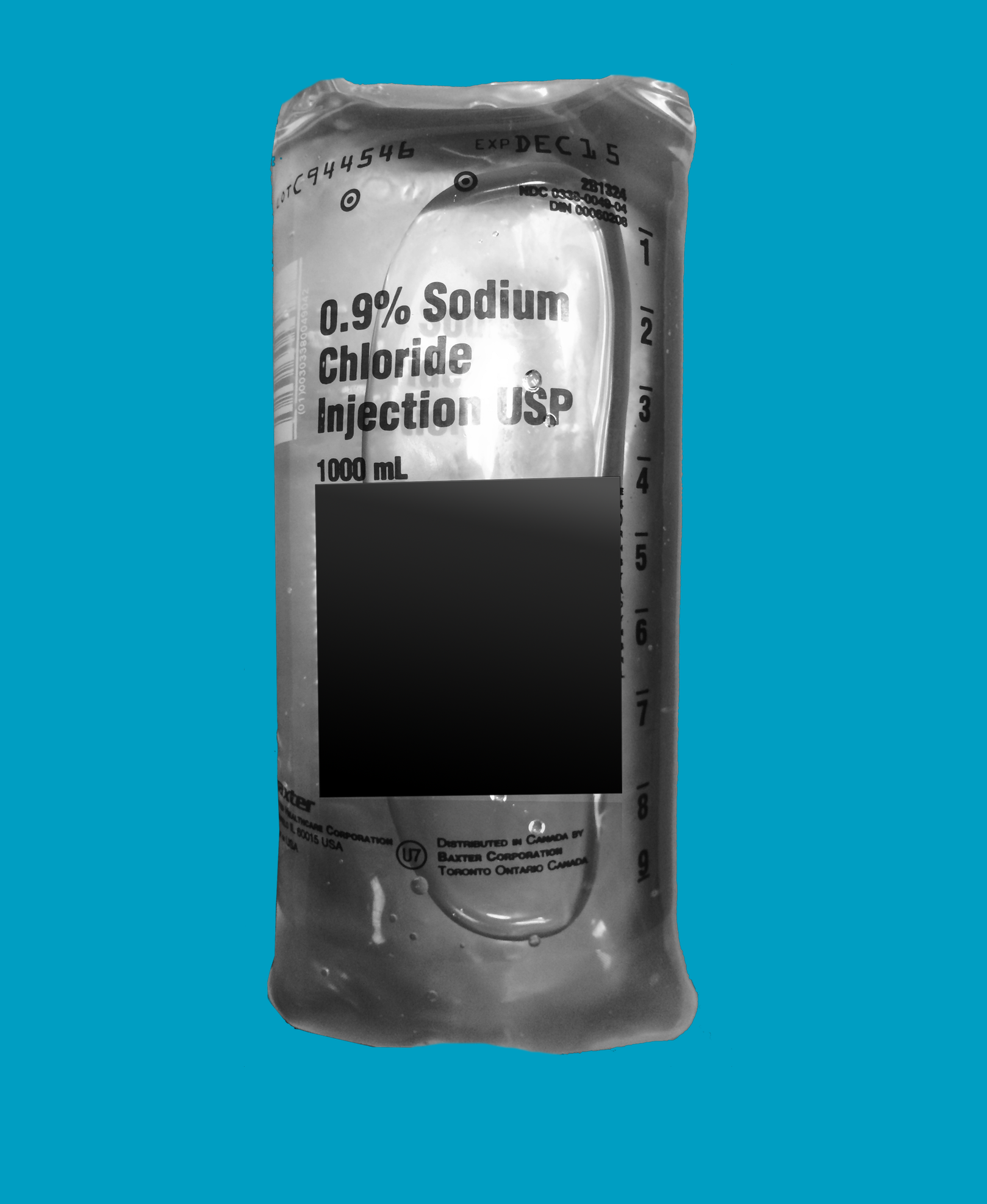
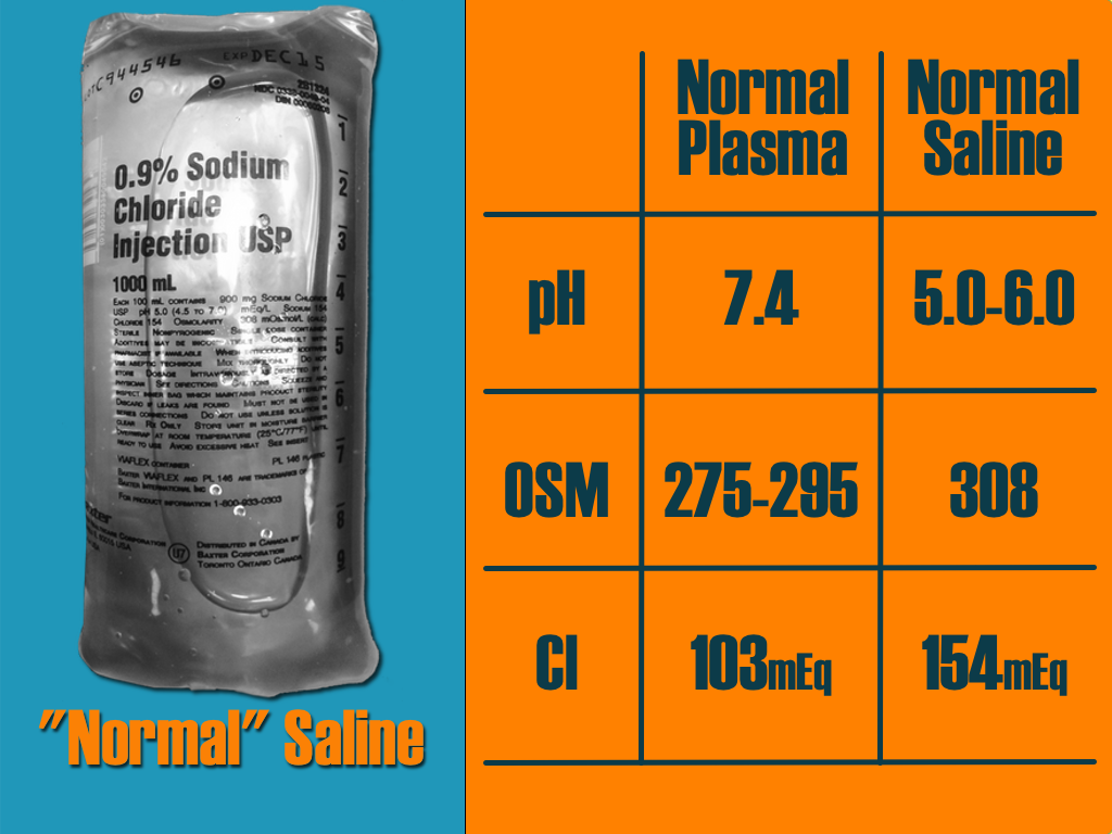
A critically-ill patient requires fluid resuscitation. Someone hands you a bag of this. What’s the pH of this fluid?
Show Answer
Show References
Nonatherosclerotic Coronary Artery Disease
- Nonatherosclerotic coronary artery disease (NACAD) is a term used to describe a category of diseases, which include: spontaneous coronary artery dissection (SCAD), coronary fibromuscular dysplasia (FMD), ectasia, vasculitis, embolism, vasospasm, or congenital anomaly.
- NACAD is an important cause of myocardial infarction (MI) in young women, but is often missed on coronary angiography.
- A small retrospective study of women <50 years of age with ACS found that 54.8% had normal arteries, 30.5% atherosclerotic heart disease (ACAD), 13% nonatherosclerotic coronary artery disease (NACAD), and 1.7% unclear etiology.
- NACAD accounted for 30% of MI’s with SCAD & Takotsubo cardiomyopathy accounting for the majority of cases.
Show References
Is there any benefit to the use of prednisone in the treatment of lower back pain? One study showed that about 5% of patients receive prednisone for the treatment of their low back pain, but does it work.
A recent study by Eskin et al published in the Journal of Emergency Medicine looked at this question. They conducted a randomized controlled trial of 18-55 year olds with moderately severe low back. Patients were randomized to receive prednisone 50mg for 5 days or placebo.
The study enrolled a total of 79 patients, and 12 were lost to follow up. At followup there was no difference in their pain, or in them resuming normal activities, returning to work, or days lost from work. To make matters worse more patients in the prednisone group sought additional medical treatment 40% versus 18%.
Conclusion: With the results of this study we should continue the treatment of low back pain with non-steroidials, muscle relaxants and exercise. There does not appear to be any role for steroids in the treatment of these patients.
Show References
6-7% of kids presenting with upper respiratory symptoms will meet the definition for ABS.
The American Academy of Pediatrics (AAP) reviewed the literature and developed clinical practice guideline regarding the diagnosis and management of ABS in children and adolescents.
The AAP defines ABS as: persistent nasal discharge or daytime cough > 10 days OR a worsening course after initial improvement OR severe symptom onset with fever > 39C and purulent nasal discharge for 3 consecutive days.
No imaging is necessary with a normal neurological exam.
Treatment includes amoxicillin with or without clauvulinic acid (based on local resistance patterns) or observation for 3 days.
Optimal duration of antibiotics has not been well studied in children but durations of 10-28 days have been reported.
If symptoms are worsening or there is no improvement, change the antibiotic.
There is not enough evidence to make a recommendation on decongestants, antihistamines or nasal irrigation.
Show References
Since synthetic cannabinoids arrived on the scene, we have become familiar with their sympathomimetic effects such as emesis, tachycardia, hypertension, agitation, hallucinations, and seizures.
Acute kidney injury is also being linked to synthetic cannabinoid use. Several clusters have been described in a handful of states, the most recent coming from Oregon with 9 patients.
AKI seems to be one more adverse effect to be on the lookout for when evaluating patients after synthetic cannabinoid use.
Show References
Should I Give My Patient with Septic Cardiomyopathy Fluids?
The incidence of acute LV dysfunction in septic shock is estimated to occur in 18 - 46% of patients within the first 24 hours of shock. Unlike the "classic" pattern of cardiogenic shock where LV filling pressure is high, in septic shock there are normal or low LV filling pressures.
Three therapeutic options should be strongly considered in the patient with a septic cardiomyopathy [CM]:
- FLUIDS: Most patients with septic CM need fluids to restore adequate preload/afterload. Severe vasoplegia requires volume resuscitation - even if the bedside ECHO suggests reduced contractility. Give fluids generously.
- Vasopressors: Catecholamine supplementation (norepi) improves patient's preload & afterload, but can often unmask septic CM. Consider epinephrine as a second line agent (over vasopressin) for inotropic support.
- Inotropes: Consider adding epinephrine (1 to 5 mcg/min) or dobutamine (start at 1-5 mcg/kg/min) to target an improved cardiac index (>2.5 L/min/m2) or ScVO2 > 70%.
Show References
Question
30 year-old female with complains of night sweats and painful lesions on her fingertips. What’s the diagnosis and list some things to have in the differential diagnosis?

Show Answer
Show References
- Toxic effects of tricyclic antidepressants (TCA) are result of the following 4 pharmacologic properties:
1. Inhibition of norepinephrine & serotonin reuptake --> resultant seizure
2. Anticholinergic activity --> resultant altered mental status, tachycardia, mydriasis, ileus
3. Direct alpha-adrenergic blockade --> resultant hypotension
4. Cardiac myocyte sodium channel blockade --> resultant widened QRS
- A QRS interval greater than 100 milliseconds has ~30% chance of developing seizures and ~15% chance of developing a life-threatening cardiac arrhythmia.
- A QRS interval greater than 160 milliseconds increases the chance of ventricular arrhythmias to greater than 50%.
- Clinical pearl: A very wide complex ventricular rhythm, concomitant hypotension and/or seizure disorder is suspicious for toxic ingestion and standard ACLS algorithm will not suffice, treatment must address the underlying culprit (i.e. TCA --> Tx. fluids, vasopressors, sodium bicarbonate, and intravenous lipid emulsion).
Show References
Return to Play After Infectious Mononucleosis (IM)
-Long incubation period make it difficult to determine source or onset
Presentation often atypical with nothing more than fatigue, decreased energy or decreased athletic performance.
DDX: Herpes simplex, HIV, CMV, toxo and strep (simultaneous infection may be seen in up to 30%)
Classic 3 to 5 day prodromal period (malaise, fatigue, anorexia)
Symptoms then progress into the classic “triad” of IM
Fever, pharyngitis, lymphadenopathy (esp. posterior cervical nodes)
May also have posterior palantine petechiae ( of cases), jaundice, exudative pharyngitis, rash and splenomegaly)
Rash (10% to 40%), transient, generalized maculopapular, petechial or urticarial)
Most commonly seen in patients treated with PCN antibiotics
Splenomegaly is an important complication in the athletic population
Mononucleosis makes the spleen susceptible to rupture (traumatic or spontaneous)
- Lymphocytic proliferation enlarges the spleen beyond protection from the ribs
- Physical examination has been shown to be unreliable for determining splenomegaly
- Highest risk is in the first 21 days (rare after 28 days)
Ultrasound is the modality of choice
-Splenomegaly peaks at 2 to 3 weeks and resolves in the majority between 4 to 6 weeks
Return to play is generally allowed after 4 weeks from diagnosis in the absence of splenomegaly and resolution of symptoms.
General Information:
Hypothermia is when the body’s core temperature is less than 35º C. Often thought as a winter disease, it can occur in nearly any climate or weather condition. However, a number of cold related conditions can occur without a drop in core body temperature.
Specific Cold Related Conditions:
- Frostnip
- Condition: Ice crystal deposited in the dermis
- Exposure: Freezing, damp exposure over hours to days
- Treatment: Warm water (37º-41ºC) immersion with movement of affected area for 15-30 minutes
- Frostbite
- Condition: Frozen skin surface with damage to dermis and deeper structures
- Exposure: Freezing, damp exposure over hours to days
- Treatment: Warm water (37º-41ºC) immersion with movement of affected area for 15-30 minutes
- Trench foot
- Condition: Tissue necrosis without freezing
- Exposure: Cold water exposure for hours to days
- Treatment: Warm water (37º-41ºC) immersion with movement of affected area for 15-30 minutes
- Chillblains
- Condition: Epidermis repeatedly partially frozen and thawed
- Exposure: Chronic cold, dry wind exposure over weeks
- Treatment: Calcium-channel blockers can provide pain relief and decrease necrosis
Bottom line:
Remember that cold related injuries can occur without core hypothermia. Don’t forget the tetanus and antibiotics, as indicated.
Show References
- Only treat strep pharyngitis after confirmed via rapid antigen test or culture
- Remember the rapid antigen test has high specificity, but low sensitivity. All negative rapid antigen tests should be followed up with a confirmatory culture
- Traditionally, strep pharyngitis was treated with penicillin V, 250mg PO tid for children and 500 mg tid for adolescents. This was then changed to bid dosing.
- Now, consider treating with amoxicillin, 50mg/kg once daily (max 1000mg). Once daily dosing and better taste improve compliance
Show References
There are many ventilator modes to choose from, but almost every mode can be distilled down to its basic principles by understanding the “Three T's of Mechanical Ventilation”
Trigger: You must determine whether the vent or patient will trigger a mechanical breath. For example, machine-triggered breaths (a.k.a. control mode of ventilation) are used for paralyzed patients and will deliver a breath after a period of time has elapsed (e.g., if RR is 10/min, then a breath is given every 6 seconds). On the other hand, if a patient’s respiratory drive is intact (a.k.a. assist-mode) than the patient triggers the breath when the vent detects a patient induced change in airflow or airway pressure. These two modes can also be mixed together.
Target: Mechanical breaths must have a specific target, either a target airway pressure or a tidal volume. Because pressure and volume are directly related, pick the variable you want to target and the other parameter will vary depending on the patient’s intrinsic physiology. For example, if you choose to target a specific tidal volume, we may get one plateau pressure in a patient with normal lungs, but a higher plateau pressure in another patient with stiffer lungs.
Terminate: You must decide when the mechanical breath (i.e., inspiration) terminates and expiration begins. Termination occurs: 1) after a set inspiratory time has elapsed in certain pressure-targeted modes, 2) when a predefined target volume has been achieved (i.e., volume-cycled modes), or 3) when airflow has been reduced by a certain percentage (as in pressure-support ventilation; to be discussed separately)
Let’s put this all together by looking at an example: pressure control ventilation (rate = 12/min and target pressure 20cm H20). Trigger: Because this is a “control”, not assist mode, the machine will trigger a breath 12 times per minute or every 5 seconds. Target: Here we chose to have pressure be the target, so when the ventilator triggers a breath it will deliver a constant airway pressure of 20 cmH2O until we tell the vent terminate that breath. Terminate: the constant airway pressure will be turned off after a fixed period of time has elapsed; for this example we will set the inspiratory time as 1 second, then expiration begins. Now, after a few vent breaths we will observe the results of our settings and reassess; if the resulting tidal volume is lower than what we wanted, we will increase the target pressure to increase the tidal volume. If the tidal volume is higher than what we wanted, we will reduce the target pressure to reduce the tidal volume. We can also tweak the inspiratory time to manipulate the tidal volume, but this does so to a lesser degree.
Try to break down your favorite modes of ventilation using the Three T’s and see if this helps you understand vent modes better.