
Question
The following is an actual case.
An ED patient without any history of asthma or COPD is intubated to facilitate an emergent MRI. After the MRI is completed (and without abnormalities). The patient is breathing synchronously and comfortably on minimal PS settings. Further sedation is weaned to facilitate extubation in the ED as the patient has no need for continued mechanical ventilation and no ICU requirements. All of a sudden, you are emergently called overhead to the patient's room because the patient is “blue” and hypoxic.
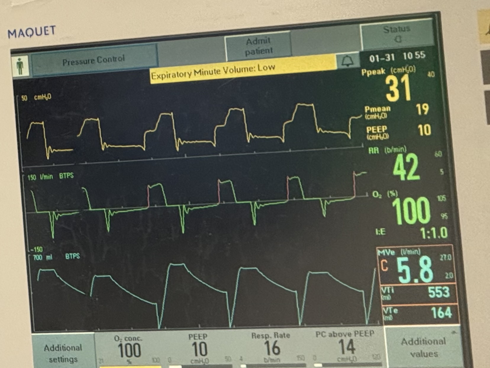
These are the ventilator waveforms.
The patient did not receive any contrast or bronchospastic medications. The ETT is clear and without obstruction.
What is going on?
Answer
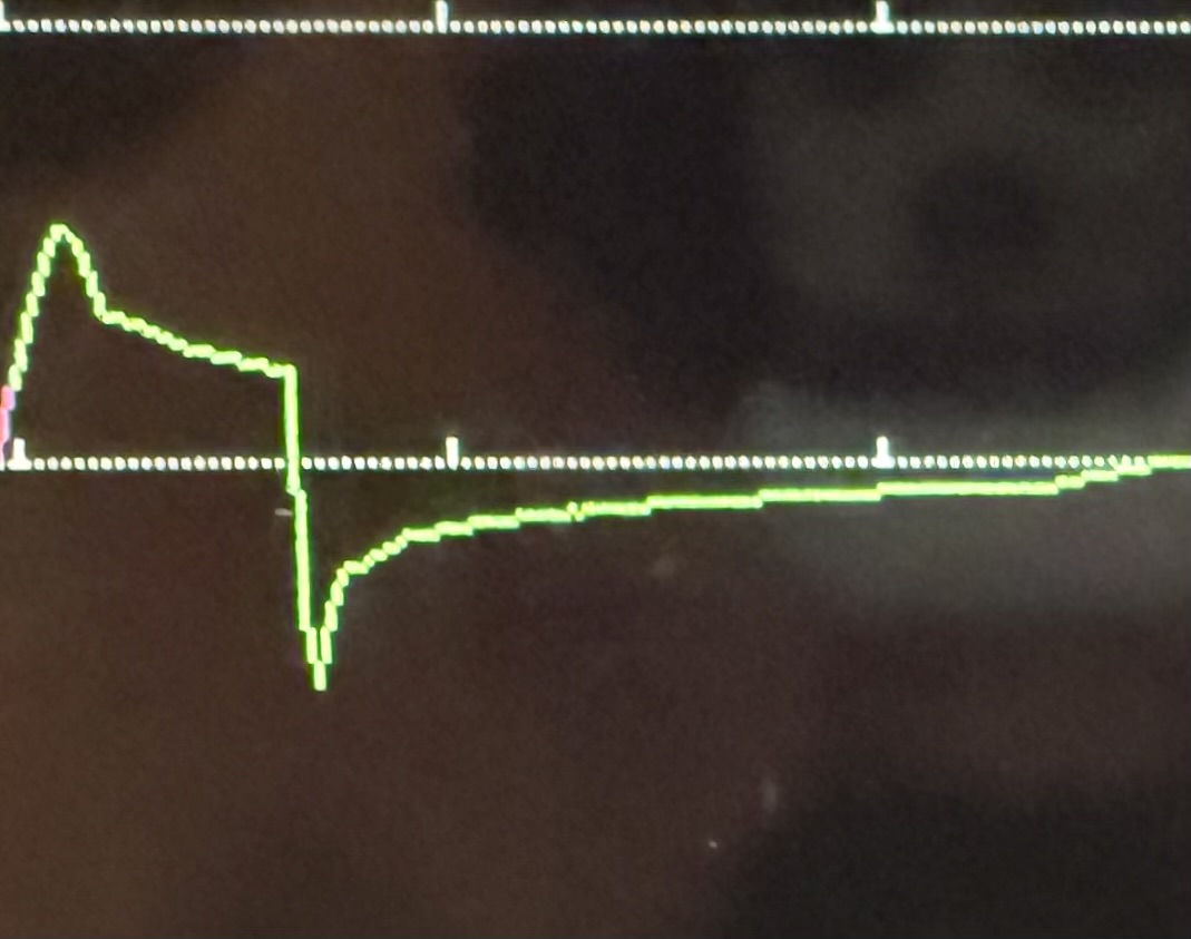
There are a lot of issues that are apparent on this ventilator waveform. However, the focus for this pearl is the expiratory waveforms which show significant expiratory obstruction (compare to normal below). The patient is getting inspiratory volumes (VTi 553 mL in light blue bottom right corner), but limited expiratory waveforms (VTe 164 mL) . Given that the patient has no history of asthma, COPD, or reason for acute obstruction, the concern was that patient effort (as patient became more awake with decreased sedation) was actually causing expiratory obstruction. Sedation was increased and the obstruction resolved.
The patient was subsequently diagnosed with excessive dynamic airway collapse, a condition where the posterior membrane of the tracheobronchial tree weakens and bulges anteriorly into the airways (this can sometimes be seen on expiratory CT chests). This movement is exaggerated during forced exhalation due to increased intrathoracic pressure and causes a more severe expiratory obstruction pattern compared to passive exhalation. Consequently, this can lead to a cycle of agitation, increased work of breathing, and breath-stacking/accumulation of intrinsic PEEP.
Of note, EDAC often gets misidentified as “COPD” or “asthma,” and given its mechanism, does not respond to bronchodilators or steroids. The weakening of the posterior membrane is a chronic process and hence EDAC does not develop acutely, it simply becomes clinically apparent in an agitated intubated patient. Hence, the treatment for this patient was a sedative to calm the patient while preserving respiratory effort (i.e. dexmedetomidine) and extubating the patient to remove the exacerbating stimulus (being intubated).
Extubating to NIPPV could be considered as a bridge granted it does not cause more agitation and increased work of breathing.
In summary, EDAC is a cause of expiratory obstruction that should be considered in patients with evidence of obstruction and increased of work of breathing, particularly if they don’t have a history of COPD/asthma or reason for bronchospasm.