University of Maryland School of Medicine logo
Emergency Medicine
All Departments
UMSOM Assistant
UMEM Educational Pearls
  • Home
  • Departments
  • Emergency Medicine
  • CME
  • UMEM Educational Pearls
  • CME
  • Courses and Conferences
  • Free Open Access Medical Education
  • UMEM Educational Pearls

Search

41-60 of 245 results with category "Visual Diagnosis"

Previous  |  1 |  2 |  3 |  4 |  5 |  6 |  7 |  8 |  ... |  13 |  Next

Title: What's the Diagnosis? Image by mi amigo, Dr. Pedro Verdugo.

Category: Visual Diagnosis

Posted: 10/6/2015 by Haney Mallemat, MD (Updated: 10/7/2015)

Question

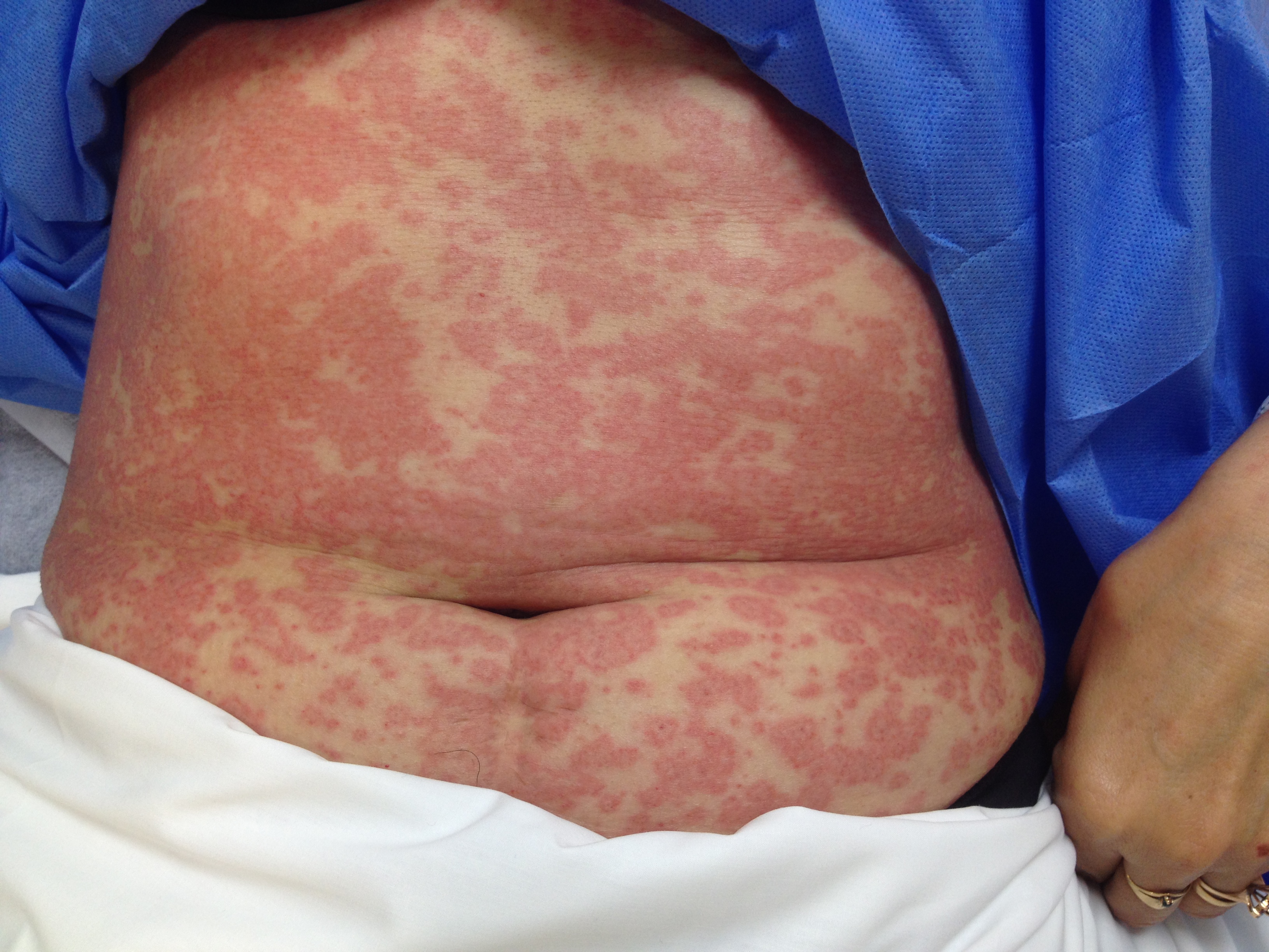
Patient presents after being started on an antibiotic for cellutlitis of lower extremity. What's the diagnosis and what are some other etiologic agents (name 3)
Show Answer

Erythema Multiforme (minor)

  • Acute, self-limited rash affecting the skin; mucus membranes are spared in the minor variant although EM major involves one mucus membrane.
  • Erythematous, macupapular, and blanching with central clearing; it is often puritic
  • Several etiologic factors that have been identified. Most commonly:
    • Infections: viral (e.g., HSV), bacterial (e.g., strep), fungal (e.g., histoplasmosis)
    • Drugs: antibiotics (e.g., sulfa drugs, PCN, etc.), NSAIDs
    • Miscellaneous: collagen vascular diseases, pregnancy, and malignancy
  • It is self-limiting and will resolve in 2-6 weeks.
  • Treat by stopping offending agent and supportive care (e.g., diphenhydramine for purtitis)

Show References

Follow me on Twitter (@criticalcarenow)


Title: What's the Diagnosis? Case by Dr. Zachary Dezman

Category: Visual Diagnosis

Posted: 9/28/2015 by Haney Mallemat, MD

Question

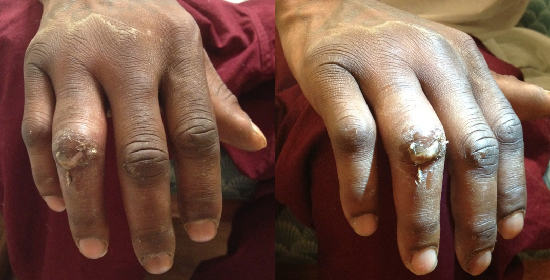
26 year-old male presents with a swollen 4th digit and pain during extension, what’s the diagnosis?
Show Answer

Answer: Infectious Flexor Tenosynovitis

Infectious Flexor Tenosynovitis

  • Closed space infection of the flexor tendon sheath; an orthopedic emergency
  • Typically an infection with skin flora secondary to penetrating trauma
  • Remember Kanavel's cardinal signs via mnemonic B.E.S.T. (the "BEST" mnemonic):
    • B: Bend finger (i.e., finger held in slight flexion)
    • E: Extension pain (i.e., pain on passive extension)
    • S: Sausage-like digit (i.e., fusiform swelling
    • T: Tenderness along tendon sheath
  • If early and not severe a course of IV antibiotics covering skin flora may be tried. Surgical intervention, however, is often necessary so early consultation with a hand surgeon is highly recommended

Show References

Follow me on Twitter (@criticalcarenow)


Title: What's the Diagnosis?

Category: Visual Diagnosis

Posted: 9/21/2015 by Haney Mallemat, MD

Question

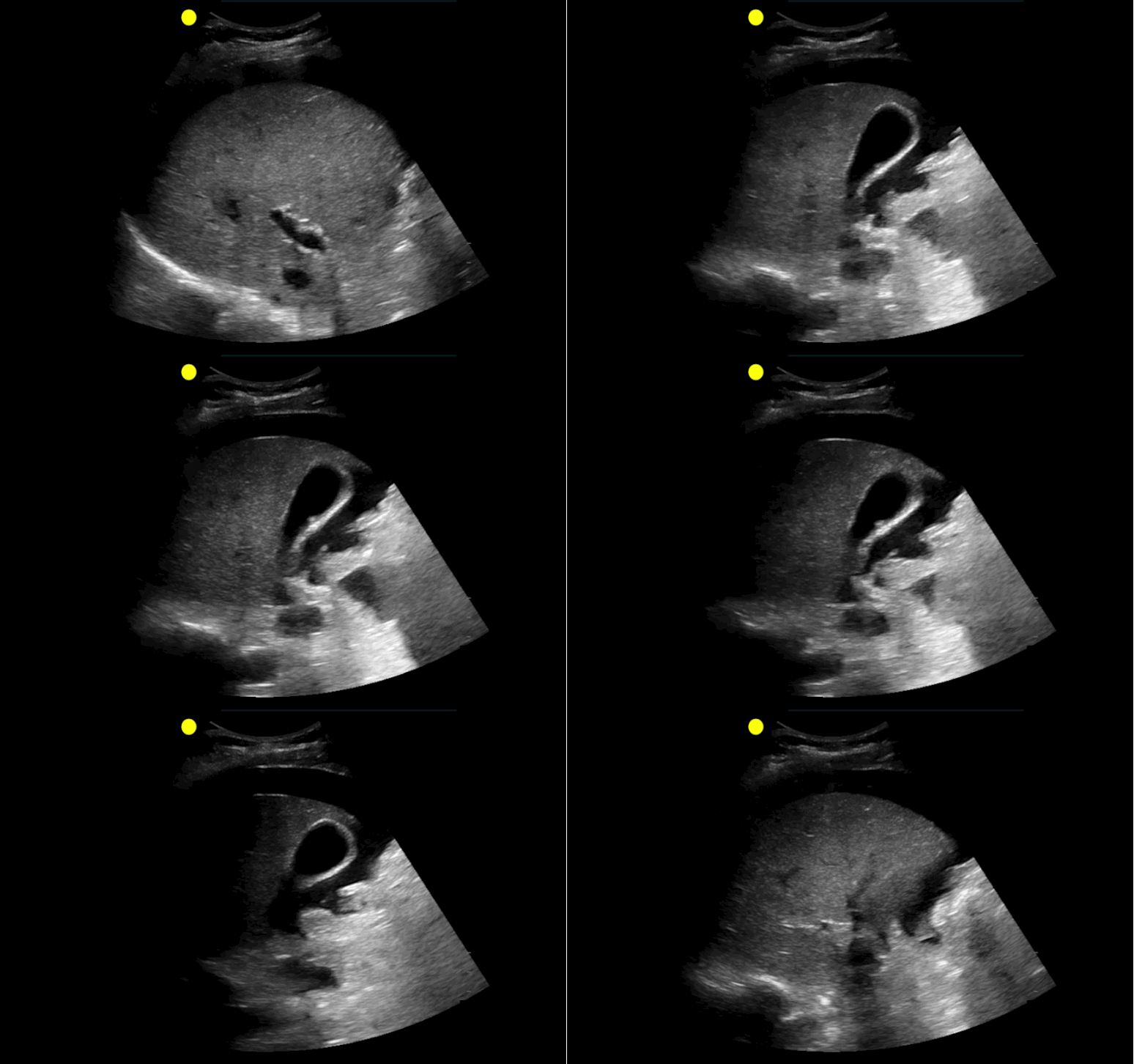
 30 year-old male with abdominal pain and diffuse tenderness on exam. Ultrasound is shown, what's the diagnosis?
Show Answer
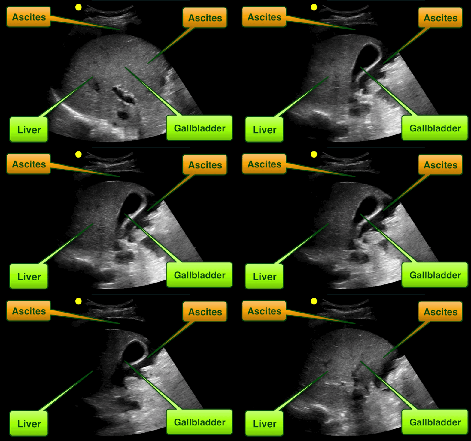
Free intra-abdominal fluid.

  • The patient had ascites in this case, with spontaneous bacterial peritonitis; this was initially thought to be pericholecystic fluid secondary to acute cholecystitis.
  • Another mimic of acute cholecystitis includes conditions with gallbladder wall thickening such as a contracted GB (e.g, post-prandial) or GB wall edema from fluid overload (e.g., CHF, renal failure, etc.)

Show References

Follow me on Twitter (@criticalcarenow)


Title: What's the Diagnosis? Case by Dr. Bennett Myers

Category: Visual Diagnosis

Posted: 9/14/2015 by Haney Mallemat, MD

Question

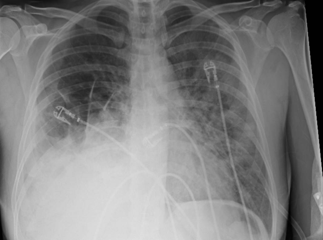
35 year-old female presents to the Emergency Room with cough and chest tightness. She was discharged from the hospital yesterday for an asthma exacerbation that was secondary to pneumonia. What's the diagnosis?
 

Show Answer
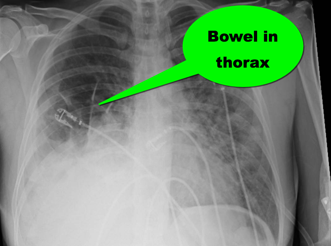
Diaphragmatic hernia

Show References

Follow me on Twitter (@criticalcarenow)


Title: What's the Diagnosis?

Category: Visual Diagnosis

Posted: 9/7/2015 by Haney Mallemat, MD (Updated: 9/7/2015)

Question

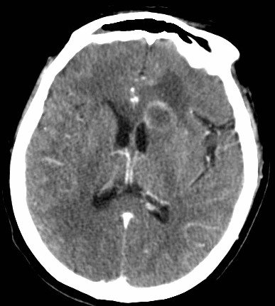
68 year-old man presents with a new-onset seizure. What's the diagnosis and what's in your differential diagnosis?
Show Answer
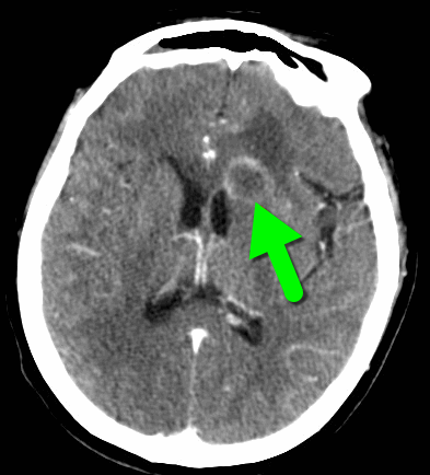
Ring-enhancing lesion

Differential diagnosis of a ring-enhancing lesion

Neoplasm Infectious Neurologic

Primary brain tumor

Pyogenic Abscess Multiple Sclerosis

Primary CNS lymphoma

Tuberculoma Sarcoidosis
Metastatic disease Syphilis Vasculitis
 

Neurocysticercosis  

Aneurysm
  Toxoplasmosis Resolving hematoma
  Nocardiosis Cerebral infarct
  Cyptococcocus Post-op changes
  Aspergillosis  


Show References

Follow me on Twitter (@criticalcarenow)


Title: What's the Diagnosis? Image by Dr. Michael Winters

Category: Visual Diagnosis

Posted: 8/31/2015 by Haney Mallemat, MD

Question

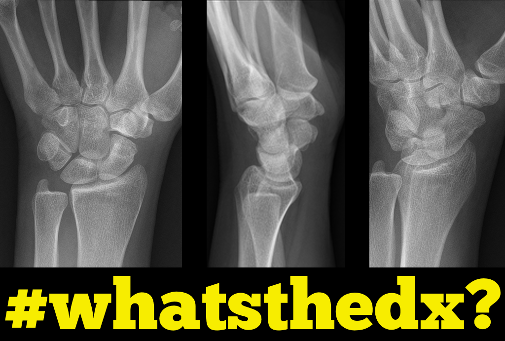
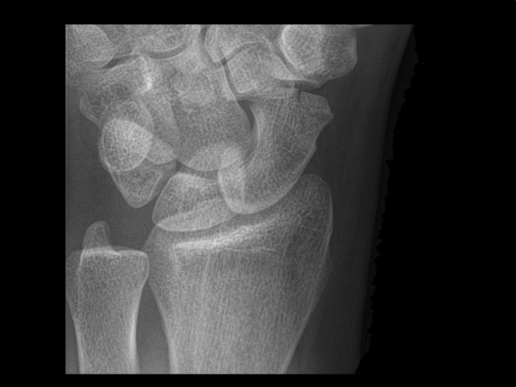
Person presents following a fall on an outstretched hand and there is snuffbox tenderness. What's the diagnosis?
Show Answer
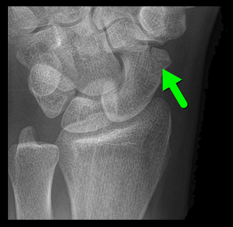
Scaphoid fracture

Show References

Follow me on Twitter (@criticalcarenow)


Title: What's the Diagnosis?

Category: Visual Diagnosis

Posted: 8/24/2015 by Haney Mallemat, MD

Question

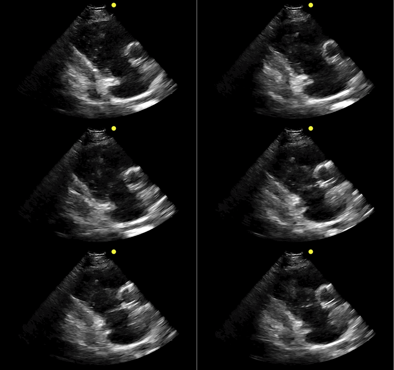
You find this interesting view while scanning a patient. Which view is this and why should you care about it? 
Show Answer
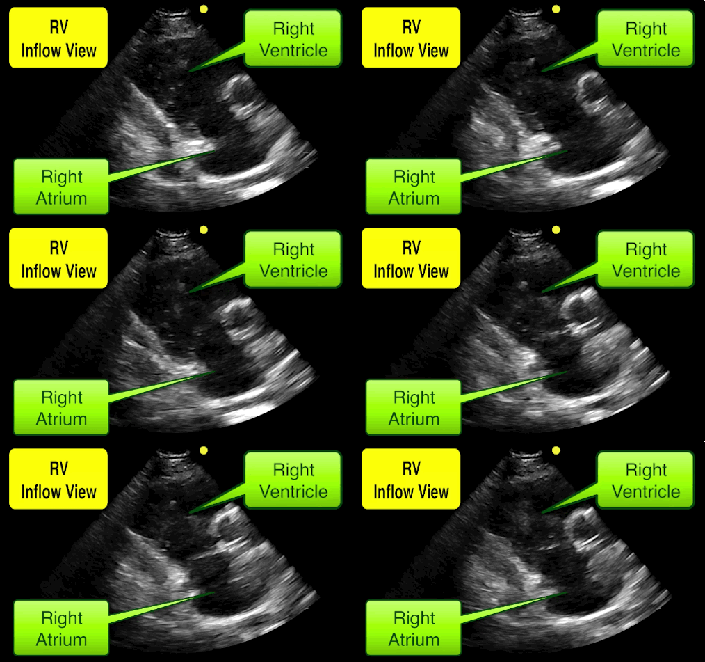
This is the right ventricular inflow view

  • RV inflow view obtained by obtaining a parasternal long-axis view and then tilting the tail of the transducer cephalad; eventually you'll see the heart as detailed below
  • It's a great view for Doppler measurements through the tricuspid valve because the flow of blood is parallel to the ultrasound beam, useful when estimating things like the pulmonary artery systolic pressure
  • Want to learn more about the RV? Click here for a whole podcast on it!

Show References

Follow me on Twitter (@criticalcarenow)


Title: What's the Diagnosis? Images by Dr. Daniel Gingold

Category: Visual Diagnosis

Posted: 8/17/2015 by Haney Mallemat, MD

Question

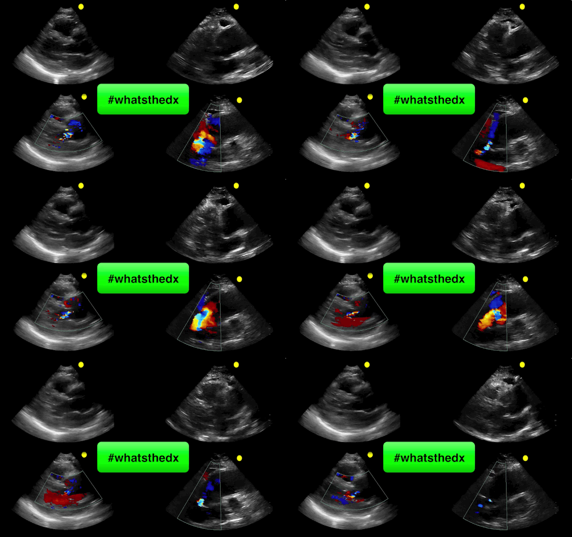
55 year-old male presents with chest pain. Echo is shown below (parasternal long-axis on the left and aortic root / ascending aorta on the right), what's the diagnosis?
Show Answer
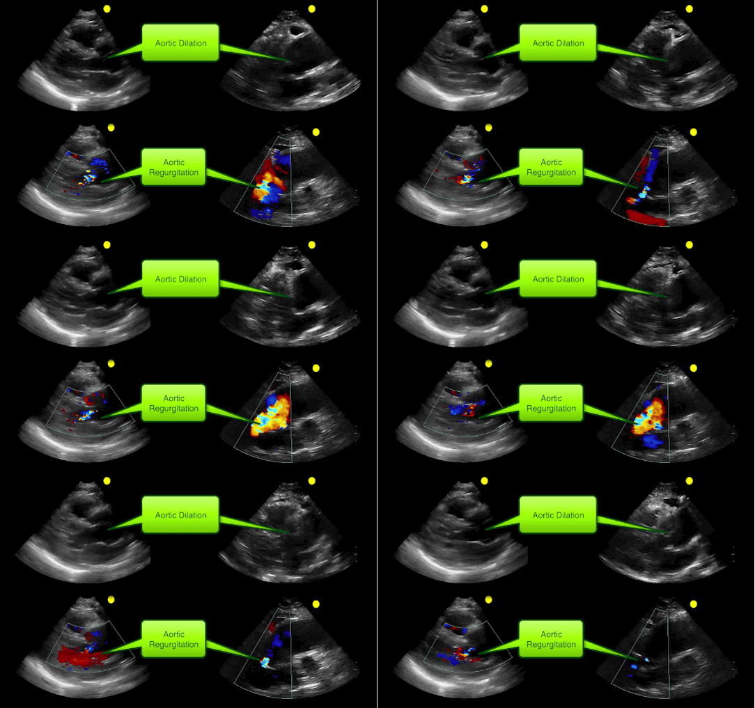
  • Ascending aortic aneurysm resulting in secondary aortic reguritation

Show References

Follow me on Twitter (@criticalcarenow)


Title: What's the Diagnosis? Case by Dr. Bethany Radin

Category: Visual Diagnosis

Posted: 8/10/2015 by Haney Mallemat, MD

Question

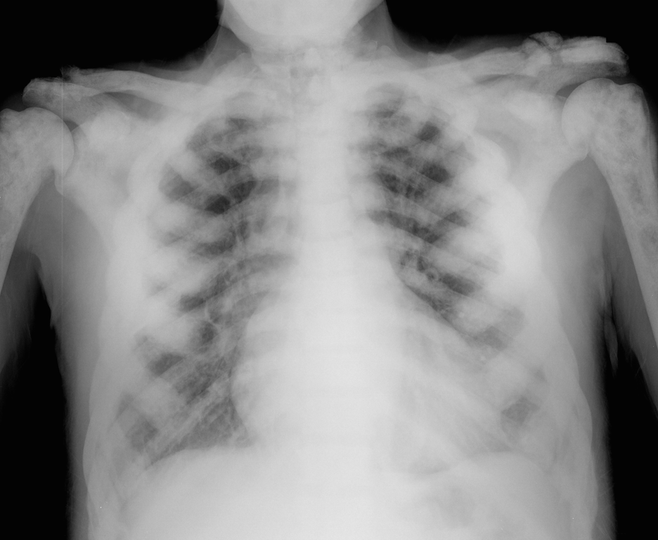
64 year-old male with no past medical history presents complaining of chronic weight-loss and diffuse chest pain; CXR is shown. What's the diagnosis and what other disease(s) may present similarly?
 

Show Answer
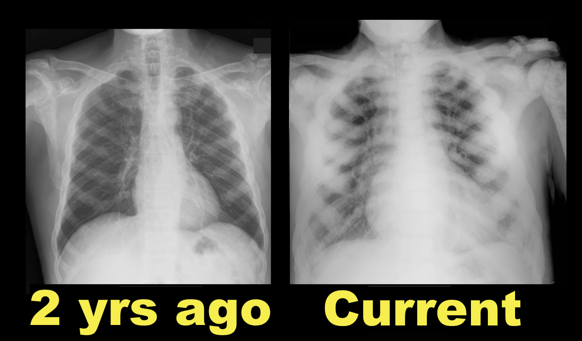
Sclerotic bone (osteoblastic) metastasis secondary to prostate cancer. The patient's CXR from 2 years prior is shown below for comparison.

Other malignancies associated with osteoblastic metastasis:

  • Breast cancer
  • Colon cancer (i.e., mucinous adenocarcinoma)
  • Lymphoma
  • Carcinoid
  • Neuroblastoma
  • Prostate cancer (#1 cause)

Show References

Follow me on Twitter (@criticalcarenow)


Title: What's the Diagnosis? Case by UMEM alumni Dr.Whitney Cesari

Category: Visual Diagnosis

Posted: 7/20/2015 by Haney Mallemat, MD (Updated: 7/20/2015)

Question

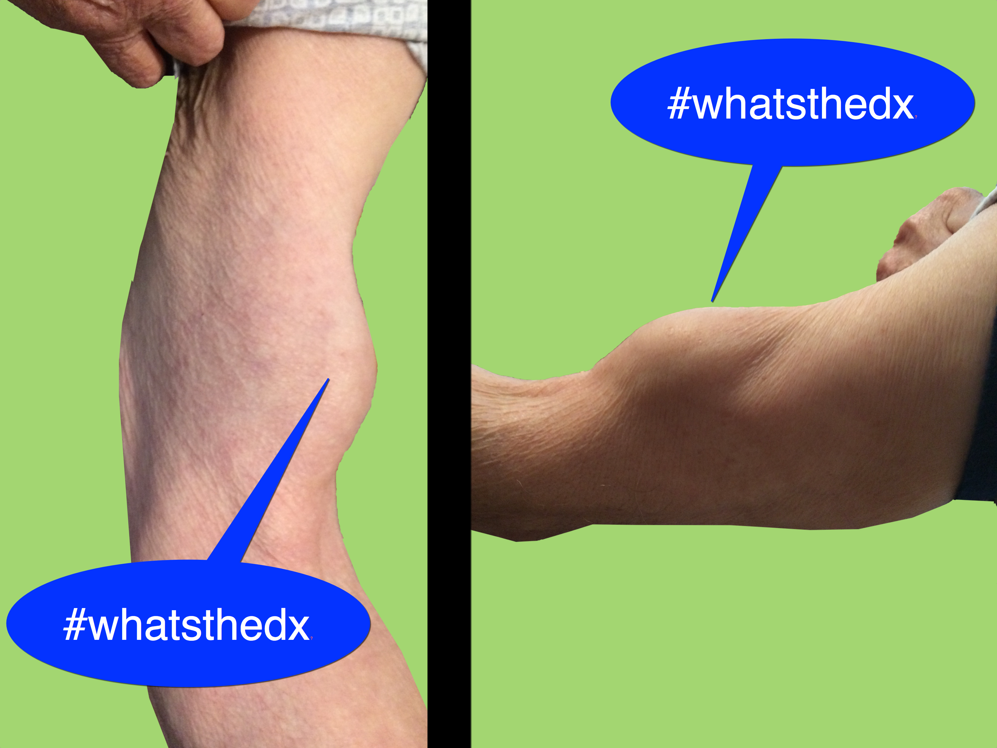
81 year-old man was mowing the lawn and then felt a sudden pop, then pain in his left arm. What's the diagnosis and what's this sign called?
Show Answer

Biceps tendon rupture (BTR) aka Popeye's Sign

  • BTR can be related to degenerative issues in the elderly or acute injuries associated vigorous bicep use (e.g., weight lifting). Patients report a "popping" sound followed by sharp, sudden pain, ecchymosis and swelling.
  • Diagnosis is clinical; a palpable mass is seen between the shoulder and the elbow. The tendon typically ruptures on the proximal end, causing coiling of the muscle distally. ROM is maintained because only one bicep tendon ruptures, the remaining one functions normally.
  • Medical management is appropriate and emergency surgery is unnecessary (except for athletes or patient who rely on muscle strength for occupational purposes) Rupture of the distal bicep tendon ruptures requires surgical referral. 

Show References

Follow me on Twitter (@criticalcarenow) or Google+ (+criticalcarenow)


Title: What' the Diagnosis? Case by Dr. Kathleen "Kat" Stephanos

Category: Visual Diagnosis

Posted: 7/13/2015 by Haney Mallemat, MD

Question

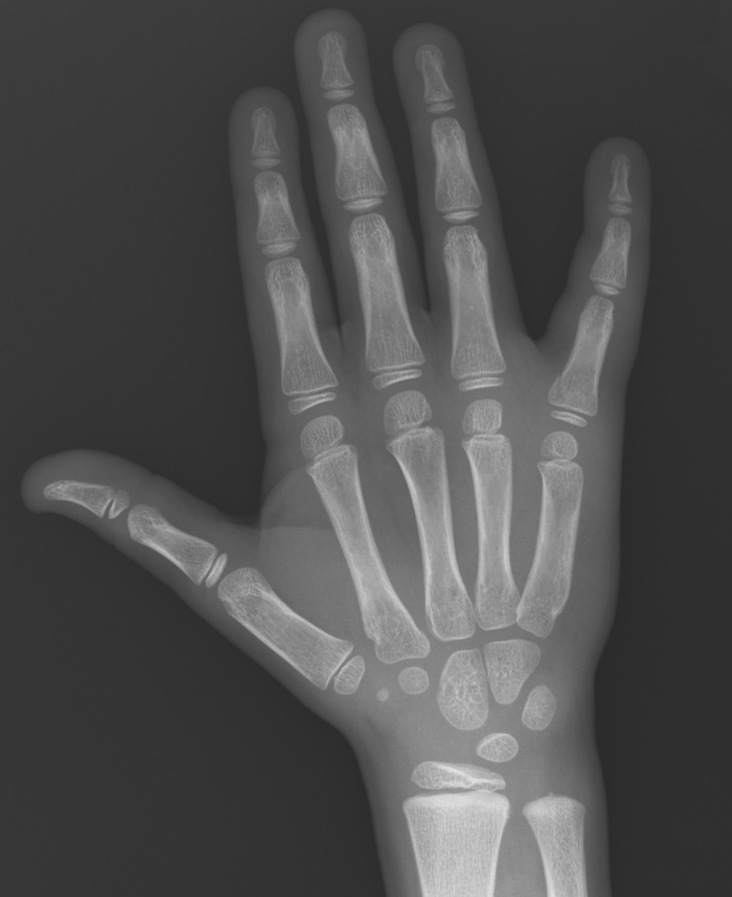
7 year-old male "jammed" 5th finger while playing basketball with pain and swelling over finger. What's the diagnosis?
Show Answer
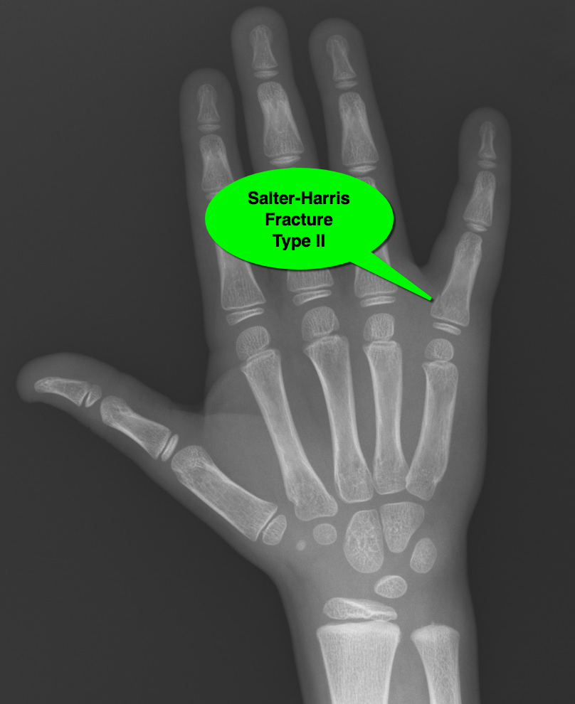
Salter-Harris fracture (type II): 

Salter-Harris Fracture (remember mnemonic SALTR)

  • Slipped (i.e., through growth plate and not involving bone) / Type I
  • Above growth plate (i.e., through metaphysis) / Type II (most common)
  • Lower growth plate (i.e., through epiphysis) / Type III
  • Through (i.e., through metaphysis growth plate and epiphysis) / Type IV
  • Rammed (i.e., Crush injury) / Type V (worst prognosis)

For more on Salter-Harris fractures, check out this and that

Show References

Follow me on Twitter (@criticalcarenow) or Google+ (+criticalcarenow)


Title: What's the Diagnosis? Case by Dr. Maite Huis in 't Veld

Category: Visual Diagnosis

Posted: 7/6/2015 by Haney Mallemat, MD

Question

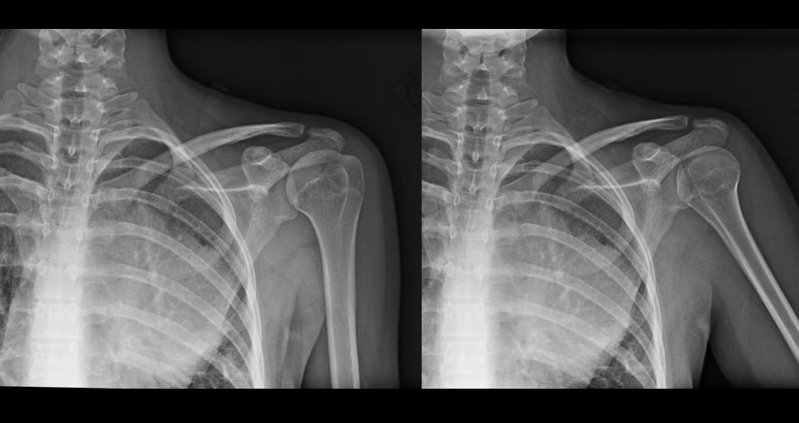
15 year-old female field hockey player presents with left shoulder pain. Besides fatigue over several weeks, she has no past medical history and there is nothing remarkable on physical exam. What's the diagnosis? 
Show Answer
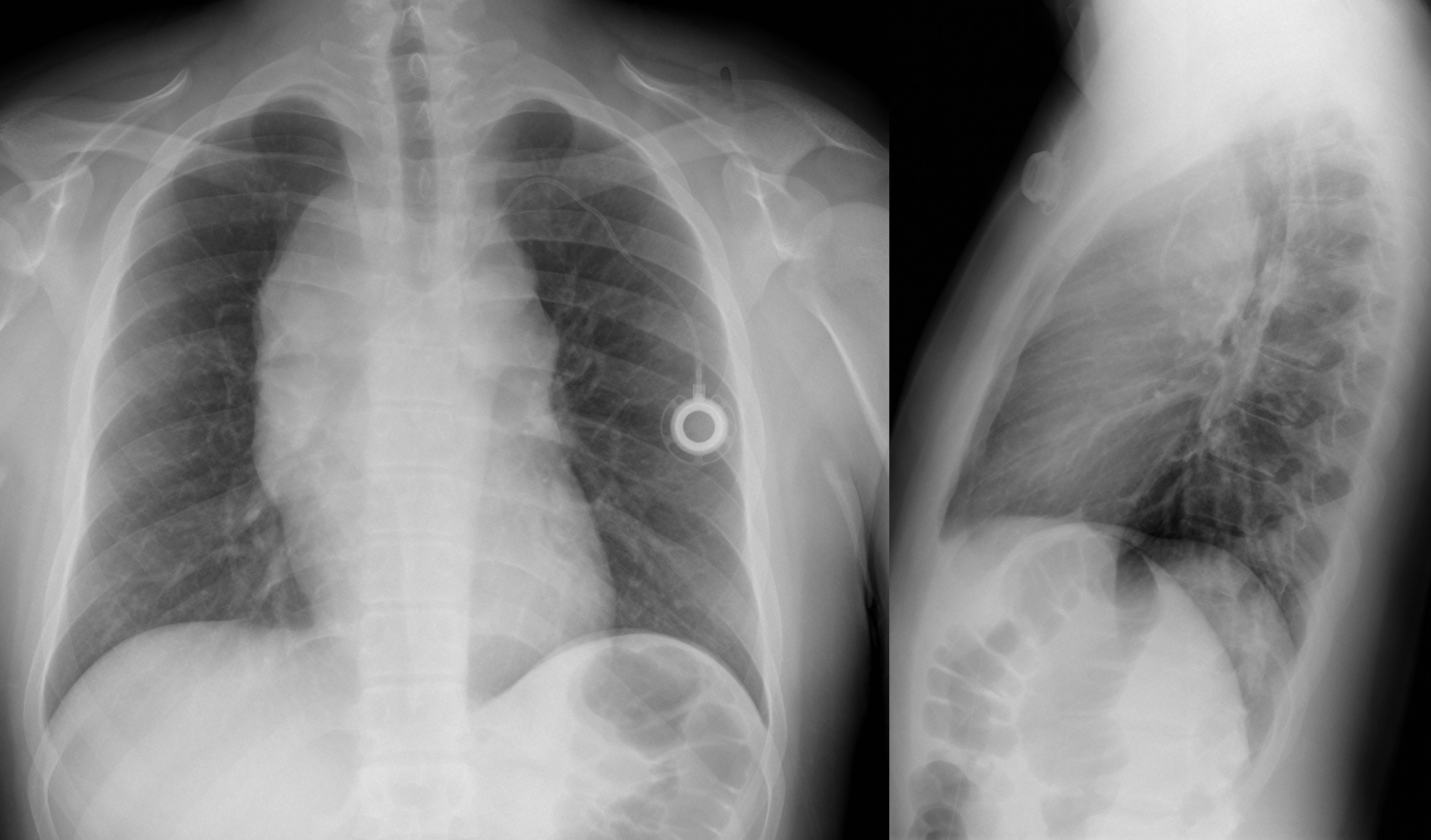
Mediastinal mass (lymphoma confirmed on biopsy)

  • There was no bony abnormality of the shoulder but the mediastinal mass was nearly missed because the focus was on the shoulder
  • Below is Xray after patient started receiving chemotherapy.
  • Bottom Line: always be sure to evaluate the entire Xray because sometimes you'll discover unrelated, but important findings 

Show References

Follow me on Twitter (@criticalcarenow) or Google+ (+criticalcarenow)


Title: What's the Diagnosis? Case by Dr. George Kochman

Category: Visual Diagnosis

Posted: 6/30/2015 by Haney Mallemat, MD (Updated: 6/30/2015)

Question

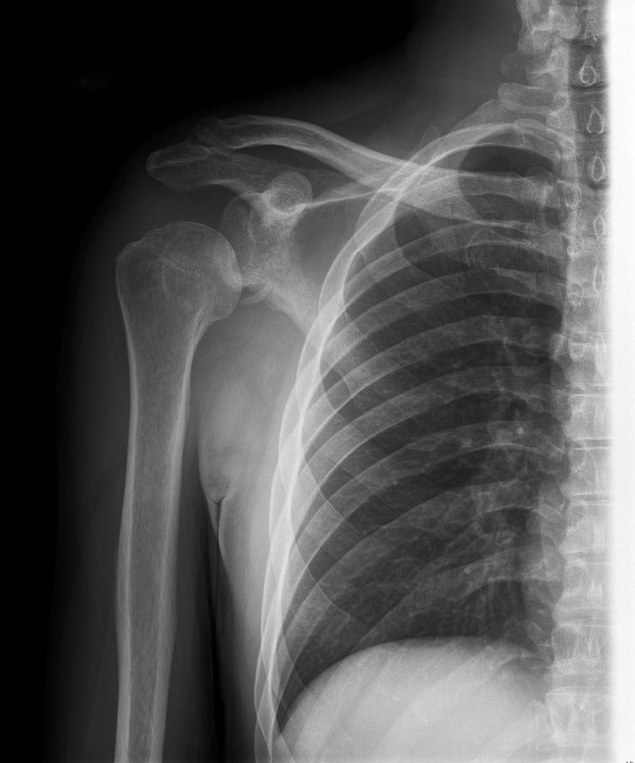
25 year-old male falls from 10 feet and lands on his right shoulder, what's the diagnosis?
 

Show Answer
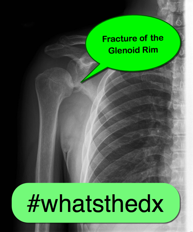
Yes, the shoulder is dislocated...but did you notice anything else (see below)?

Bankart lesion (a.k.a. fracture of the glenoid fossa)

  • Fractures of the glenoid fossa occur when the humeral head collides with the glenoid margin by a laterally directed force.
  • Although less than 10% of glenoid fossa fractures result in significant displacement, surgical repair is indicated if:
    • Fracture displacement >10mm
    • >33% involvement of posterior glenoid fossa
    • >25% involvement of anterior glenoid fossa
    • Persistent humeral head subluxation

Show References

Follow me on Twitter (@criticalcarenow) or Google+ (+criticalcarenow)


Title: What's the Diagnosis? Case by Dr. Ben Smith (@ultrasoundjelly)

Category: Visual Diagnosis

Posted: 6/23/2015 by Haney Mallemat, MD (Updated: 6/23/2015)

Question

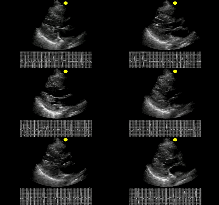
30 year-old patient presents with palpitations. A parasternal long-axis clip is shown below along with the rhythm strip. What's the diagnosis and what drug was given during this clip? 
Show Answer

This was SVT and adenosine was given; notice how the heart pauses in the echo and in the rhythm strip.

Show References

Follow me on Twitter (@criticalcarenow) or Google+ (+criticalcarenow)


Title: What's the Diagnosis? Image by Dr. Semhar Tewelde

Category: Visual Diagnosis

Posted: 6/15/2015 by Haney Mallemat, MD

Question

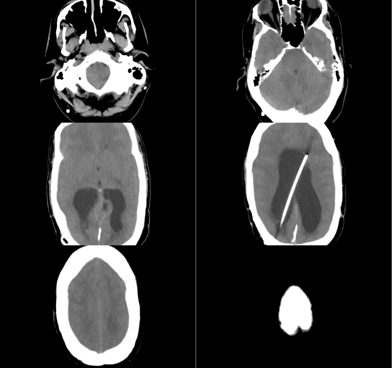
Patient presents with headache and papilledema. What's the diagnosis?
Show Answer
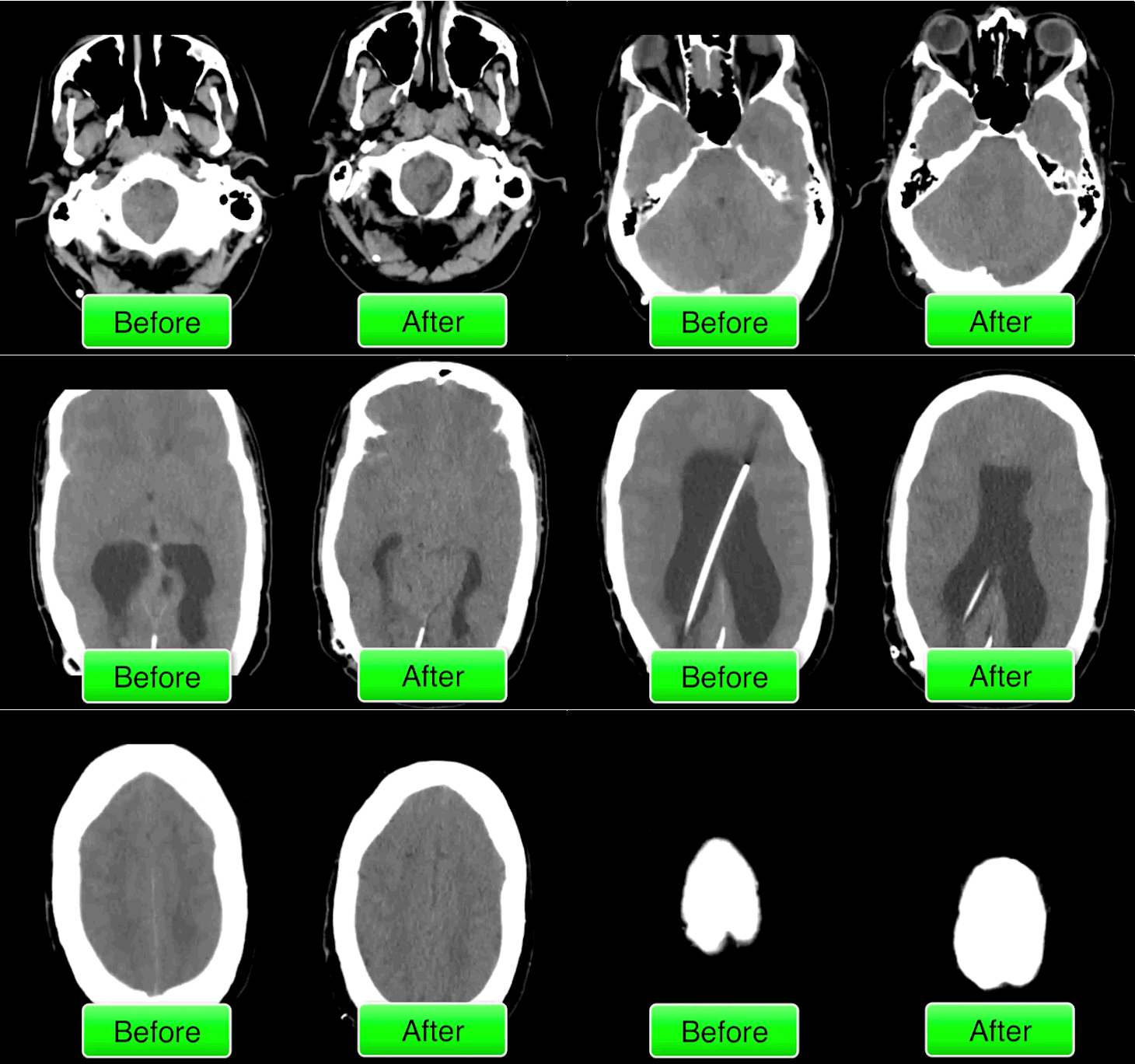
Hydrocephalus secondary to malfunctioning VP shunt. 

The patient went to the operating room for a shunt repair and the CT after the repair is shown; although not dramatically different it does show an improvement of the hydrocephalus.  

Show References

Follow me on Twitter (@criticalcarenow) or Google+ (+criticalcarenow)


Title: What's the Diagnosis? Clip by Dr. Denise Fraga and Dr. Adeleke Oni

Category: Visual Diagnosis

Posted: 6/8/2015 by Haney Mallemat, MD

Question

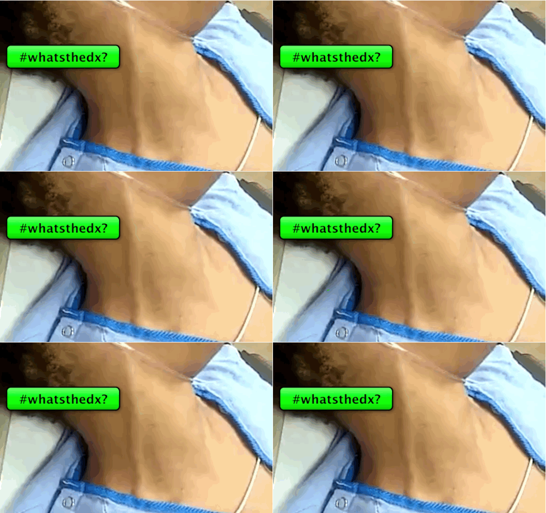
The following clip is one of three findings found in Beck’s triad. Name all three findings and how often are all 3 signs present for patients with pericardial tamponade?
Show Answer

  • Beck's triad
    • Jugular venous distention
    • Muffled (or distant) heart sounds
    • Low blood pressure
  • None of these findings is sensitive or specific for the diagnosis of pericardial tamponade
  • Only 10-40% of patients with pericardial tampaonde will have all three signs present

Show References

Follow me on Twitter (@criticalcarenow) or Google+ (+criticalcarenow)

Spodick, D. Cardiac Tamponade Revisited: A Postmortem Look at a Cautionary Case: Tex Heart Inst J. 2007; 34(3): 347–351.


Title: Learn to how to place a pigtail catheter

Category: Visual Diagnosis

Posted: 5/25/2015 by Haney Mallemat, MD

Do you need to get stuff out of the thorax (like fluid or air) and don't want to place a HUGE chest tube? Consider a pigtail catheter; don't know how to place one? Well check out this video and learn how.

Show References

Follow me on Twitter (@criticalcarenow) or Google+ (+criticalcarenow)


Title: What's the Diagnosis? Case by Dr. Ibrahim H. Abuduruk

Category: Visual Diagnosis

Posted: 5/19/2015 by Haney Mallemat, MD (Updated: 5/19/2015)

Question

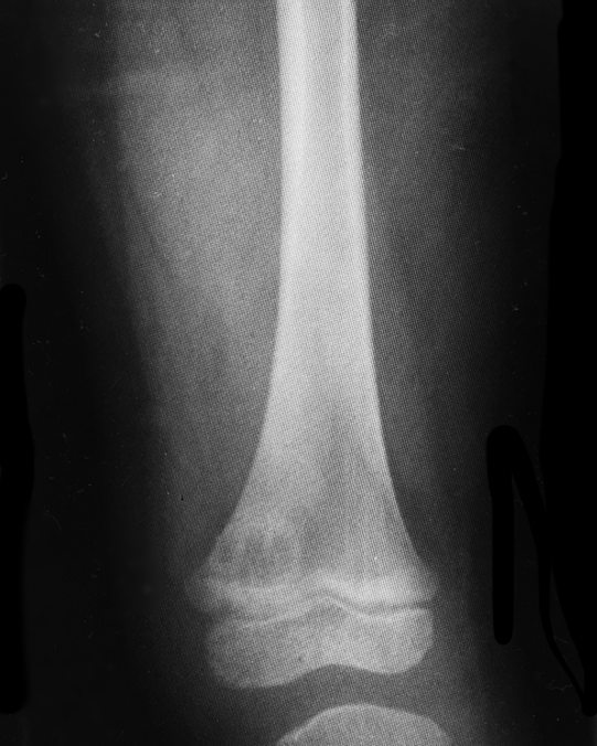
5 year-old with no past medical history, complains of a limp and mild left knee pain. No history of trauma. Physical exam is significant for a low-grade fever and is otherwise normal. What’s the diagnosis?
Show Answer
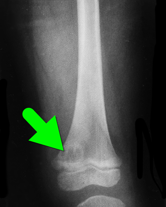
Brodie's abscess a.k.a subacute osteomyelitis

  • Form of osteomyelitis, which is difficult to diagnose, and is often mistaken for a tumor.
  • Males are affected more than females and the range of presentation is 2-15 years.
  • Work up is difficult as CBC, ESR, CRP, and blood cultures are inconclusive. Xrays may mimic tumors.
  • Treatment is antibiotics if the diagnosis is obvious; if not, biopsy to make the diagnosis. Surgery an option if either the lesion is aggressive or there is pus extending into the synovial fluid. 

Show References

Follow me on Twitter (@criticalcarenow) or Google+ (+criticalcarenow)


Title: What's the Diagnosis? Case by Dr. Nicholas Santavicca and Dr. Semhar Tewelde

Category: Visual Diagnosis

Posted: 5/11/2015 by Haney Mallemat, MD

Question

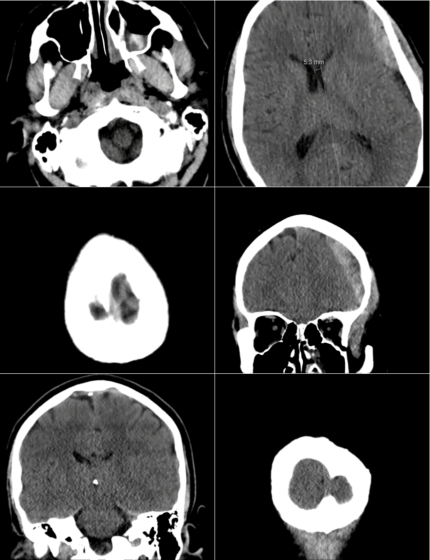
40 year-old male sustains a blunt force injury the left side of his lead. What's the diagnosis and what structure was injured?
Show Answer
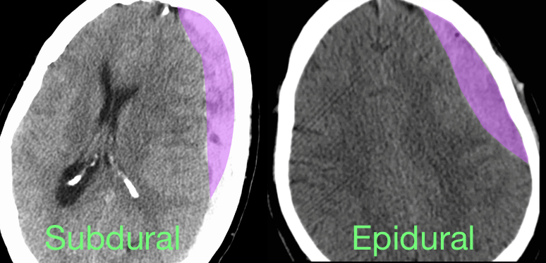
Epidural Hematoma with mid-line shift from middle meningeal artery injury; see image below for comparison of subdural and epidural

Epidural hematoma

  • Blood accumulates between dura and calvarium secondary to trauma; concurrent skull fractures are common
  • Middle menial artery is usually involved in the temporal region; other arteries may be involved depending on which area of the skull receives the impact
  • Up to one-third of cases present with the "classic" lucid interval from injury and loss of consciousness to a decline in mental status. Diagnosis should be suspected, however, based on mechanism
  • Treatment is surgical evacuation, however aggressive medical management of increased intracranial pressure should not be delayed:
    • Head of bed elevated and keep neck mid-line
    • Anti-emetics and analgesics
    • Osmotic agents such as mannitol or hypertonic saline
    • Avoid hypoxia, hypotension, and hyperthermia

Show References

Follow me on Twitter (@criticalcarenow) or Google+ (+criticalcarenow)


Title: What's the Diagnosis? Case by Dr. Ahmed Alrasheedi

Category: Visual Diagnosis

Posted: 5/4/2015 by Haney Mallemat, MD

Question

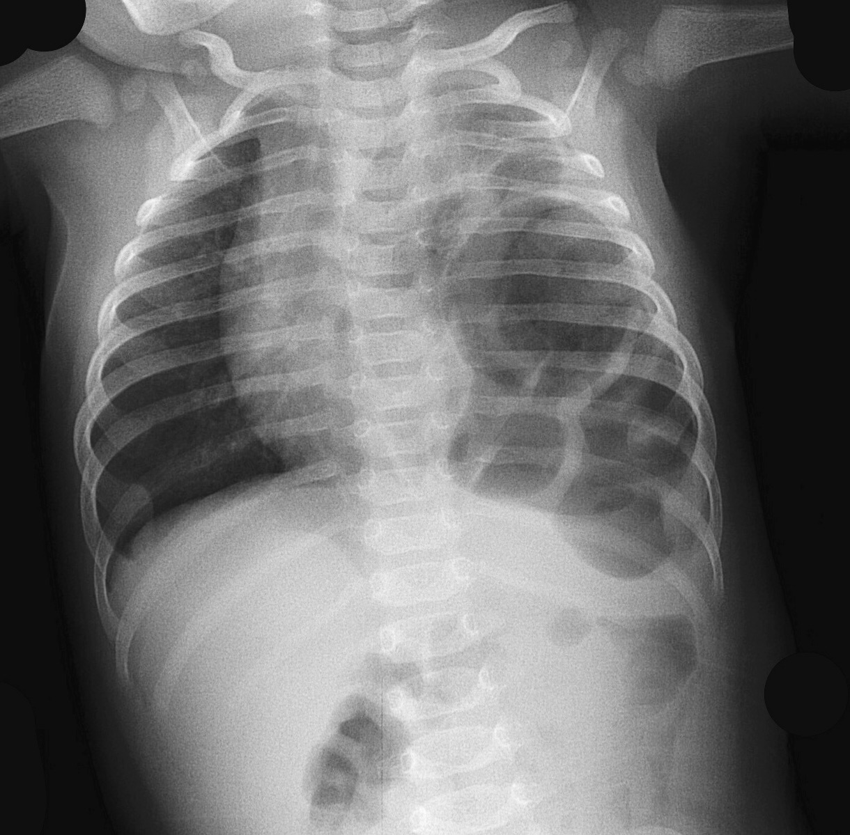
3 month-old male presents with severe respiratory distress; oxygen saturation is 81% (on room air), he is grunting, and there are no breath sounds on the left. What's the diagnosis?

Show Answer

Congenital diaphragmatic hernia
Management:
  • Aggressive fluid and vasopressor support
  • Early intubation and airway protection should be considered in severe cases; minimize BVM given the risk of gastric distention.
  • NG tube decompressing of the stomach will help the ventilation/oxygenation process.
  • ECMO may be considered if conventional therapy has failed and any one of the following criteria is met:
  1. Inability to maintain pre-ductal PaO2 saturations >85 percent or post-ductal PaO2 >30 mmHg
  2. Peak inspiratory pressure >28 cm H2O or mean airway pressure >15 cm H2O
  3. Hypotension that is resistant to fluid and inotropic support
  4. Inadequate oxygen delivery with persistent metabolic acidosis

Show References

Follow me on Twitter (@criticalcarenow) or Google+ (+criticalcarenow)


Previous  |  1 |  2 |  3 |  4 |  5 |  6 |  7 |  8 |  ... |  13 |  Next
University of Maryland School of Medicine logo
  • Departments
  • Programs
  • Research Centers
  • Institutes
  • Graduate Education
  • CIBR Cores
  • Offices of the Dean
    • University of Maryland, Baltimore
    • University of Maryland Medical Center
    • Medical Alumni Association of the University of Maryland
    • Health Sciences and Human Services Library
University of Maryland School of Medicine
655 W. Baltimore Street
Baltimore, MD 21201
Contact Us

© University of Maryland School of Medicine

    • Webmaster
    • Web Accessibility
    • Site Index
    • UMB Hotline
    • Employment
    • Map