
Search
Subscribe to daily UMEM Pearls (FREE!) via:
401-420 of 479 results with category "Orthopedics"
Patellofemoral Syndrome (Chondromalacia Patella)
- Due to degeneration of the cartilage underneath the patella
- Patients often present with:
- A grinding sensation when the knee is extended
- Pain in the front of the knee that typically worsens after sitting for a long period of time
- Knee pain that worsens with using stairs, running or when needing to bend the knee deeply (i.e.: squats)
- Commonly thought to be due to overuse (i.e.: new running program, or marching as in military recruits), but can also be due to anatomic abnormalities like pes planus or a large Q angle. Ultimate cause is likely to be multifactorial
-

- Can be treated with NSAIDs, and limiting activity
- Physical Therapy that helps to strengthen the quadriceps can help prevent the patella from grinding on the femoral condyles.
Show References
Injury was originally described as an occupational hazard in Scottish gamekeepers (from breaking the necks of rabbits against the ground). Today, skiing is now the most common cause and injury is now the second most common orthopedic injury in skiers (MCL injury #1).
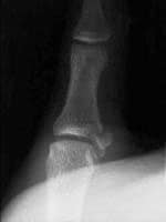
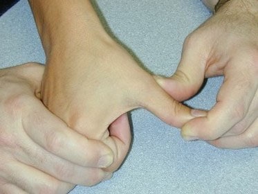
Injury to the ulnar collateral ligament (UCL) results from a sudden forced abduction (radial deviation) stress at the MCP joint of the thumb, commonly due to a fall against a ski pole or the ground.

http://blog.fitter1.com/wp-content/uploads/2010/04/b_14_1_2a.jpg
The most frequent site of rupture is the insertion into the proximal phalanx. The UCL may even avulse a small portion of the proximal phalanx at its insertion site.
http://img.medscape.com/pi/emed/ckb/sports_medicine/84611-97564-98460-1652013.jpg
Consider imaging before stress testing (to avoid further displacing a fracture)
http://img.medscape.com/pi/emed/ckb/sports_medicine/84611-97564-98460-1652060.jpg
Stabilize in a thumb spica splint and refer to hand surgery.
Calling this entity a “simple sprain” may result in chronic disability (chronic pain, instability, loss of pinch strength)
Show References
Pain Control in the Elderly
- Narcotic pain relievers are often avoided in the elderly due to the concern of sedation, risk of falls and the concern of them causing delirium.
- Delirium can cause significant morbidity and mortality and can be difficult to differentiate between the sedation and mild confusion that often occurs with opioid dose escalation.
- However, delirium has been shown to occur more commonly as a result of the under treatment of pain rather than as an opioid adverse effect.
So the take home lesson for this pearl is that the elderly have a lower risk of delirium if their pain is treated appropriately.
Show References
Supraspinatus: “Empty can” test. Have the patient abduct the shoulders to 90 degrees in forward flexion with the thumbs pointing downward. The patient attempts to lift the arms against the examiner’s resistance.
http://bjsportmed.com/content/42/8/628/F2.large.jpg

Infraspinatus and teres minor: These muscles are responsible for external rotation of the shoulder. Have the patient flex both elbows to 90 degrees while the examiner provides resistance against external rotation.
http://www.physio-pedia.com/images/4/4b/Infraspinatus_test.jpg
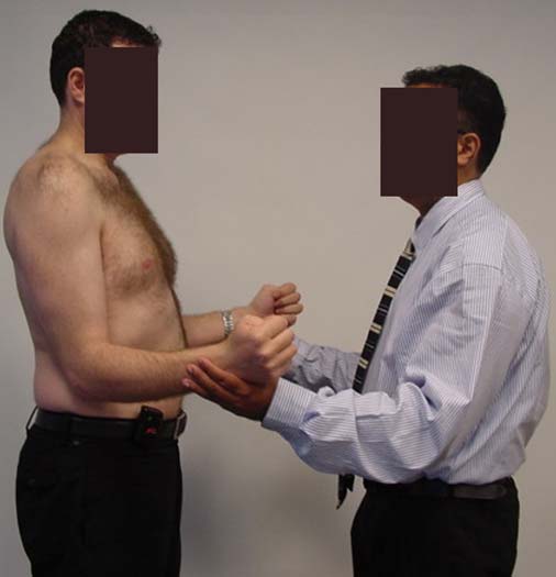
Subscapularis: “Lift-off” test. The patient rests the dorsum of the hand on the lower back (palm out) and then attempts to move the arm and hand off the back. Patients with tears may be unable to complete test due to pain.
http://www.aafp.org/afp/2008/0215/afp20080215p453-f4.jpg

Show References
Radiologic evaluation of the elbow (Part 2)
Helpful clues in the evaluation of elbow trauma:
- The Anterior humeral line and the Radiocapitellar line
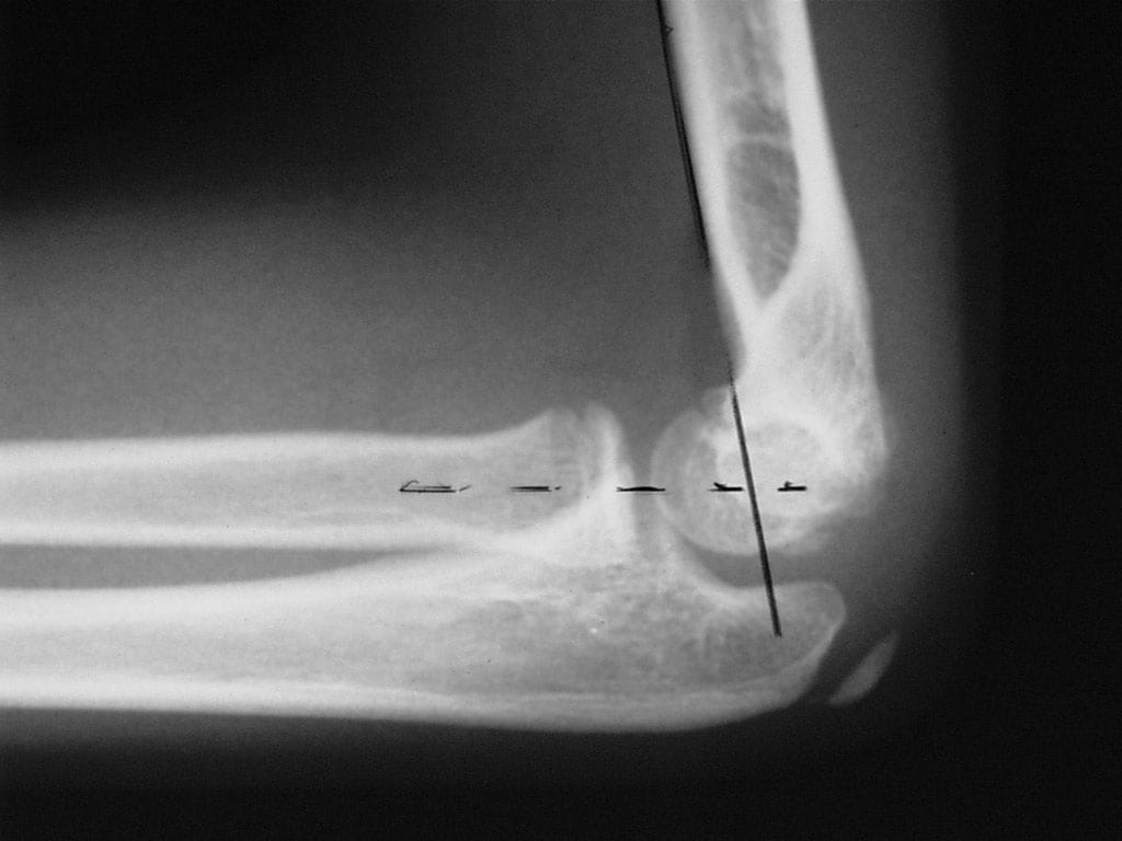
- The anterior humeral line: On a true lateral film, this line is drawn along the anterior aspect of the humeral shaft on the lateral radiograph This line passes through the middle one third of the capitellum in bones that are not injured. It is very useful for detecting subtle fractures.
- Fractures (i.e. supracondylar) usually result in displacement of the capitellum posteriorly.
- Thus, the anterior humeral line passes through the anterior one third or entirely anterior to the capitellum.
- The Radiocapitellar line: Since the radius articulates with the capitellum, a line is drawn through the middle of the radius shaft and extended proximally through the joint should bisect the capitellum on all views (AP & lateral).
- http://img.medscape.com/pi/emed/ckb/radiology/336139-415822-5412.jpg
- http://nypemergency.org/images/v2c18n.jpg
-
Improper alignment indicates a radial head dislocation (which may be very subtle)




Show References
Adhesive Capsulitis -- Frozen Shoulder
- Characterized by pain and loss of motion or stiffness in the shoulder.Normally not seen below the age of 40, affects ~2% of the population and diabetics are at increased risk.
- Due to thickening and contracture of the capsule surrounding the shoulder joint.
- Can occur after trauma to the shoulder if the shoulder is not moved early enough, but is also know to occur idiopathically.
- X-rays are only helpful to rule out other causes of the shoulder pain and are typically normal in Adhesive capsulitis.
- Typically will get better on its own over 2-3 years.
- Physical Therapy and home exercises aimed at restoring ROM can shorten the duration of pain and stiffness.
- Surgery can be done if there is no improvement with medical management and physical therapy.
- Prevention strategies include early ROM exercises in those with shoulder injuries especially in the elderly diabetic.
Show References
Rotator Cuff Tears:
Four muscles make up the rotator cuff (SITS) which control internal and external rotation of the shoulder and abduct the shoulder.
- Supraspinatus
- Infraspinatus
- Teres Minor
- Subscapularis
Tears can be due to acute injuries (falls, heavy lifting, forceful abduction), though the majority (>90%) of rotator cuff tears are chronic in nature and due to subacromial impingement and decreased blood supply to the tendons.
Most patients can be treated with sling immobilization, NSAIDs and referral to sports medicine or orthopaedic surgeons. Elderly patients should be referred quickly as prolonged immobilization can lead to a frozen shoulder.
Show References
Helpful clues in the evaluation of elbow trauma
Fat pads: The fat pad sign can be seen with any joint effusion (infection, inflammation) but in the setting of trauma, effusions are indicative of fractures about the elbow (even if no fracture line can be identified).
There are two fat pads within the elbow. Normally, on a true lateral radiograph only the anterior fat pad is seen as a small triangular radiolucent shadow anterior to the distal humeral diaphysis. The posterior fat pad is ordinarily not visualized on a lateral radiograph because it is tucked away within the olecranon fossa.
Normal lateral view: http://nypemergency.org/images/ElbowNormal.jpg

With fractures, the joint becomes distended with blood. The anterior fat pad becomes displaced superiorly and outward from the humerus giving the so called "sail sign." Similarly, the posterior fat pad gets displaced out of the olecranon fossa and becomes visible on the lateral radiograph.
Anterior (sail) and posterior fat signs: http://nypemergency.org/images/Elbowsfatpadarrow.jpg

Show References
Some common injuries and their board review associated complications
- Anterior Shoulder Dislocation = Axillary nerve or artery injury
- Supracondylar Fracture = Brachial Artery injury
- Posterior Elbow Dislocation = Brachial Artery injury
- Knee Dislocation = Popiteal Artery Injury and Peroneal and tibial nerve injury
- Humeral shaft fracture = radial nerve injury
- Posterior hip dislocation = sciatica nerve injury
- Anterior hip dislocation = femoral nerve injury
Show References
- Back pain is the most common musculoskeletal complaint that results in visits to the ED.
- It has a benign course in more than 90% of patients, so we must be vigilant and comfortable looking for warning signs of a neurologically impairing or life-threatening cause.
- We rely on the presence of so-called "red flags" or alarm symptoms to guide further diagnostic tests, specialty evaluation, and treatment.
- Additionally, always consider 2 important extra-spinal causes of back pain: aortic dissection (sudden onset back pain) and abdominal aortic aneurysm (patients >50, esp. those who you think have a kidney stone- isolated back and groin pain is a common presentation).
| History and Physical Examination Red Flags | |
| Historical Red Flags | Physcial Red Flags |
| Age under 18 or over 50 Pain lasting more than 6 weeks History of cancer Fever and chills Night sweats, unexplained weight loss Recent bacterial infection Unremitting pain despite rest and analgesics Night pain Intravenous drug users, immunocompromised Major trauma Minor trauma in the elder | Fever Writhing in pain Bowel or bladder incontinence Saddle anesthesia Decreased or absent anal sphincter tone erianal or perineal sensory loss Severe or progressive neurologic defect Major motor weakness |
The Salter Harris Classification System is used in pediatric epiphyseal fractures. The higher the type of fracture the poorer the prognosis
Some common exam facts about Salter Harris Fractures are:
- The type II fracture is the most common.
- The small metaphyseal fragment in Salter Harris type II and IV fractures is called the Thurston Holland Sign.
- Type III and IV fractures often require open reduction and internal fracture due to the fracture extending into the joint.
- Type V fractures may appear normal, but the epiphyseal plate is crushed and the blood supply is interrupted.
The Classification system as listed by Type:
- Type I: A fracture through the physeal growth plate. Typically can not be seen on x-ray unless they growth plate is widened.
- Type II: A fracture through the physeal growth plate and metaphysis.
- Type III: A fracture through the physeal growth plate and epiphysis.
- Type IV: A fracture through the physis, physeal growth plate and metaphysis.
- Type V: A crush injury of the physeal growth plate.
A image of the fractures can be found on FP Notebook at http://www.fpnotebook.com/_media/OrthoFractureSalterHarris.jpg
Show References
- Spondylolysis is a unilateral or bilateral defect in the pars interarticularis portion of the vertebrae.
- It is a stress fracture mostly seen in the lumbar vertebrae, and most commonly L5.
- Pain is relieved with rest and worsened by lateral bending or extension (NOTE: most back pain is worsened by flexion).
- If neurologic symptoms and/or radiculopathy are present, an alternative diagnosis should be considered, because they are rarely associated with spondylolysis.
- Diagnostic imaging should start with plain radiographs with added oblique views.
- Classically, oblique views show the Scotty dog sign with a crack on the dog’s neck/collar, the pars.
http://www.gentili.net/signs/images/400/spinescottyparsdefectdrawing.JPG
The Scotty dog’s head (superior articular facet), nose (transverse process), eye (pedicle), neck (pars interarticularis), and body (lamina) should be easily identified on the oblique radiograph.
Show References
Odontoid Fractures:
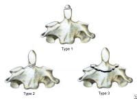
There are three types of C2 odontoid fractures:
- Type I is an oblique fracture through the upper part of the odontoid process. This fracture is normailly stable and can be treated with a hard cervical collar.
- Type II is a fracture occurring at the base of the odontoid as it attaches to the body of C2. These fractures can be treated surgically, or conservatively with hard collar or a halo brace
- Type III fractures occurs when the fracture line extends through the body of the axis. These fractures are normally treated surgically with or without a halo brace.
Show References
Calcaneus Fractures:
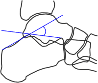
Calcaneus fractures can easily be missed on plain films and the true extent of the injury might not be appreciated until a CT is done. However, you can increase your change of picking up a calcaneal fracture by evaluating Bohler's Angle.
Lateral radiographs of the foot are needed to evaluate the Bohler angle. This is the angle made by drawing a line from anterior process of the calcaneus to the peak of the posterior articular surface and a second one drawn from the peak of the posterior articular surface to the peak of the posterior tuberosity. (See Picture) The average angle is 25-40°. Angles less than 25' are strongly suggestive of a fracture and the patient should probably get a CT of their foot if there is clinical suspicion.
Show References
Wound Care:
Patients and many providers want to irrigate or wash a wound with an antiseptic solution in order to decrease the risk of infection. Most studies have shown that irrigation whether with tap water or sterile water is effective enough in reducing bacterial counts in a wound so does adding an antiseptic solution offer any additional benefit.
It turns out that hydrogen peroxide, and iodine based solutions can actually hinder wound healing as they causes delays in the migration and proliferation of fibroblasts at concentrations that are not even bactericidal. Chlorhexidine, and silver containing antiseptics [i.e.: silver sulfadiazine and silver nitrate] are bactericidal at concentrations that do not affect fibroblasts.
So in the end, if you feel the need to use an antiseptic, use chlorhexidine or a silver containing antiseptic. The use of hydrogen peroxide and iodine based solutions should be abandoned as they are not even bactericidal at concentrations that have profound affects on the fibroblasts.
Show References
Septic Arthritis versus Arthritis:
Though CRP and ESR levels are significantly higher in patients that have septic arthritis, a 1998 study showed that there is extensive overlap between patients with septic arthritis crystal assoicated arthritis that both CRP and ESR have low sensitivity, specificity and predictive values. Peripherial WBC counts did not differ between the two disease processes..
The morale of the story: If you are suspecting septic arthritis you need to perform an arthorcentesis to analysis the synovial fluid. Systemic biomarkers can not support one diagnosis over the other.
Show References
Osteomyelitis:
- An acute or chronic inflammatory, infectious process of bone. Can occur via hematogenous spread or direct innoculation of bone.
- Can be diagnosed on plain radiographs but bony changes might not be evident for 14-21 days. By 28 days 90% of patients will demonstrate a bony abnormality.
- Initially plain radiographs will show periosteal elevation. Later cortical or medullary lucencies are seen.
- Additional tests to help make the diagnosis include:
- Three phase bone scan: often not practical for the ED.
- CT Scan: better in areas with complex anatomy [i.e.:spine, pelvis, ,mid and hind foot]
- MRI: most effective in early detection and to guide surgical approaches. Sensitivity is estimated at 90-100%.
Show References
Radial Head Fractures:
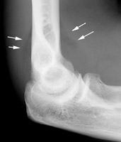
Radial head fractures can often be difficult to visualize on plain films especialing Mason Type 1 fractures (see prior pearl on classification system) which are nondisplaced. Often the only sign of a fracture will be a posterior fat pad sign which is always considered to be pathologic. The posterior fat pad lies outside the synovium of the elbow joint and is normally hidden in the fossa of the distal humerus preventing it from being seen on lateral films of a normal elbow. Trauma to the elbow that results in a intraarticular fracture (typically a radial head fracture) produces an intra-articular hemorrhage that distends the synovium and displaces the fat out of the fossa, producing the typical triangular radiolucent shadow posterior to the distal end of the humerus.
Show References
Conservative Treatment of Back Pain:
Muscle relaxanats and benzodiazipnes are often used in the non-operative management of sciatica and non-specific low back pain. In fact, a 2003 Cochrane review concluded that muslce relaxanats were effective in the management of non-specific low back pain. However, a recent analysis of randomized trials reported little efficacy or only minor benefits with the use of benzodiazapines in treatment of low back pain.
A recent prospective, randomized, placebo-controlled, double-blinded trial conducted in Germany that enrolled a total of 60 patients found that the use of diazepam was equivilant to placebo in the reduction of distance of referred pain at day 7 of treatment. Diazepam was also noted on average to increase the length of stay of those patients hospitalized by 2 days (median hospital days of 8 for placebo versus 10 for diazepam), and the probablility of pain reduction on a visual analog scale by more than 50% was twice as high in the placebo group (p< 0.0015). Placebo reduced the patients pain more than diazepam.
Though the sample size was small; this study should really make one reevaluate the use of diazepam in the treatment of back pain. Early movement and discouraging bed rest have been associated with decreased back pain, so one mechanism by which benzodiazepines may make things work is by causing enough sedation to prevent early movement.
Show References
Carpal Tunnel Syndrome (CTS):
- A compressive neuropathy of the median nerve at the wrist as it travels through the carpal tunnel.
- Median nerve is bound on three sides by carpal bones and anteriorly by the transverse carpal ligament. Surgical repair typically consists of cutting this ligament to allow decompression of the nerve.
- The neuropathy results in:
- parasethesia of the thumb, index and middle fingers
- weaknesss of the thumb and thenar muscles.
- NO physical exam test has great senstivity or specificity for CTS. The two most common are:
- Phalen's test: hyperflexion of the wrist. Need to hold for 60 seconds. Sensitivity ~68% and Specificity ~73%
- Tinel Sign: tapping over cubital tunnel to produce parasthesia along the median nerve. Sensitivity ~50% and Specificity ~77%.
- Increased risk in those patients with:
- Diabetes
- Rheumatoid arthritis
- hypothyroidism
- amyloidosis