University of Maryland School of Medicine logo
Emergency Medicine
All Departments
UMSOM Assistant
UMEM Educational Pearls
  • Home
  • Departments
  • Emergency Medicine
  • CME
  • UMEM Educational Pearls
  • CME
  • Courses and Conferences
  • Free Open Access Medical Education
  • UMEM Educational Pearls

Search

261-280 of 321 results by Haney Mallemat

Previous  |  1 |  ... |  9 |  10 |  11 |  12 |  13 |  14 |  15 |  16 |  17 |  Next

Title: Blunt Vascular Injury

Category: Critical Care

Keywords: blunt trauma, vascular inury, anticoagulation, thrombosis, emboli (PubMed Search)

Posted: 1/3/2012 by Haney Mallemat, MD

Carotid or vertebral artery injury following blunt trauma is a rare (%1 of blunt trauma), but a potentially serious injury potentially causing stroke and long-term disability.

Injury leads to an intimal tear becoming a nidus for platelet aggregation; thrombosis and/or distal emboli may subsequently develop.

Mechanisms of injury include:

  • Blunt trauma to the neck
  • Hyper-extension of neck with contralateral rotation of the head
  • Intra-oral trauma
  • Arterial laceration secondary to adjacent sphenoid or petrous bone fracture.

Symptoms of carotid injury may include contralateral sensorimotor deficits; Symptoms of vertebral injury may include ipsilateral facial pain and numbness, headache, ataxia, or dizziness.

Angiography is the diagnostic “gold standard” but these days a 16-slice CT angiography (or greater) is a reliable screening tool.

Anticoagulation with heparin is the treatment of choice for severe injury, if there are no contraindications (e.g., intracranial bleeding). Anti-platelet drugs may be acceptable in certain cases.

Show References

Kim YK, Schulman S. Cervical artery dissection: pathology, epidemiology and management. Thromb Res. Apr 2009;123(6):810-21.

Schievink WI. Spontaneous dissection of the carotid and vertebral arteries. N Engl J Med. Mar 22 2001;344(12):898-906. 

Follow me on Twitter (@criticalcarenow) or Google+ (+haney mallemat)


Title: What's the diagnosis? Case submitted by Dr. Bethany Radin

Category: Visual Diagnosis

Posted: 12/26/2011 by Haney Mallemat, MD

Question

64 year old male with emphysema and stage 4 lung cancer presents in respiratory distress. What's the diagnosis?

Show Answer

Answer: Pneumothorax (left chest) and bullous disease (right chest).

In questionable cases like this, bedside ultrasound can help distinguish between bullae and pneumothorax. Bullae should have a positive lung sliding sign whereas pneumothorax does not.

See the referenced case report for more information:

Simon, B. et al. Two cases where bedside ultrasound was able to distinguish pulmonary bleb from pneumothorax. J Emerg Med. 2005 Aug;29(2):201-5.

Show References

Follow me on Twitter (@criticalcarenow) or Google+ (+haney mallemat)


Title: Amiodarone-Induced Lung Toxicity

Category: Critical Care

Keywords: amiodarone, lung toxicity, ARDS, infection, critical care (PubMed Search)

Posted: 12/20/2011 by Haney Mallemat, MD

Amiodarone-induced lung toxicity (ALT) is a serious and sometimes fatal complication of amiodarone use.

Symptoms range from mild (e.g., dyspnea with exertion) to acute respiratory distress syndrome and risk of death.

ALT is secondary to either release of toxic oxygen radials that are directly toxic to the lung or the reaction is secondary to an indirect immunologic reaction.

Risk factors for ALT: use > 2 months, dose > 400mg/day, advanced age, or pre-existing lung injury

ALT is typically a diagnosis of exclusion so suspect ALT through a detailed history; physical exam and radiology are non-specific. Lung biopsy is the only confirmatory test.

Treat ALT by discontinuing the drug, steroids, and supportive care. In rare cases where amiodarone cannot be safely discontinued (i.e., life-threatening arrhythmia), dosage should be reduced and steroids added immediately.

Generally, ALT is reversible with a good prognosis.

Show References

Mahajan, V. et al Amiodarone induced acute interstitial pneumonitis.BMJ Case Rep. 2009;2009.

Follow me on Twitter (@criticalcarenow) or Google+ (+haney mallemat)


Title: What's the Diagnosis?

Category: Visual Diagnosis

Posted: 12/12/2011 by Haney Mallemat, MD (Updated: 8/28/2014)

Question

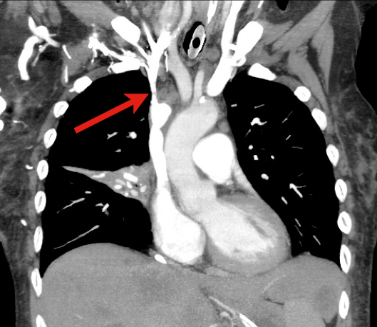
60 year old male with 6 months of weight loss and recent epistaxis. Diagnosis?

Show Answer

Answer: Partial obstruction of the Superior vena cava (SVC)


SVC Syndrome

SVC syndrome is caused by compression or obstruction of the superior vena cava blocking anterograde flow.

Most cases are secondary to extrinsic compression by malignancy. Other causes are secondary to thrombosis and internal obstruction (e.g., central venous catheter placement).

Symptoms present sub-acutely, worsen with bending over, and are secondary to increased venous pressure in the head and neck (e.g., epistaxis, headache, tinnitus, conjunctival injection, neck swelling, etc.).

Treatment focuses on reversing the underlying cause (e.g., radiation or chemotherapy if due a sensitive tumor) and treatment of symptoms:

  • Steroids, diuretics, and hyperosmolar agents to reduce edema
  • Elevate the head of bed to reduce brain edema
  • Anti-emetics to prevent vomiting-associated increases in intracranial pressure

Rice TW, Rodriguez RM, Light RW. The superior vena cava syndrome: clinical characteristics and evolving etiology. Medicine (Baltimore). Jan 2006;85(1):37-42.

Nunnelee JD. Superior vena cava syndrome. J Vasc Nurs. Mar 2007;25(1):2-5; quiz 6.

Follow me on Twitter (@criticalcarenow) or Google+ (+haney mallemat)


Title: An alternative to CPAP?

Category: Critical Care

Posted: 12/6/2011 by Haney Mallemat, MD

Up until recently, a tight-fitting mask was one of the only ways to deliver non-invasive positive-pressure ventilation.

High-flow nasal cannulas (HFNC) have been adapted from use in neonates to adults to deliver continuous positive airway pressure (CPAP).

HFNC provides continuous, high-flow (up to 60 liters), and humidified-oxygen via nasal cannula providing positive pressure to the pharynx and hypopharynx. Patients tolerate it well and it is less claustrophobic than tight-fitting masks.

HFNC does not generate the same amount of pressure as CPAP so it may be best utilized as an intermediate step between low-flow oxygen (i.e., traditional nasal cannula) and non-invasive positive pressure ventilation with tight-fitting masks.

Check with your respiratory department if these devices are locally available.

Show References

Kernick, j. What is the evidence for the use of high flow nasal cannula oxygen in adult patients admitted to critical care units? A systematic review. Aust Crit Care. 2010 May;23(2):53-70. Epub 2010 Mar 5.

Parke, R. A preliminary randomized controlled trial to assess effectiveness of nasal high-flow oxygen in intensive care patients. Respir Care. 2011 Mar;56(3):265-70. Epub 2011 Jan 21.

Follow me on Twitter (@criticalcarenow) or Google+ (+haney mallemat)


Title: What's the Diagnosis? Case submitted by Dr. Zachary Dezman

Category: Visual Diagnosis

Posted: 11/28/2011 by Haney Mallemat, MD (Updated: 11/28/2011)

Question

9 year-old boy with sudden onset of unilateral facial swelling. What’s the diagnosis?

Show Answer

Answer: Acute Parotitis

  • Viral parotitis
    • Usually bilateral swelling without warmth or erythema; typically a benign course
    • 85% of cases occur in children <15 years old
    • Common causes: Influenza, Para-influenza, and Coxsackie; Rarely Paromyxovirus (Mumps) due to vaccination programs
    • Treat conservatively; sialagogues to stimulate gland drainage (lemon drops or orange juice), warm compresses, or local massage.
  • Bacterial parotitis
    • Commonly presents with unilateral pain, induration, erythema, and tenderness; 20-50% mortality!
    • Risk factors: chronic illness, age > 60, recent surgery, and dehydration
    • Treatment: Antibiotics (covering oral flora) and certain cases may require surgical intervention
    • Lack of improvement within 48 hours requires CT scan to exclude abscess and IV antibiotics

Bonus Trivia: U.S. President Garfield died from parotitis after becoming dehydrated following abdominal surgery

Shelly J. McQuone MD, Acute Viral and Bacterial Infections of the Salivary Glands, Otolaryngologic Clinics of North America, Volume 32, Issue 5 (October 1999)

Show References

Follow me on Twitter (@criticalcarenow) or Google+ (+haney mallemat)


Title: Ultrasound for a HI MAP

Category: Critical Care

Keywords: hypotension, shock, ultrasound, hi map (PubMed Search)

Posted: 11/22/2011 by Haney Mallemat, MD

Determining the exact etiology of hypotension / shock can sometimes be difficult in the Emergency Department.

The Rapid Ultrasound for Shock / Hypotension (RUSH) exam is a sequential, 5 step-protocol (typically requiring less than 2 minutes) that can be used to determine the cause(s) of hypotension.

The mnemonic for the exam is “HI MAP”, and is easy to remember because a "HI MAP" is our goal with hypotensive patients.

H - Heart (parasternal and four-chamber views)
I  - Inferior Vena Cava (for volume responsiveness)
M - Morrison’s pouch (i.e., FAST exam) and views of thorax (looking for free fluid)
A - Aortic Aneurysm (ruptured abdominal aneurysm)
P - Pneumothorax (i.e., Tension PTX)

Refer to the link for a more detailed discussion and podcast from the creators of this exam: emcrit.org/rush-exam


Title: The risks of intubation with pericardial tamponade

Category: Critical Care

Keywords: tamponade, critical care, intubation, positive pressure, PEA arrest (PubMed Search)

Posted: 11/8/2011 by Haney Mallemat, MD

Positive-pressure ventilation (e.g., mechanical ventilation) increases intrathoracic pressure potentially reducing venous return, right-ventricular filling, and cardiac output.

Pericardial tamponade similarly causes hemodynamic compromise through increased pericardial pressure which reduces right-ventricular filling and cardiac output.

When mechanically ventilating a patient with known or suspected pericardial tamponade the mechanisms above may be additive, causing cardiovascular collapse and possibly PEA arrest.

For the patient with known or suspected pericardial tamponade consider draining the pericardial effusion prior to intubation or delaying intubation until absolutely necessary.

If intubation is unavoidable, consider maintaining the intrathoracic pressure as low as possible (by keeping the PEEP and tidal volumes to a minimum) to ensure adequate cardiac filling and cardiac output.

Show References

Ho, A. et. al. Timing of tracheal intubation in traumatic cardiac tamponade: A word of caution. Resuscitation, 80(2), 272–274.

Follow me on Twitter (@criticalcarenow) or Google+ (+haney mallemat)


Title: What's the diagnosis? Pearl submitted and written by Dr. Zachary Dezman

Category: Visual Diagnosis

Posted: 10/31/2011 by Haney Mallemat, MD (Updated: 10/31/2011)

Question

72 year-old man, one-week post right fem-pop bypass presents with painful blue and black toe. Diagnosis?


Show Answer

Answer: Blue-Toe Syndrome

  • Blue-toe syndrome is acute digital-ischemia, presenting as one or more cold, painful, and cyanotic toes (or other digits).
  • Multiple causes:
    • Emboli (most common)
    • Cholesterol emboli from recent endovascular procedure or atherosclerotic plaque rupture
    • Vascular aneurysm
    • Infectious (e.g., endocarditis)
    • Medications (e.g., warfarin)
    • Drugs (e.g., cocaine, methamphetamine, etc.)
  • Emergency Department evaluation should focus on determining the underlying cause to prevent limb loss.
  • Patients should have emergent vascular consult and admission for potential revascularization.

Show References

Hirschman, J. et al. Blue (or purple) toe syndrome. J Am Acad Dermatol.2009 Jan;60(1):1-20

O'Keeffe S, et al. Blue toe syndrome: Causes and management.Arch Intern Med. 1992 Nov;152(11):2197-202.

Follow me on Twitter (@criticalcarenow) or Google+ (+haney mallemat)


Title: Xigris no more.

Category: Critical Care

Keywords: xigris, activated protein C, sepsis, multi-organ failure, resuscitation (PubMed Search)

Posted: 10/25/2011 by Haney Mallemat, MD

  • On October 25, 2011, Eli Lilly announced a voluntary-recall of activated drotrecogin alfa (Xigris) following a recent trial (PROWESS-SHOCK), which demonstrated no survival benefit when using the drug when compared to placebo.

  • Activated drotrecogin alfa is a recombinant form of human activated protein C previously recommended for adults with severe sepsis and a high-risk of death (APACHE II > 25 or multi-organ failure); it is included in the 2008 International Sepsis Guidelines (Grade 2b recommendation).

  • The PROWESS-SHOCK trial reported an all-cause mortality rate of 26.4% in the drotrecogin alfa group compared with 24.2% in the placebo group; this difference was not statistically significant.

  • Interestingly, the study also found that severe bleeding (the drug's main side-effect) was found to be 1.2% in the activated drotrecogin alfa group compared to 1.0% for the placebo group (also non-significant) suggesting it does not increase the risk of bleeding as it had previously been reported.

  • Hospitals should revise their sepsis guidelines based on this recent news.

Show References

www.medscape.com/viewarticle/752169?sssdmh=dm1.728719&src=nl_newsalert

Dellinger, R. P., et al. Surviving Sepsis Campaign: International guidelines for management of severe sepsis and septic shock: 2008. Critical Care Medicine, 36(1), 296–327. doi:10.1097/01.CCM.0000298158.12101.41

Follow me on Twitter (@criticalcarenow) or Google+ (+haney mallemat)


Title: What s the Diagnosis? (Case submitted and written by Dr. Rich Lichenstein)

Category: Visual Diagnosis

Posted: 10/17/2011 by Haney Mallemat, MD

Question

5 year-old male with developmental delay presents with intractable non-bloody and non-bilious vomiting over 10 days; bowel movements are normal. Four weeks ago he was placed in a hip-spica cast following a motor vehicle crash. Abdominal x-ray is below. Diagnosis?

Show Answer

Answer: CAST syndrome (also known as Superior Mesenteric Artery Syndrome)

  • Caused by extrinsic compression of the SMA with ensuing partial gastric outlet obstruction
  • Commonly seen in the second decade of life when there is increased spinal flexibility and truncal casting increases lumbar lordosis. This changes the take-off of the SMA which crosses the horizontal part of the duodenum.
  • Those at risk include those with patients with spinal disease, hip and pelvic fractures.  Children with disabilities who have poor fat mass, hypotonicity, and skeletal distortion may also be affected.
  • Clinical symptoms include nausea, vomiting, abdominal distention which may lead to dehydration, metabolic alkalosis and shock and sometimes death.
  • Treatment includes IV replacement therapy, NG tube to decompress the stomach and duodenum and replacement of the cast.

Show References

Wheeless Textbook of Orthopedics. Updated August 29,2011

Lichenstein, R. Radiology Cases in Pediatric Emergency Medicine, Volume 5, Number 16

Follow me on Twitter (@criticalcarenow) or Google+ (+haney mallemat)


Title: Listeria infections of the central nervous system

Category: Critical Care

Keywords: listeria, food borne illness, cns infection (PubMed Search)

Posted: 10/11/2011 by Haney Mallemat, MD

Lisiteria Monocytogenes is typically transmitted from ingestion of contaminated food such as unpasteurized milk or cheese, raw foods, and recently cantaloupes; transmission from veterinary exposure, infected soil and water have also been reported.

Listeria has a predilection for the central nervous system (CNS) causing several infections including meningioencephalitits, brain or spinal abscess, cerebritis (infection of brain parenchyma), and rhomboencephalitis (encephalitis of the brainstem).

Risk factors include immunosuppression, advanced age, newborns, and pregnancy.

There is no clinical way to distinguish CNS infection with Listeria from other pathogens, therefore blood and cerebrospinal fluid (CSF) culture is required.

CSF analysis demonstrates pleocytosis, elevated protein, and low glucose. CSF gram stain has a low sensitivity (~33%), but consider Listeria in the differential if "diptheroid-like" bacteria are reported on gram stain.

Ampicillin is the drug of choice and should be continued for at least three weeks (sometimes longer). Adding gentamycin is sometimes recommended for synergy in severe infection.

Show References

Mylonakis E, Hohmann EL, Calderwood SB. Central nervous system infection with Listeria monocytogenes. 33 years' experience at a general hospital and review of 776 episodes from the literature. Medicine (Baltimore). Sep 1998;77(5):313-36.

http://emedicine.medscape.com/article/220684-overview

http://www.cdc.gov/listeria

Follow me on Twitter (@criticalcarenow) or Google+ (+haney mallemat)


Title: What's the Daignosis? (submitted and written by Dr. Katherine Baugher)

Category: Visual Diagnosis

Posted: 10/3/2011 by Haney Mallemat, MD

Question

Question: 50-year-old diabetic female s/p foot burn several weeks ago, now presenting with pain and discharge from a poorly healing wound. Diagnosis?

Show Answer

Answer:  Osteomyelitis

Osteomyelitis 

· Acute or chronic bone infection

· Risk factors: Immunosuppression (diabetes, chronic steroid use, AIDS, and sickle-cell dz.)

· Secondary to direct trauma, contiguous spread from local infection, or hematogenous spread (in children)

· Common bacteria: S. Aureus, Pseudomonas, Salmonellae (classically in Sickle cell dz.)

· X-ray (limited sensitivity):

- 3-5 days post-infection: Soft-tissue swelling

- 14-21 days: Some patients demonstrate bony changes (e.g., periosteal elevation, bone lucencies, etc.)

- >28 days: >90% with Xray findings

· MRI is the imaging gold standard

· Two of the following needed for diagnosis:

- Purulent aspiration

- Positive blood or tissue culture

- Positive imaging

- Tenderness + erythema / edema

· Antibiotic coverage based on culture results. When immediate empiric therapy required (sepsis), cover most likely pathogen plus MRSA.


References

Carek PJ, Dickerson LM, Sack JL. Diagnosis and management of osteomyelitis. Am Fam Physician. 2001 Jun 15;63(12):2413-20.

Pruthi S, Thapa MM. Infectious and inflammatory disorders. Radiol Clin North Am. Nov 2009;47(6):911-26.

Zink BJ, Raukar NP. Bone and Joint Infections. In: Marx JA, Hockberger RS, Walls RM, Adams JG, Barsan WG, Biros MH, Danzl DF, Gausche-Hill M, Ling LJ, Newton EJ, eds. 7th ed. Emergency Medicine: Concepts and Clinical Practice.Volume 2. Philadelphia, PA: Mosby; 2010:1821-1830.

Follow me on Twitter (@criticalcarenow) or Google+ (+haney mallemat)


Title: Simply saline for cardiac arrest?

Category: Critical Care

Keywords: Epinephrine, adrenaline, cardiac arrest, return of spontaneous circulation, ROSC, critical care, ICU, saline (PubMed Search)

Posted: 9/27/2011 by Haney Mallemat, MD

·  The use of epinephrine in cardiac arrest is currently standard of care.

·  Several observational and non-randomized trials have demonstrated the efficacy of epinephrine in cardiac arrest, but there has never been a randomized double-blind placebo-controlled trial in humans.

·  A recently published Australian trial randomized cardiac patients (of any type) to receive either 1 mg of epinephrine (n=272) or 0.9% normal saline (n=262); the primary end-point was survival to hospital discharge. Secondary end-points were pre-hospital return of spontaneous circulation (ROSC) and neurological outcomes at hospital discharge.

·  Significantly more patients had pre-hospital ROSC in the epinephrine group (regardless of the underlying rhythm), however, there was no statistically significant difference in survival to discharge (the primary outcome) between groups.

·  This randomized double-blinded placebo-controlled trial raises many new and interesting questions about epinephrine, but more study is needed before changing current practice.

Show References

Jacobs IG, et al. Effect of adrenaline on survival in out-of-hospital cardiac arrest: A randomized double-blind placebo-controlled trial. Resuscitation 2011;82:1138-1143.

Follow me on Twitter (@criticalcarenow) or Google+ (+haney mallemat)


Title: What s the diagnosis? Case submitted by Dr. Ari Kestler

Category: Visual Diagnosis

Posted: 9/19/2011 by Haney Mallemat, MD (Updated: 8/28/2014)

Question

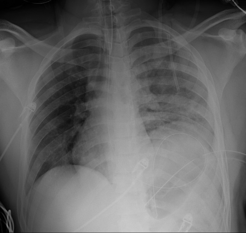
19 year-old male s/p high-speed MVC with hypotension and diminished breath sounds on left. Diagnosis?
Show Answer
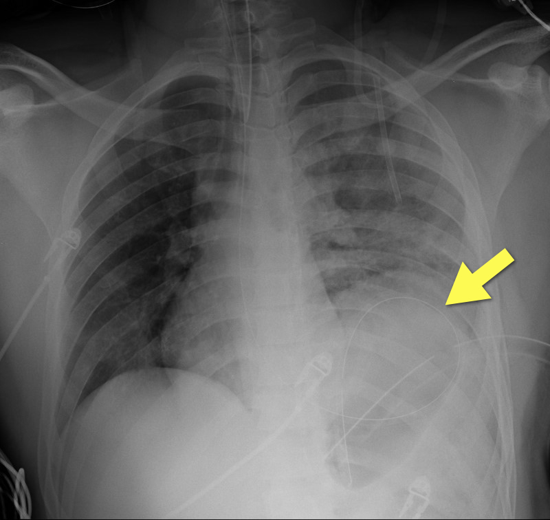
Answer: Diaphragmatic rupture (Note position of NG tube on image below).

·Uncommon; less than 1% of all traumatic injuries

·Diagnosis may be obvious on CXR (as in example) or subtle (e.g., diaphragmatic elevation, basilar atelectasis, etc.)

·Requires a high-index of suspicion because delayed diagnosis increases the risk of abdominal organ herniation or strangulation.

·Mortality depends on the mechanism and presence of associated injuries; mortality is highest for blunt injuries.

Show References

Bhavnagri S, et al. When and how to image a suspected broken rib. Cleveland Clinic J Med. 2009;76(5):309.

Williams M, et al. Predictors of mortality in patients with traumatic diaphragmatic rupture and associated thoracic and/or abdominal injuries. Am Surg. 2004;70(2):157.

National Trauma Data Base. American College of Surgeons 2000-2004.

Follow me on Twitter (@criticalcarenow) or Google+ (+haney mallemat)


Title: Axillary Arterial-Lines

Category: Critical Care

Keywords: Procedures, Arterial lines, Axillary, hemodynamic monitoring (PubMed Search)

Posted: 9/13/2011 by Haney Mallemat, MD

Radial and femoral arteries are common sites for arterial-line placement, but are not without complications (e.g., Radial artery: malfunction with positioning and Femoral artery: contamination and infection); an alternative site to consider is the axillary artery.

The axillary artery's superficial location and large size make it a desirable choice for cannulation.

The "anatomical-landmark" and "palpation" methods have been the traditional techniques of axillary arterial cannulation, however these methods may be difficult for to a variety of reasons (e.g., obesity, anasarca, arterial disease, etc.)

Ultrasound allows visualization of the axillary artery and avoids unintended injury to structures in close proximity (e.g., brachial plexus, pleura, axillary vein, etc.); please see figures 1 and 2 in the referenced Sandhu article and http://www.youtube.com/watch?v=Z31YiyV7cNQ.

A recent study (Killu, 2011) found that ultrasound increases success rates when compared to the traditional landmark approach.

Show References

Killu, K. et al. Utility of Ultrasound Versus Landmark-Guided Axillary Artery Cannulation for Hemodynamic Monitoring in the Intensive Care Unit. ICU Director; 2011. 2(3), 54–59.

Sandhu, N. The Use of Ultrasound for Axillary Artery Catheterization Through Pectoral Muscles: A New Anterior Approach. Anesthesia and analgesia. 2004; 562–565.

Follow me on Twitter (@criticalcarenow) or Google+ (+haney mallemat)


Title: What's the diagnosis? Images by Dr. Mak Moayedi

Category: Visual Diagnosis

Posted: 9/5/2011 by Haney Mallemat, MD (Updated: 8/28/2014)

Question

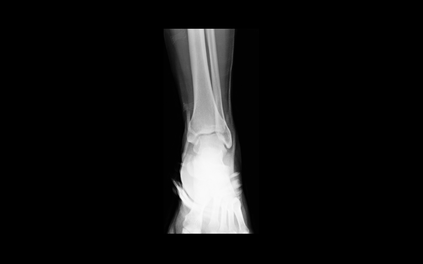
26 year old male presents s/p basketball dunk. Diagnosis?

Show Answer

Answer: Talar dislocation

Below, a post-reduction Xray, as well as a link to a video demonstrating a few ankle reductions (the video's background music makes all the difference): http://www.youtube.com/watch?v=AzqCsLX1K1E

Show References

Follow me on Twitter (@criticalcarenow) or Google+ (+haney mallemat)


Title: Tracheal Rapid Ultrasound Exam (T.R.U.E.)

Category: Critical Care

Keywords: ultrasound, tracheal intubation, esophageal intubation, critical care, airway (PubMed Search)

Posted: 8/30/2011 by Haney Mallemat, MD

  • Multiple methods of confirming endotracheal tube placement exist, however quantitative waveform capnography is the most reliable method. Unfortunately this may not be immediately available at all medical centers.

  • Recent studies demonstrate that bedside ultrasound may assist in the detection of proper endotracheal tube placement.

  • The T.R.U.E. (Tracheal Rapid Ultrasound Exam) was demonstrated to be 99% sensitive, 94% specific, 99% PPV, and 94% NPV during intubation.

  • The basic exam involves placing a high-frequency linear-array probe on the anterior neck above the sternal notch and identifying the trachea and esophagus during intubation.

  • The following video is an example of what you DO NOT want to see during an intubation: http://www.youtube.com/watch?v=LvfThxhQ93A

Show References

Chou, H. et al. Tracheal rapid ultrasound exam (T.R.U.E.) for confirming endotracheal tube placement during emergency intubation. Resuscitation. Jun 2011

Werner SL,et al. Pilot study to evaluate the accuracy of ultrasonography in confirming endotracheal tube placement. Ann Emerg Med 2007;49:75–80.

Follow me on Twitter (@criticalcarenow) or Google+ (+haney mallemat)


Title: What's the diagnosis? Dr. Michael Santiago

Category: Visual Diagnosis

Posted: 8/22/2011 by Haney Mallemat, MD

Question

79 y.o. male lung cancer patient with tachypnea, tachycardia, and normal blood pressure. Click here: http://vimeo.com/27973006

Possible diagnosis?

Show Answer

Answer: Right ventricular (RV) dysfunction secondary to submassive pulmonary embolism (PE).


Ultrasound for suspected PE

Consider bedside echo with PE and elevated troponin or BNP.

Recall the classes of PE:

  1. Non-massive PE: Normal RV function with normotension
  2. Submassive PE: RV dysfunction (see below) with normotension
  3. Massive PE: RV dysfunction with hypotension
  4. Cardiac arrest (Pulseless electrical activity)

Ultrasound “clues” of submassive / massive PE:

  1. Right ventricular enlargement: RV diastolic volume is >60% of the LV; normally  RV is 60% the size of the LV.
  2. Interventricular septal shift ("D-sign"): The normally concentric LV cavity will now look like the letter “D” from diastolic septal flattening; "D" sign indicates elevated pulmonary pressure (e.g., pulmonary embolism)
  3. McConnell’s sign: Hypokinesis in RV free wall with normal apical motion; originally though to be specific for PE, but sensitivity and specificity have been questioned because other disorders also have a "McConnel'-type" appearance (severe pulmonary hypertension, RV infarction); can still help add to pre-test probability.
  4. Free-floating thrombus: clot visualized in pulmonary artery and/or in right-side of heart (rare)

Show References

Lodato JA, Parker Ward RP, Lang RM. Echocardiographic Predictors of Pulmonary Embolism in Patients Referred for Helical CT. Echocardiography 2008;25:584-590.

McConnell MV, Solomon SD, Rayan ME, et. al. Regional Right Ventricular Dysfunction Detected by Echocardiography in Acute Pulmonary Embolism. Am J Cardiol. 1996; 78: 469-473.

ACEP. Critical Issues in the Evaluation and Management of Adult Patients Presenting to the Emergency Department With Suspected Pulmonary Embolism. http://www.acep.org/content.aspx?id=30060. [July 24, 2011].

John Griffiths. Respiratory: Management of small, submassive and massive pulmonary embolism. http://www.frca.co.uk/article.aspx?articleid=100750. [July 24, 2011].


Follow me on Twitter (@criticalcarenow) or Google+ (+haney mallemat)


Title: Bougie-Assisted Cricotyrotomy

Category: Critical Care

Keywords: bougie, cricothyrotomy, trauma, critical care, intubation, failed airway (PubMed Search)

Posted: 8/16/2011 by Haney Mallemat, MD

The open cricothyrotomy technique is taught as the trauma airway standard when one “cannot intubate and cannot ventilate” however, it is not without difficulty and limitations. The B.A.C.T. (Bougie-Assisted Cricothyrotomy Technique) may improve the procedure by using a bougie to assist.

Steps for the B.A.C.T. (as described in the paper):
1. Stabilize the larynx with the thumb and middle finger, then identify the cricothyroid membrane.
2. Make a transverse stabbing incision with a scalpel through both skin and cricothyroid membrane.
3. Insert tracheal hook at the inferior margin of the incision and pull up on the trachea.
4. Insert a bougie through the incision with curved tip directed towards the feet
5. Pass 6-0 endotracheal tube or Shiley over bougie into trachea.

Advantages of a bougie:
1. Thin and easy to insert into incision
2. Tactile feedback from tracheal rings confirms proper placement
3. Ensures that stoma will not be lost during procedure

EMRAP.tv has a great video of Dr. Darren Braude demonstrating the procedure;
http://bit.ly/nB3BMG

Show References

Hill, C., et al. Cricothyrotomy Technique Using Gum Elastic Bougie Is Faster Than Standard Technique: A Study of Emergency Medicine Residents and Medical Students in an Animal Lab. Academic Emergency Medicine17(6), 666–669.

Follow me on Twitter (@criticalcarenow) or Google+ (+haney mallemat)


Previous  |  1 |  ... |  9 |  10 |  11 |  12 |  13 |  14 |  15 |  16 |  17 |  Next
University of Maryland School of Medicine logo
  • Departments
  • Programs
  • Research Centers
  • Institutes
  • Graduate Education
  • CIBR Cores
  • Offices of the Dean
    • University of Maryland, Baltimore
    • University of Maryland Medical Center
    • Medical Alumni Association of the University of Maryland
    • Health Sciences and Human Services Library
University of Maryland School of Medicine
655 W. Baltimore Street
Baltimore, MD 21201
Contact Us

© University of Maryland School of Medicine

    • Webmaster
    • Web Accessibility
    • Site Index
    • UMB Hotline
    • Employment
    • Map