
Search
Subscribe to daily UMEM Pearls (FREE!) via:
21-40 of 268 results by Michael Bond
Take Home Points:
- A radial head subluxation that is common in 1-3 year olds
- Often secondary to a longitudinal traction on an extended arm
- With a classic story radiographs are not required
- The hyperpronation technique has been show to be more effective and less painful in reducing it
Show Additional Information
It is common teaching that a Segond Fracture is associated with ACL tears. A reverse Segond fracture, avulsion fracture of the knee due to avulsion of the deep fibers of the medial collateral ligament, has also been described that was initially reported as associated with PCL tears. However, a more recent study has not been able to collaborate the PCL connection, but has shown that a reverse Segond fracture is associated with multiple ligamentous injuries to the knee.
Take home point: If you note a Reverse Segond fracture on your plain flips have the patient followup with orthopedics for a possible MRI, as they probably have other ligamentous injuries that might need treatment.
Show Additional Information
Show References
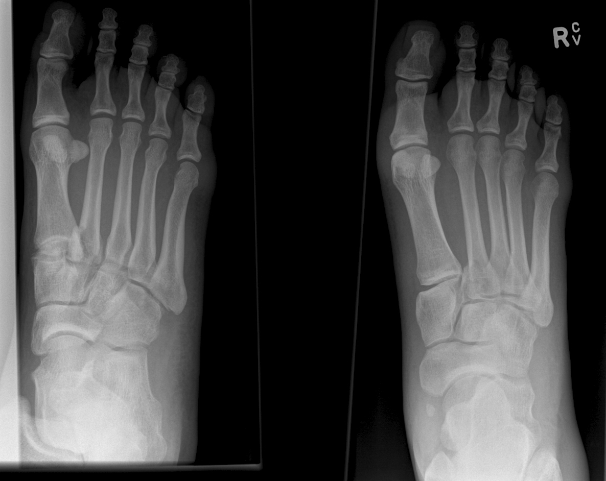
Lisfranc Fracture: Typically consists of a fracture of the base of the second metatarsal and dislocation, though it can also be associated with fractures of a cuboid.
- Fracture findings on plain films may be subtle.
- If in doubt obtain weight bearing AP views of the foot to demonstrate dislocation/fracture.
- If weight bearing films are negative and you are still suspicious consider a CT scan of the foot.
Click below see image of fracture
Show Additional Information
We all wish there was a great treatment regimen for our patients with back pain. However, most studies have shown that it really does not matter what you do, as most patients will get better in 6 weeks.
A recent study published in JAMA looked at the role of spinal manipulation to improve pain and function in adults with low back pain. They looked at 26 randomized controlled trails and found that there was modest benefit for spinal manipulation and it was similar to using NSAIDs.
So spinal manipulation may or may not work for some patients. Something to consider along with physical therapy if patients are not getting relief with home remedies.
Show References
Treatment of Low Back Pain
A recent recommendation from the American College of Physicians (Internal Medicine) now recommends nonpharmacologic therapies as the first line treatment of acute or subacute lower back pain lasting 12 weeks or less. This might bring more people to our Emergency Departments so it is important that we know their current recommendations.
Some nonpharmacologic therapies recommended are:
- Moderate Evidence: Superficial heat
- Low quality evidence: Massage, Spinal manipulation, or accupuncture
For acute back pain they recommend:
- NSAIDs or muscle relaxants
- Acetominophen is NOT recommended. No evidence it is beneficial
For chronic back pain:
- Start with NSAIDs—>tramadol—>duloxetine.
- Opioids are only recommended for treatment failures.
Show References
Take Home Point:
- According to a recent article in the NEJM there does not seem to be any difference in the rate of symptomatic venous thromboembolism (VTE) in patients given low molecular weight heparin that underwent arthorscopy or had lower leg casting at 3 months.
- Overall, the rates of VTE were really low ( casting: 1.4% vs. 1.8%; arthroscopy: 0.7% vs. 0.4%), so there is probably not need for prophalaxis in these patients.
Show Additional Information
Show References
Take home points:
- Cellulitis is overdiagnosed
- 1/3 of patients diagnosed with cellulitis in the ED are ultimately given a different diagnosis
- The most common final diagnoses are vascular or inflammatory conditions.
- The over treatment of cellulitis increases healthcare costs, increases risk of adverse reactions, and can contribute to the development of drug resistant organisms.
Show Additional Information
Davos Shoulder Reduction Technique
Take Home Points
- Uses the patients own weight to reduce their anterior shoulder dislocation.
- No sedation is required
- Provider exerts no effort and only sits on the patients foot.
Interested, well find out more by watching this video by Larry Mellick https://www.youtube.com/watch?v=u2MsnjVNoPM or clicking the link below.
Show Additional Information
Patellofemoral Syndrome Treatment options
Patients do best with a combined intervention (ie, exercise therapy, education, manual therapy and taping) plan or patellofemoral bracing may improve outcomes for people with patellofemoral syndrome and the subtype of patellofemoral osteoarthritis.
For for the ED, we can start NSAIDs, and then have them follow up with Physical Therapy, A sports trainer if in organized sports, or with a sports medicine physician/PCP. Physical therapy is targeted at strengthening the quadricep muscle particularly vastus medialis, which improves the patella’s tracking with knee flexion.
According to the 4th International Patellofemoral Pain Research Retreat recently published in British Journal of Sports Medicine, the core criterion required to define Patelofemoral Pain (PFP) syndrome is pain around or behind the patella, which is aggravated by at least one activity that loads the patellofemoral joint during weight bearing on a flexed knee (eg, squatting, stair ambulation, jogging/running, hopping/jumping).
Additional criteria (not essential):
- Crepitus or grinding sensation emanating from the patellofemoral joint during knee flexion movement
- Tenderness on patellar facet palpation
- Small effusion
- Pain on sitting, rising on sitting, or straightening the knee following sitting
PFP is common in young adolescents, with a prevalence of 7–28%, and incidence of 9.2%.
Stay tuned for recommendations on treatment and diagnosis.
Show References
Non-Musculoskeletal Causes of Neck Pain
Neck pain is a common complaint of people presenting to the ED. Most of the cases will be musculoskeleteal in origin and will respond to conservative therapy with NSAIDs or acetominophen. However, other non-musculoskeletal causes of pain could be lurky behind this benign complaint.
Don't forget to consider:
- Early mengingitis (84% of patients with meningitis will complain of neck stiffness)
- Myocardial infarction/angina. Women are known to have atypical symptoms and might just have dull pain in their neck. Be sure to ask about whether exertion increases the pain.
- Epidural Abscess- fever and neuro symptoms are often missing early on. Make sure to ask about risk factors for spinal epidural abscess.
- Vertebral Artery Discection - most common identifiable cause of stroke in your people. <50% are associated with trauma and <8% of patients have connective tissue disorder. Patients are at increased risk if they have had
- Cervical trauma (remember seen in < 50% of cases)
- Recent infection
- Hypertension
- h/o migraines
The PATCH trail, recently published in the Lancet, looked at whether giving platelets to patients, that were on anti-platelet therapy (e.g.: aspirin, clopedrigrel, or dipyridamole) for at least 7 days at the time of their spontaneous intracerebral hemorrhage, improved neurologic outcomes and mortality.
This was a large (60 hospitals) multicener, open-label, masked endpoint, randomized trial that enrolled a total of 190 patients (97 platelet transfusion and 93 standard care).
The outcomes were surprising. Patient in the Platelet group had a higher rat of death or dependence at 3 months (Adjusted OR 2.05; 95% CI 1.18 3.56; p = 0.0114).
The authors concluded "Platelet transfusion seems inferior to standard care for people taking anti-platelet therapy before a spontaneous intracerebral hemorrhage"
Though this is the first study to look at this, the studies design and outcomes should really make use reconsider whether we give these patients platelets. The thought is that ICB or hemorrhagic strokes also have a component of ischemic stroke and a watershed area that's blood flow becomes compromised with the platelet transfusion.
TAKE HOME POINT: We should not routinely transfuse platelets in our patients that were on antiplatelet therapy prior to their ICB.
Show References
A meta-analysis of 74 randomized trials with a total of 58,556 patients was recently published in the Lancet that looked at the effectiveness of NSAIDs in the treatment of osteoarthritis (OA) pain.
Briefly, their conclusion was that:
- Acetaminophen is ineffective as a single-agent in the treatment of OA.
- Diclofenac 150 mg/day had best evidence to support it as the most effective NSAID available presently with respective to its effectiveness in relieving pain and improving function.
- They found no evidence that treatment effects varied over the duration of treatment ( no tolerance)
You can find the article here http://www.thelancet.com/journals/lancet/article/PIIS0140-6736%2816%2930002-2/abstract
Show References
Borrella mayonii a new species
There is a new bacteria that is causing Lyme disease. Borrella burgdorferi is the typical bacteria associated with lyme disease, but now several cases of Borrelia mayonii have been isolated from patients and ticks that live in Minnesota, Wisconsin and North Dakota. What is unique about this new species is that it is associated with nausea, vomiting, diffuse macular rashes, and neuro symptoms [e.g.: confusion, visual disturbance, and somnolence) along with the typical lyme disease symptoms of arthralgias and headaches.
Current lyme tests should detect this new species and treatment is the same as Borrella burgdorferi. The take home pearl is that we may see patients with "atypical" lyme disease symptoms so this should be on our differential for patients presenting with rashes, nausea, vomiting and neurologic complaints.
Diverticulitis
It seems like the standard treatment course for patients with suspected diverticulitis in the ED is to obtain a CT of the Abdomen and pelvis and then to start antibiotics. A CT scan is really only needed if you suspect that they have an abscess, micro perforation, are not responding to conventional treatment, or you suspect an alternative diagnosis.
However, what should the conventional treatment be? Several recent studies from Sweden, Iceland and the Netherlands have shown that patients treated with antibiotics did not fair any better then patients who were just observed. There was no difference in time to resolution of symptoms, complications, recurrence rate, or duration of hospitalization.
Several national societies (Dutch, Danish, German, and Italian) now recommend withholding antibiotics in patients free of risk factors who have uncomplicated disease, but these patients will need close follow up.
TAKE HOME POINT: Patients with diverticulitis can be treated supportively and probably do not require antibiotics unless you suspect they have complicated disease or are immunosuppressed.
Show References
The Salter Harris Classification System is used in pediatric epiphyseal fractures. The higher the type of fracture the greater the risk of complications and growth disturbance.
Some common exam facts about Salter Harris Fractures are:
- The type II fracture is the most common.
- The small metaphyseal fragment in Salter Harris type II and IV fractures is called the Thurston Holland Sign.
- Type III and IV fractures often require open reduction and internal fixation due to the fracture extending into the joint.
- Type V fractures may appear normal, but the epiphyseal plate is crushed and the blood supply is interrupted.
The Classification system as listed by Type:
- Type I: A fracture through the physeal growth plate. Typically can not be seen on x-ray unless they growth plate is widened or displaced..
- Type II: A fracture through the physeal growth plate and metaphysis.
- Type III: A fracture through the physeal growth plate and epiphysis.
- Type IV: A fracture through the epiphysis, physeal growth plate and metaphysis.
- Type V: A crush injury of the physeal growth plate.
For Maite, a helpful mnemonic is SALTR , Slipped (Type I), Above (Type II), Lower (Type III), Through (Type IV), and Ruined or Rammed (Type V)
A image of the fractures can be found on FP Notebook at http://www.fpnotebook.com/ortho/fracture/ephyslfrctr.htm
Quick pearl for those that are trying to complete their holiday shopping.
Mulder's sign is not a sign that there is an extra-terrestial in your ED, But rather a sign that your patient is suffering from a Morton's Neuroma (see pearl from 2012)
Patients will often complain of pain in 3rd and 4th intermetatarsal space and if you can reproduce the pain by compressing the metatarsal heads together then you have a Positive Mulder's sign. Check out the original pearl at https://umem.org/educational_pearls/1684/
Steroids and Back Pain:
This pearl, https://umem.org/educational_pearls/2805/, by Dr. Corwell reported on the trail published in JAMA that showed that Steroid use does NOT help in the treatment of acute sciatica. But what about just general back pain. Do steroids help with that?
An article published in January in the Journal of Emergency Medicine, http://dx.doi.org/10.1016/j.jemermed.2014.02.010, reported on a randomized controlled trial of prednisone 50mg daily for 5 days versus placebo for the treatment of Emergency Department patients with Low Back Pain.
The study showed that at follow-up there was no difference between the groups in respect to pain, resuming normal activities, returning to work, or days lost from work. More patients in the prednisone group then the placebo group sought additional medical treatment (40% vs 18%).
CONCLUSION: The authors detected no benefit from oral corticosteroids in ED patients with musculoskeletal back pain, and it might actually increase their chance of returning for additional medical care. Just say NO to steroids in back pain.
Show References
Happy Halloween!!
I hope you have had a safe and fun Halloween. Thank you to all the people that are staffing the EDs on a Saturday Night Halloween.
Prostate-Selective Alpha Antagonists have been tied to Falls and increased risk of fractues in elderly men. These medications can lead to syncope and hypotension putting patients at increased risk of falls. A recent canadian study showed that at 90 days of use; individuals on alpha antagonists were at increased risk of hospital visits for falls (1.45% vs. 1.28%) or fractures (0.48% vs. 0.41%). There was also an increased risk of head trauma.
Please warn patients that are on these medications of the risks, so that injuries can be minimized. They should take specific care when changing postural positions, and report episodes of lightheadedness to their PCPs.
The article can be found at http://www.bmj.com/content/351/bmj.h5398
Show References
Shoulder Dislocation Reduction
Do you have a chronic dislocated that frequents your ED? Are you interested in teaching them a way to relocate their shoulder without looking like Mel Gibson from Lethal Weapon, https://youtu.be/Igrdi_lhhW4, then the newly described GONAIS method might be what you are looking for.
This technique has the patient grab the top of a chair with the hand on the affected side, and then slowly equating, effectively bringing the hand and arm above their head. Once in the full squat position the patient can step backwards which should reduce the shoulder. If not they can use the opposite hand to apply pressure to push the humerus backward and reduce the location.
The full article can be found at http://bit.ly/1iZ8a9z