
University of Maryland School of Medicine logo
Emergency Medicine
All Departments
UMSOM Assistant
UMEM Educational Pearls
  • Home
  • Departments
  • Emergency Medicine
  • CME
  • UMEM Educational Pearls
  • CME
  • Courses and Conferences
  • Free Open Access Medical Education
  • UMEM Educational Pearls

Search

1-7 of 7 results by Zach Wynne


Title: Can Lactate Lie?: Common Pitfalls with Lactate Interpretation

Category: Critical Care

Keywords: lactate, fluids, resuscitation, sepsis, septic shock (PubMed Search)

Posted: 6/12/2026 by Zach Wynne, MD

Bottom Line: Lactate is a useful but imperfect marker of critical illness. Below are some key points to consider when interpreting lactate.

  1. Lactated ringers should generally not significantly increase your measured lactate unless there is poor clearance (liver injury).
  2. Many medications can cause an elevation in lactate through multiple mechanisms that do not improve with fluid resuscitation.
  3. Lactate should be interpreted within the company it keeps (history, exam, vitals, urine output, hemodynamics).

Show Additional Information

Lactate is one of the most commonly used lab markers used in the emergency department to identify critically ill patients. However, the lactate is often used in isolation which leads to potentially excessive fluid administration (previously identified by cited physicians as the “Lacto-Bolo reflex”). Some studies have demonstrated signal towards harm in excessive fluid resuscitation. Additionally, an elevated lactate can sometimes be representative of an etiology that requires alternative treatment to fluids (mesenteric or limb ischemia, severe anemia, and others). Therefore, it is important to use the lactate in context with other clinical data (vitals, urine output, hemodynamics, etc) to determine need for fluid resuscitation.

Below are some common pitfalls with lactate management to consider on your next shift:

“I can't give Lactated Ringer's solution if I'm measuring lactates!?”

The human body makes 20 mmol/kg/day of lactate under normal conditions. A liter of lactated ringers contains 28 mmol/liter of lactate. This means three liters of lactated ringer's would be only about 5% of normal lactate in a 70 kg person. With normal clearance this is unlikely to have a clinically significant effect unless there is issue with clearance (liver injury). This was demonstrated in a study by Zitek et al that showed lactated ringer's and normal saline boluses had similar effects on lactate concentration.

"Medications rarely cause lactate elevation"

While lactate elevations are often ascribed to anaerobic metabolism (leading to pyruvate being metabolized into lactate), there are other mechanisms through which lactate elevation occurs including (with a few associated medications):

  • Increased sympathetic stimulation (leads to excess pyruvate, causing lactate elevation) - albuterol, epinephrine, sympathomimetics
  • Increased NADH/NAD ratio - ethanol, toxic alcohols
  • Blockage of electron transport chain in mitochondria (prevents aerobic metabolism) - metformin, propofol (esp with propofol related infusion syndrome)

While lactate is a useful screening marker, it is not helpful in isolation alone and requires trending as well as clinical context. So next time you see an elevated lactate, think BEFORE you bolus.

Show References

  1. Spiegel R, Gordon D, Marik PE. The origins of the Lacto-Bolo reflex: the mythology of lactate in sepsis. J Thorac Dis. 2020 Feb;12(Suppl 1):S48-S53.
  2. Wardi G, Brice J, Correia M, Liu D, Self M, Tainter C. Demystifying Lactate in the Emergency Department. Ann Emerg Med. 2020 Feb;75(2):287-298. doi: 10.1016/j.annemergmed.2019.06.027. Epub 2019 Aug 29. Erratum in: Ann Emerg Med. 2020 Apr;75(4):557.
  3. Garcia-Alvarez M, Marik P, Bellomo R. Sepsis-associated hyperlactatemia. Crit Care. 2014 Sep 9;18(5):503.
  4. Zitek T, Skaggs ZD, Rahbar A, Patel J, Khan M. Does Intravenous Lactated Ringer's Solution Raise Serum Lactate? J Emerg Med. 2018 Sep;55(3):313-318.


Title: More Protein or More Water?: Albumin vs. Crystalloid in Septic Shock

Category: Critical Care

Keywords: albumin, sepsis, septic shock, crystalloid, resuscitation (PubMed Search)

Posted: 4/14/2026 by Zach Wynne, MD

Summary:

The recent ARISS (Albumin Resuscitation in Septic Shock) trial showed no difference in 90-day mortality or other secondary outcomes, similar to other trials comparing albumin and crystalloid. Notably however, the trial did not meet its predetermined enrollment requirement of patients (in the setting of the COVID-19 pandemic) and had a large portion of its intervention group failing to meet goal serum albumin level. 

The Bottom Line:

There remains no evidence-based mortality benefit of albumin over crystalloid in patients with septic shock that do not have additional indications for albumin (such as hepatorenal syndrome). Crystalloid resuscitation remains a staple of appropriate and cost-effective care in septic shock. Albumin can be considered on a case-by-case basis after standard crystalloid resuscitation in this clinical setting.

Show Additional Information

Background:

What is the ideal fluid for resuscitation in septic shock? Crystalloids or colloids, such as albumin?

Many trials have sought to prove albumin would be beneficial in septic shock. Some data has suggested an immune modulatory role of albumin. Additionally, albumin is thought to help maintain serum oncotic pressure to prevent further capillary leak in vasodilatory shock. A summary of some trials before ARISS are summarized below:

  • SAFE trial (2003) compared 4% albumin and normal saline in the ICU setting and found no mortality difference at 28 days except a higher mortality rate in subgroup of patients with TBI. 
  • CRISTAL trial (2013) looked at all crystalloids vs all colloids in septic shock in the ICU setting and found no difference in 28 day mortality. There was a non-statistically significant trend showing decreasing 90 day mortality with colloids.
  • ALBIOS trial (2014) compared 20% albumin (targeting albumin level of >3g/dL) to crystalloids in the ICU setting. There was no difference in 28 and 90 day mortality but a non-statistically significant trend showing early albumin having decreased mortality.
  • ICARUS-ED trial (2025) was a pilot RCT in the ED setting, comparing single 400 mL 20% albumin vs. crystalloids alone. There was no difference in SBP at 24 hours or mortality at 72 hours but a trend towards lower fluid volume and vasopressor use.

With this background, researchers in Germany sought to further evaluate albumin's role in septic shock resuscitation.

ARISS (Albumin Resuscitation in Septic Shock) Trial - Feb 2026

Patients: Adults admitted to ICUs in Germany from 10/2019 to 5/2022 that had probable or definitive evidence of infection for septic shock,  required vasopressors for at least one hour (MAP > 65 mmHg)?, had a lactate less than 18 mg/dL (2.0 mmol/L)?, and were enrolled within 24 hours of onset of septic shock. Exclusion criteria included patients that had a disease process that albumin is particularly harmful or advantageous (CHF, TBI, hepatorenal)?, pregnancy/lactation, alternative etiology of shock, and end of life care.

Intervention: All patients in intervention group received a 60-g loading dose of 20% albumin over 2-3 hours within 6-24 hrs after diagnosis of septic shock. Remainder of albumin administration was done by a resuscitation scheme to target an albumin greater than 3 g/dL while they remained alive and in the ICU.

Control: All patients in control group received crystalloid resuscitation but could receive albumin in certain situations deemed necessary (such as albumin < 1.5 g/dL).

Outcome: Primary outcome was 90-day all-cause mortality. Secondary outcomes included 28-day and 60-day mortality, ICU and hospital mortality, SOFA score change, ICU and hospital length of stay, ventilator-free and vasopressor-free days, and occurrence of adverse events.

Results: 440 patients were randomized, with 419 included in analysis. Albumin was administered in the intervention group for a median of 5 days. 15 patients received the full 28-day limit of protocol treatment with albumin. More than 50% of patients in the intervention group failed to achieve the target albumin level of greater than 3 g/dL. 90-day mortality by intention to treat analysis was 43.4% in the albumin group versus 45.9% in the control group (RR of 0.94 [95% CI, 0.76-1.17]) with no differences in subgroup analyzes. No secondary outcomes showed a statistically significant difference. There was no statistically significant difference in adverse events between groups.

Internal Validity: Enrollment did not meet need based on power calculation (estimated 1662 patients by their power calculation for a relative risk reduction of 15%. Factors affecting this included COVID-19 pandemic and a high exclusion rate for the trial enrollment of 72%. Additionally, many patients in the control group received albumin. The researchers additionally did a per-protocol analysis which also showed no statistically significant difference.

Ending Thoughts: This was a well designed trial combining elements of trials comparing albumin to crystalloids previously and using albumin to reach a defined target, similar to the ALBIOS trial. However, the lack of enrollment and not meeting their predetermined power calculation likely contributed to the results found in this trial. The trial leaves unanswered questions about albumin's role in septic shock, particularly with earlier timing and a clear concentration target.

Show References

  1. Finfer S, Bellomo R, Boyce N, French J, Myburgh J, Norton R; SAFE Study Investigators. A comparison of albumin and saline for fluid resuscitation in the intensive care unit. N Engl J Med. 2004 May 27;350(22):2247-56. doi: 10.1056/NEJMoa040232. PMID: 15163774.
  2. Annane D, Siami S, Jaber S, Martin C, Elatrous S, Declère AD, Preiser JC, Outin H, Troché G, Charpentier C, Trouillet JL, Kimmoun A, Forceville X, Darmon M, Lesur O, Reignier J, Abroug F, Berger P, Clec'h C, Cousson J, Thibault L, Chevret S; CRISTAL Investigators. Effects of fluid resuscitation with colloids vs crystalloids on mortality in critically ill patients presenting with hypovolemic shock: the CRISTAL randomized trial. JAMA. 2013 Nov 6;310(17):1809-17. doi: 10.1001/jama.2013.280502. Erratum in: JAMA. 2013 Mar 12;311(10):1071. Régnier, Jean [corrected to Reignier, Jean]; Cle'h, Christophe [corrected to Clec'h, Christophe]. PMID: 24108515.
  3. Caironi P, Tognoni G, Masson S, Fumagalli R, Pesenti A, Romero M, Fanizza C, Caspani L, Faenza S, Grasselli G, Iapichino G, Antonelli M, Parrini V, Fiore G, Latini R, Gattinoni L; ALBIOS Study Investigators. Albumin replacement in patients with severe sepsis or septic shock. N Engl J Med. 2014 Apr 10;370(15):1412-21. doi: 10.1056/NEJMoa1305727. Epub 2014 Mar 18. PMID: 24635772.
  4. Williams JM, Greenslade JH, Hills AZ, Ray MT. Intervention With Concentrated Albumin for Undifferentiated Sepsis in the Emergency Department (ICARUS-ED): A Pilot Randomized Controlled Trial. Ann Emerg Med. 2025 Jul;86(1):59-69. doi: 10.1016/j.annemergmed.2024.12.016. Epub 2025 Jan 23. PMID: 39846907.
  5. Sakr Y, Nierhaus A, Schumacher U, Utzolino S, Jaschinski U, Petros S, Fichtner F, Eimer C, Putensen C, Tanev I, Kreienbühl L, Kluge S, Kousoulas L, Kuhn SO, Jarczak D, Quintel M, Bauer M; SepNet Critical Care Trials Group and Albumin Replacement Therapy in Septic Shock (ARISS) investigators. Albumin Replacement Therapy in Septic Shock: A Randomized Clinical Trial. JAMA Netw Open. 2026 Feb 2;9(2):e2559297. doi: 10.1001/jamanetworkopen.2025.59297. PMID: 41712212; PMCID: PMC12921518.


Title: Baking Soda for the Brain?: Sodium Bicarbonate as a Hyperosmolar Therapy in TBI

Category: Critical Care

Keywords: Sodium, ICP, neurocritical care, sodium bicarbonate, bicarb, hyperosmolar (PubMed Search)

Posted: 2/17/2026 by Zach Wynne, MD

Bottom Line: Hypertonic sodium bicarbonate (8.4%) can be used judiciously as an alternative hyperosmolar therapy in the setting of increased intracranial pressure (ICP) or cerebral edema with impending herniation, particularly in setting of concomitant metabolic acidosis. Two 50 mL ampules of hypertonic sodium bicarbonate is the equivalent of approximately 200 mL of 3% sodium chloride (hypertonic saline).

Show Additional Information

Scenario: 

The CT scan on your patient presenting with altered mental status shows a large intraparenchymal hemorrhage with 8 mm of midline shift. Suddenly, the patient becomes bradycardic with irregular respirations. Examination shows aniscoria with a non reactive right pupil. You call for 3% sodium chloride (hypertonic saline) and mannitol but neither will arrive from pharmacy for the next 10 minutes. What can you do in the meantime?

Background: 

Sodium bicarbonate (commonly known as baking soda, NaHCO3) is a salt that acts as a weak base when dissolved in water. Clinically, it comes in two forms: hypertonic sodium bicarbonate (8.4% in 50 mL ampules) and isotonic sodium bicarbonate (1.3%, made with 3 ampules of hypertonic bicarbonate in one liter of D5 water).

Hyperosmolar therapy is often used to temporize patients in the setting of cerebral edema/increased ICP with concern for herniation syndrome (Cushing triad, aniscoria with non reactive pupil, posturing). This therapy will temporize patients for CT imaging and definitive management. Usual choices include 3% hypertonic saline or mannitol. The administration of these agents increases intravascular osmolality and theoretically causes solute drag to pull water out of organs, such as the brain, decreasing edema.

Hypertonic sodium bicarbonate can also function in this manner.  To compare osmolality:

  • Hypertonic sodium bicarbonate (8.4% NaHCO3) - 2000 mOsm/kg (think of as basically 6% hypertonic saline)
  • Typical hypertonic saline (3% NaCl) - 1000 mOsm/kg

Hypertonic sodium bicarbonate can be given by two 50 mL ampules given in rapid succession in the setting of elevated ICP. This is the osmotic equivalent to giving approximately 200 mL of 3% hypertonic saline. Hypertonic sodium bicarbonate is often found in code carts in the emergency department and can sometimes be easier to access quickly in case of an acute clinical change like our above scenario. Hypertonic sodium bicarbonate can also be considered in patients that have received multiple rounds of hypertonic saline and thus have developed a hyperchloremic metabolic acidosis. There is limited data from the Neurocritical Care literature that has shown decreased ICP in the setting of TBI with hypertonic sodium bicarbonate administration (references below).

Hypertonic sodium bicarbonate side effects include metabolic alkalosis which can be detrimental in the patient with elevated ICP; normocapnea/normocarbia is critical to maintain cerebral blood flow and excess sodium bicarbonate administration should be avoided in patients that already have a metabolic alkalosis. Additionally, the metabolic alkalosis from sodium bicarbonate can also precipitate hypocalcemia if a patient is at risk. Additionally, hypertonic sodium bicarbonate can also cause some irritation to peripheral veins.

References:

  1. Emergent Treatment of Hyponatremia or Elevated ICP with Bicarbonate Ampules. https://emcrit.org/pulmcrit/emergent-treatment-of-hyponatremia-or-elevated-icp-with-bicarb-ampules/
  2. Bourdeaux C, Brown J. Sodium bicarbonate lowers intracranial pressure after traumatic brain injury. Neurocrit Care. 2010 Aug;13(1):24-8. doi: 10.1007/s12028-010-9368-8. PMID: 20422466.
  3. Bourdeaux CP, Brown JM. Randomized controlled trial comparing the effect of 8.4% sodium bicarbonate and 5% sodium chloride on raised intracranial pressure after traumatic brain injury. Neurocrit Care. 2011 Aug;15(1):42-5. doi: 10.1007/s12028-011-9512-0. PMID: 21298358.


Title: Bored of ICU Boarding?: When to Consider ED Extubation

Category: Critical Care

Keywords: ventilator, extubation, critical care, respiratory, SBT (PubMed Search)

Posted: 12/23/2025 by Zach Wynne, MD (Updated: 12/23/2025)

The emergency department serves many critically ill patients that require airway management and mechanical ventilation. Most of these patients go on to require ICU care. However, some patients require only brief intubation and should be appropriate candidates considered for emergency physician-driven extubation. Early extubation can minimize the risks associated with mechanical ventilation for patients such as ventilator associated pneumonia (VAP), ventilator induced lung injury (VILI), and others. Additionally, in setting of high levels of ED boarding and limited ICU resources, extubating appropriate candidates in the ED can reduce boarding times and improve patient flow.

Who?

  1. Patients with temporary neurologic dysfunction (alcohol/drug intoxication)
  2. Need for brief procedural sedation that cannot be accomplished without a definitive airway (endoscopy)
  3. Patients transitioning to a palliative, comfort-focused approach to treatment

Screening Checklist

  • Returned to baseline mental status, able to follow commands
  • Appropriate vital signs on minimal ventilator support
  • Breathing spontaneously with RR <30, FiO2 of 30-40%, PEEP 5-8 cmH2O, achieving TV > 6-8 cc/kg
  • May be on low-dose vasopressor to manage sedation-related hypotension
  • No history of difficulty intubation (in case emergent reintubation is required)

Testing

  • Perform spontaneous breathing trial (SBT):
    • IPAP 10 cmH2O over EPAP of 5 cmH2O, also described as pressure support of 5 cmH2O over PEEP of 5 cmH2O
    • 30 minutes
    • Assess the RSBI (Rapid Shallow Breathing Index — available on MDCalc)
  • Patient fails for EP-driven extubation if one or more of the following is present:
    • respiratory distress
    • severe anxiety
    • hypoxemia (SaO2 < 90%)
    • tachypnea (usually RR > 30)
    • somnolence
    • RSBI > 105 breaths/min/L

Prepare - depending on institution, may require consultation with the hospital intensivist

  • Notify the respiratory therapist (extubation ideally performed by the RT, if available)
  • Have standard AND difficult airway equipment at bedside
  • These specifically selected patients can usually be extubated to temporary standard nasal cannula
    • Optimal respiratory support post-extubation for palliative patients depends on patient-specific care plan
  • For patients with respiratory distress with plan for compassionate extubation, we advise palliative opiate and anxiolytic administration closely titrated to patient comfort, adjusted as ventilator support is weaned down to a pressure support of 0 over PEEP of 0-5. This ensures the patient remains comfortable with minimal distress and air hunger when ventilator support is removed. Other palliative patients with no tachypnea or distress do not necessarily require this measure.
  • Some of these patients may be anxious when transitioning off mechanical ventilation; consider use of dexmedetomidine in the peri-extubation period to facilitate patient comfort while maintaining respiratory drive

Perform - see this video courtesy of Respiratory Skills - LSC on performing extubation

  • Make sure to monitor for post-extubation hypoxemia and post-extubation stridor
  • Always be prepared for the potential need for re-intubation

Show References

  1. Weingart SD, Menaker J, Truong H, Bochicchio K, Scalea TM. Trauma patients can be safely extubated in the emergency department. J Emerg Med. 2011 Feb;40(2):235-9. doi: 10.1016/j.jemermed.2009.05.033. Epub 2009 Aug 22. PMID: 19703744.
  2. Nwakanma CC, Wright BJ. Extubation in the Emergency Department and Resuscitative Unit Setting. Emerg Med Clin North Am. 2019 Aug;37(3):557-568. doi: 10.1016/j.emc.2019.03.004. Epub 2019 May 21. PMID: 31262421.
  3. Extubation Assessment in the ED. https://litfl.com/extubation-assessment-in-the-ed/.


Title: Not Just Background Noise: Watch out for autoPEEP!

Category: Critical Care

Keywords: Ventilator, autoPEEP, asthma, COPD, obstructive lung disease (PubMed Search)

Posted: 10/28/2025 by Zach Wynne, MD

Bottom line:

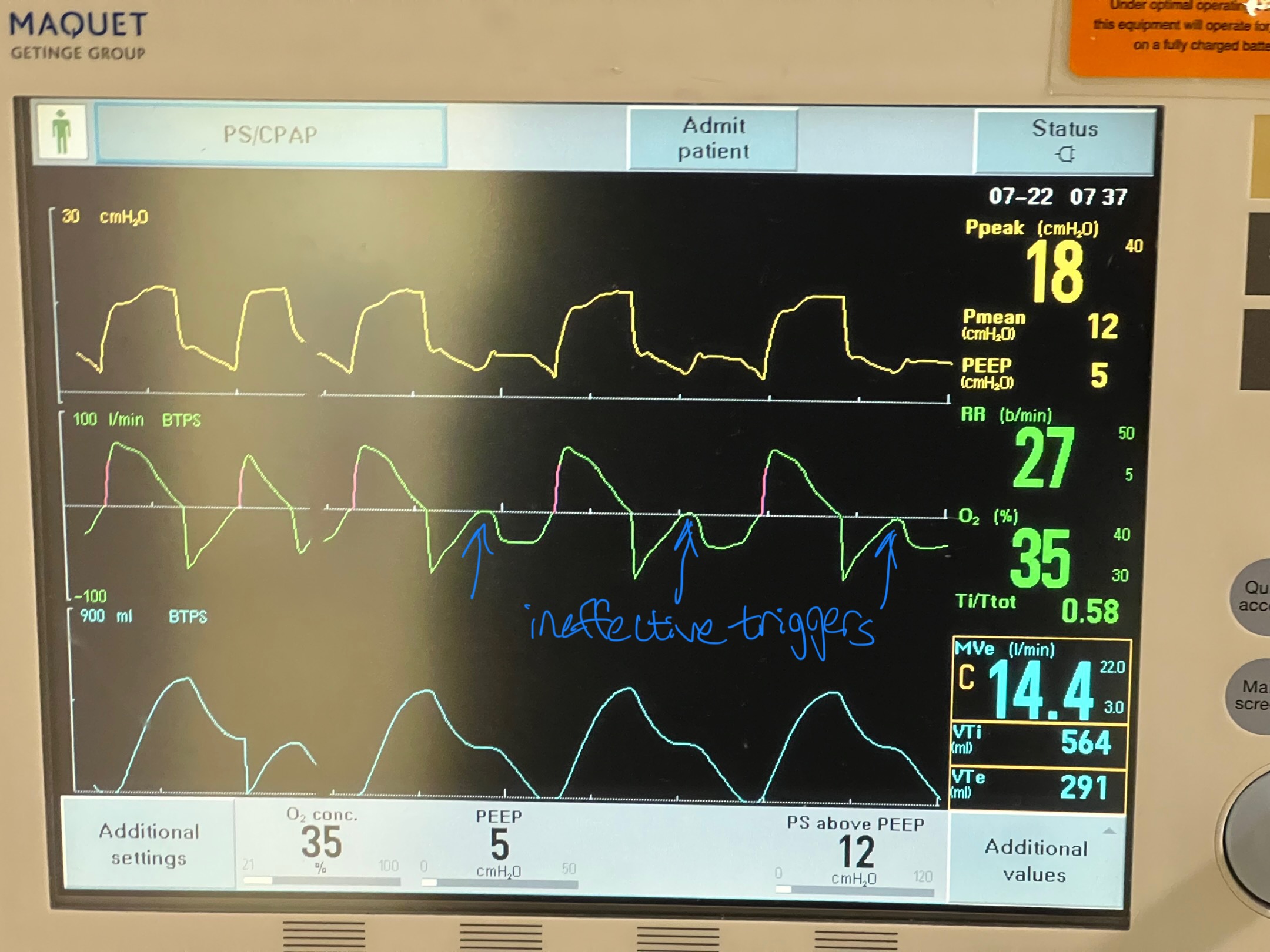
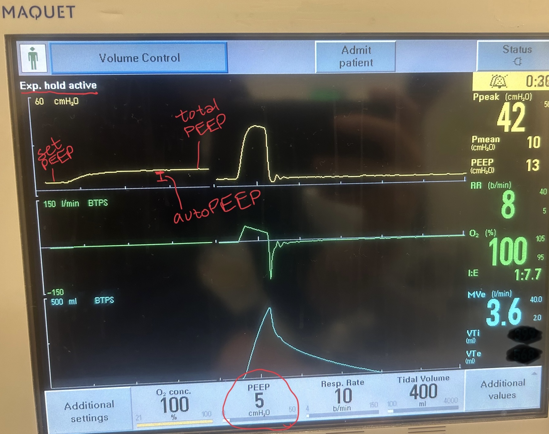
If a ventilated patient exhibits at least one of: persistent end expiratory flow, unequal inspiratory and expiratory flow-time areas, or ineffective breath triggers; autoPEEP must be evaluated by performing an end-expiratory hold.

If present, ventilator settings should be changed to maximize exhalation time.

In critically ill patients with obstructive lung disease, intubation and mechanical ventilation is often a last resort as it does not fix the underlying pathology of small airway disease. While many complications can arise, the most feared complication is autoPEEP.

What is autoPEEP?

AutoPEEP is excess air trapping in the lungs because the patient has insufficient time to fully exhale. Patients at highest risk include those with obstructive lung pathology due to their increased resistance (from bronchospasm) and sometimes increased compliance (such as in emphysema). 

However, it is possible for any patient to develop autoPEEP depending on the amount of time they have to exhale. As respiratory rate increases, the expiratory time decreases proportionally if inspiratory time is kept constant. Ultimately, autoPEEP can lead to rapidly increasing intrathoracic pressures causing decreased preload leading to hemodynamic instability and potentially cardiac arrest. These elevated pressures also place the patient at significant risk of barotrauma/volutrauma.

How do I find it?

There are several signs on the ventilator waveforms for autoPEEP. Some patients may only exhibit one of the following signs of autoPEEP. They are demonstrated in the attached pictures in various ventilator modes.

Image A. Persistent end expiratory flow on the flow-time curve (middle curve) - demonstrated by the expiratory limb of the flow curve not returning to zero (remains negative)
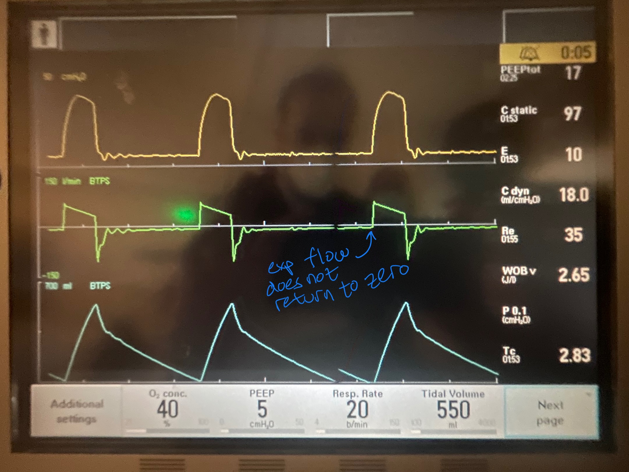
Image A

Image B. Unequal inspiratory and expiratory volumes on the flow-time curve (area of flow curve inspiratory limb does not equal area of flow curve expiratory limb)
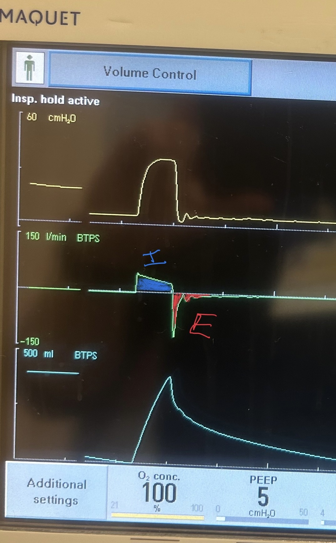
Image C. Ineffective triggering (seen on flow-time curve; patient has to perform more work to reach trigger threshold when autoPEEP is present; they are sometimes unable to trigger a breath)
If any of these are present, an end-expiratory hold maneuver should be performed.

Image D - End-expiratory hold maneuver (done if patient is passive on the ventilator) - the pressure-time curve will begin at ventilator set PEEP and reach total PEEP at the end of the maneuver. The difference between total PEEP and set PEEP is autoPEEP.
If autoPEEP is present, ventilator changes to allow for more exhalation time should be made. The most effective change is by decreasing the respiratory rate though small improvements can be made by changing the inspiratory time and tidal volume. Appropriate bronchodilator therapy, sedation, and treatment of underlying pathology is also critical in these patients.

For more information on autoPEEP, check out this post by Dr. John Greenwood discussing autoPEEP on MarylandCCProject with video demonstrations!

Show References

  • Finding the AutoPEEP. Dr. John Greenwood. MarylandCCProject. https://maryland.ccproject.com/2015/05/13/finding-the-auto-peep/
  • Image C courtesy of Dr. Caleb Chan, University of Maryland Medical Center
  • Image D courtesy of Dr. James Brady, University of Maryland Medical Center


Title: How low can the O2 go? Choosing a SaO2 goal in the mechanically ventilated patient

Category: Critical Care

Keywords: Critical Care, oxygen, ventilator, SaO2 (PubMed Search)

Posted: 9/2/2025 by Zach Wynne, MD

What is the ideal oxygen saturation goal for a mechanically ventilated patient? Literature over the past decade has led away from the perfect 100% oxygen saturation due to its association with worse patient outcomes across many disease states. It is theorized that excess oxygen leads to free radical production causing a lung injury pattern. However, there is no clear guidance for the ideal range of oxygen saturation goals, particularly in the mechanically ventilated patient, despite a meta-analysis and several recent trials.

UK-ROX Trial - JAMA - June 2025

Question: Does an oxygen saturation goal of 88-92% lead to a lower 90-day mortality compared to usual care?

Population: 16,500 mechanically ventilated adult patients in 97 ICU’s across the UK, excluded patients on ECMO

Intervention: Goal oxygen saturation of 88-92%, using the lowest possible FiO2

Control: Usual care, defined as oxygen supplementation at the discretion of the treating physician (no limits set to FiO2 or SaO2)

Outcomes:

  • Conservative and usual therapy groups were randomized 1:1 and had similar characteristics
  • 90 day all-cause mortality - 35.4% in conservative group vs. 34.9% in usual care group (p=0.28)
  • Time at 88-92% SaO2 -  62.6 hrs in conservative group vs. 27.2 hrs in usual care group (did not look at oxygen exposure peri-intubation) 
  • No difference in secondary outcomes - duration of ICU stay, days alive and free of organ support, duration of acute hospital stay, and others

Bottom Line:

Ideal oxygenation targets remain elusive. UK-ROX adds to the growing literature of oxygenation targets in mechanically ventilated patients but does not clearly show that lower oxygen saturation targets lead to improved ICU outcomes. In your emergency department ICU boarder, avoid a 100% oxygen saturation to prevent oxygen toxicity associated lung injury and consider an oxygen saturation goal of 90-96% (88-92% if history of COPD).

Show References

Martin DS, Gould DW, Shahid T, Doidge JC, Cowden A, Sadique Z, Camsooksai J, Charles WN, Davey M, Francis-Johnson A, Garrett RM, Grocott MPW, Jones J, Lampro L, Mackle DM, O'Driscoll BR, Richards-Belle A, Rostron AJ, Szakmány T, Warren A, Young PJ, Rowan KM, Harrison DA, Mouncey PR; UK-ROX Investigators. Conservative Oxygen Therapy in Mechanically Ventilated Critically Ill Adult Patients: The UK-ROX Randomized Clinical Trial. JAMA. 2025 Aug 5;334(5):398-408. doi: 10.1001/jama.2025.9663. PMID: 40501321; PMCID: PMC12163715.


Title: Another ICU boarder…What sedative should I use?

Category: Critical Care

Keywords: Sedation, propofol, dexmedetomidine, RASS (PubMed Search)

Posted: 7/8/2025 by Zach Wynne, MD

The presence of an endotracheal tube by itself does not mandate sedation and many patients require no sedatives while intubated in the ICU. However, patients intubated in the emergency department usually require initial sedation while still paralyzed from RSI. Sedation can also help facilitate procedures and imaging in critically ill patients during initial management. 

Current literature has found increased mortality and length of ventilator requirement in oversedated ED patients. The target sedation level for the general population remains a goal RASS (Richmond Agitation-Sedation Scale) of 0 to -1. Society of Critical Care Medicine guidelines from early 2025 recommend dexmedetomidine over propofol as the preferred sedative for light sedation and reducing delirium risk in intubated critically ill patients. A recent trial re-examined other clinical outcomes between these two common sedative agents.

A2B Randomized Clinical Trial - JAMA 2025

Clinical Question: Does alpha 2 adrenergic receptor agonist sedation (dexmedetomidine or clonidine) reduce duration of mechanical ventilation in mechanically ventilated patients compared to a propofol based regimen (usual care)?

Where: 41 UK ICU’s from December 2018 to October 2023

Who: 1438 adults receiving mechanical ventilation for less than 48 hours, receiving propofol and opioid for sedation/analgesia, expected to require mechanical ventilation for greater than 48 hours

Intervention: protocol driven sedation to reach a RASS score of -2 to +1 (either dexmedetomidine, clonidine, or propofol). Of note, propofol could be added to achieve deeper sedation goal if deemed necessary by care team.

Outcomes:

  • No significant difference in time to extubation between dexmedetomidine vs. propofol (HR of 1.09, p=0.2) OR clonidine vs. propofol (HR of 1.05, p=0.34)
  • Higher rates of agitation in the dexmedetomidine group (HR of 1.54, CI 1.21-1.97) and clonidine group (HR of 1.55, CI 1.22-1.97) compared to propofol group
  • Mortality at 180 days similar between all groups
  • Severe bradycardia seen more frequently in dexmedetomidine and clonidine groups compared to propofol group although unclear if ongoing propofol administration had any effect on these groups
  • Subgroup analysis showed a weak interaction with age as a continuous variable showing reduced benefit on time to extubation with dexmedetomidine vs. propofol at later decades of life (i.e. dexmedetomidine showing potential benefit at younger ages)

Bottom Line:

While either dexmedetomidine or propofol, with appropriate use of opiates for pain management, are appropriate agents in non-paralyzed mechanically-ventilated patients, propofol may be a more appropriate choice in patients with greater agitation while boarding in the emergency department. However, close attention is needed to avoid the overly deep analgosedation associated with increased mortality. Maintain a goal RASS of 0 to -1 with frequent re-evaluation of your ICU boarders.

Show References

Walsh TS, Parker RA, Aitken LM, McKenzie CA, Emerson L, Boyd J, Macdonald A, Beveridge G, Giddings A, Hope D, Irvine S, Tuck S, Lone NI, Kydonaki K, Norrie J, Brealey D, Antcliffe D, Reay M, Williams A, Bewley J, Creagh-Brown B, McAuley DF, Dark P, Wise MP, Gordon AC, Perkins GD, Reade MC, Blackwood B, MacLullich A, Glen R, Page VJ, Weir CJ; A2B Trial Investigators. Dexmedetomidine- or Clonidine-Based Sedation Compared With Propofol in Critically Ill Patients: The A2B Randomized Clinical Trial. JAMA. 2025 Jul 1;334(1):32-45. doi: 10.1001/jama.2025.7200. PMID: 40388916; PMCID: PMC12090071.

Lewis K, Balas MC, Stollings JL, et al. A focused update to the clinical practice guideline for the prevention and management of pain, anxiety, agitation/sedation, delirium, immobility, and sleep disruption in adult patients in the ICU. Crit Care Med. 2025 Mar 1;53(3):e711-e727.

Stephens RJ, Ablordeppey E, Drewry AM, Palmer C, Wessman BT, Mohr NM, Roberts BW, Liang SY, Kollef MH, Fuller BM. Analgosedation Practices and the Impact of Sedation Depth on Clinical Outcomes Among Patients Requiring Mechanical Ventilation in the ED: A Cohort Study. Chest. 2017 Nov;152(5):963-971. doi: 10.1016/j.chest.2017.05.041. Epub 2017 Jun 21. PMID: 28645462; PMCID: PMC5812748.


University of Maryland School of Medicine logo
  • Departments
  • Programs
  • Research Centers
  • Institutes
  • Graduate Education
  • CIBR Cores
  • Offices of the Dean
    • University of Maryland, Baltimore
    • University of Maryland Medical Center
    • Medical Alumni Association of the University of Maryland
    • Health Sciences and Human Services Library
University of Maryland School of Medicine
655 W. Baltimore Street
Baltimore, MD 21201
Contact Us

© University of Maryland School of Medicine

    • Webmaster
    • Web Accessibility
    • Site Index
    • UMB Hotline
    • Employment
    • Map