
University of Maryland School of Medicine logo
Emergency Medicine
All Departments
UMSOM Assistant
UMEM Educational Pearls
  • Home
  • Departments
  • Emergency Medicine
  • CME
  • UMEM Educational Pearls
  • CME
  • Courses and Conferences
  • Free Open Access Medical Education
  • UMEM Educational Pearls

Search

Title: Visual diagnosis-pelvis

Category: Trauma

Posted: 12/9/2024 by Robert Flint, MD (Updated: 12/9/2024)

Question

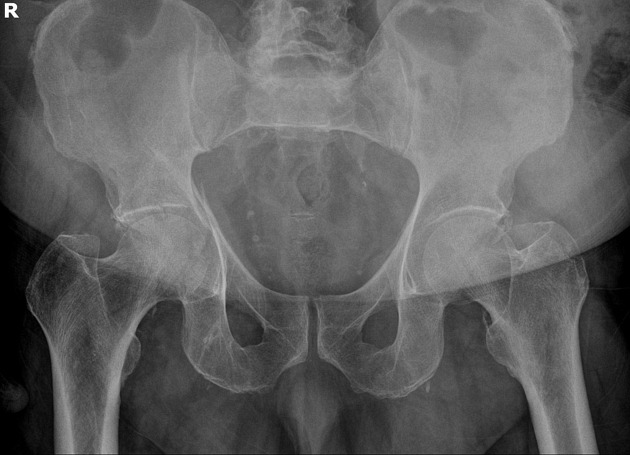
What is the diagnosis? Treatment? Other imaging indicated? 
Answer

Acetabulum fracture. 
Usually seen in high mechanism situations such as MVCs. Can be seen in elderly fall patients. 
Treatment is usually surgical (ORIF) unless non-displaced or if it involves less than 20% of posterior column. These can be managed with 6 weeks of minimal weight bearing. CT scan is almost universally used for better visualization and operative management. 
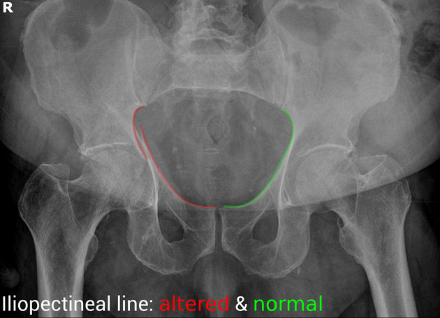
“Some landmarks of the hip are compared on the right and left sides of the pelvis:

  • iliopectineal line - disruption indicates anterior column fracture
  • ilioischial line - disruption indicates posterior column fracture
  • acetabular roof“          Radiopedia

References

https://radiopaedia.org/cases/acetabular-fracture-anterior-and-posterior-columns

https://www.orthobullets.com/trauma/1034/acetabular-fractures


University of Maryland School of Medicine logo
  • Departments
  • Programs
  • Research Centers
  • Institutes
  • Graduate Education
  • CIBR Cores
  • Offices of the Dean
    • University of Maryland, Baltimore
    • University of Maryland Medical Center
    • Medical Alumni Association of the University of Maryland
    • Health Sciences and Human Services Library
University of Maryland School of Medicine
655 W. Baltimore Street
Baltimore, MD 21201
Contact Us

© University of Maryland School of Medicine

    • Webmaster
    • Web Accessibility
    • Site Index
    • UMB Hotline
    • Employment
    • Map